. 2023 Apr 26;13(1):6857.
doi: 10.1038/s41598-023-33941-2.

Scalable continuous-flow electroporation platform enabling T cell transfection for cellular therapy manufacturing

Affiliations

Scalable continuous-flow electroporation platform enabling T cell transfection for cellular therapy manufacturing

Jacob A VanderBurgh et al. Sci Rep. .

Abstract

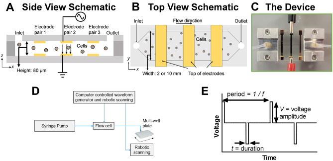
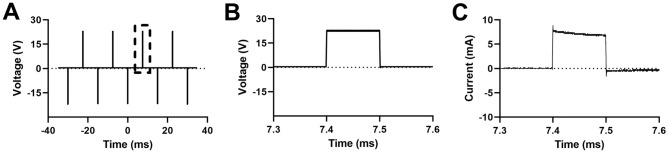
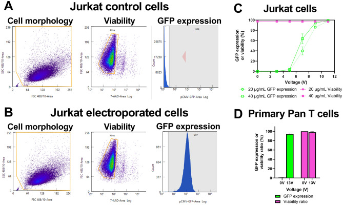
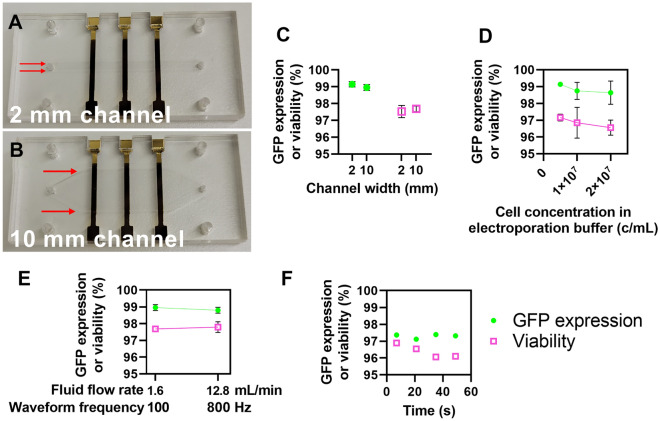
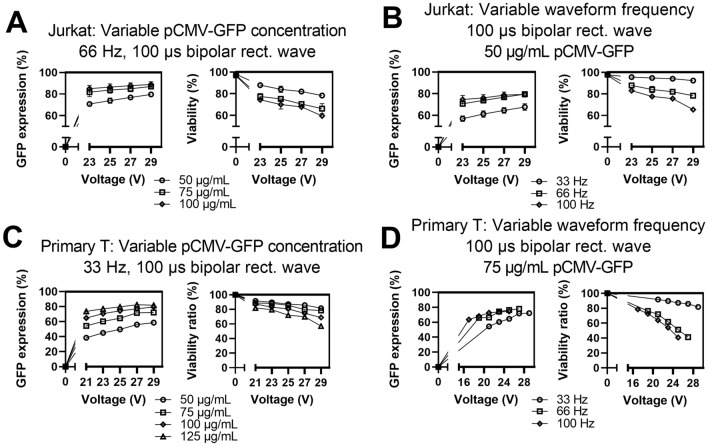
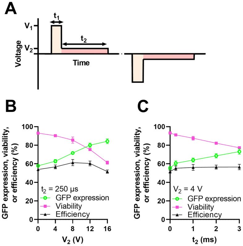
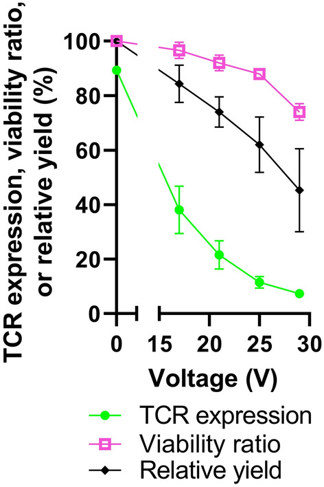
Viral vectors represent a bottleneck in the manufacturing of cellular therapies. Electroporation has emerged as an approach for non-viral transfection of primary cells, but standard cuvette-based approaches suffer from low throughput, difficult optimization, and incompatibility with large-scale cell manufacturing. Here, we present a novel electroporation platform capable of rapid and reproducible electroporation that can efficiently transfect small volumes of cells for research and process optimization and scale to volumes required for applications in cellular therapy. We demonstrate delivery of plasmid DNA and mRNA to primary human T cells with high efficiency and viability, such as > 95% transfection efficiency for mRNA delivery with < 2% loss of cell viability compared to control cells. We present methods for scaling delivery that achieve an experimental throughput of 256 million cells/min. Finally, we demonstrate a therapeutically relevant modification of primary T cells using CRISPR/Cas9 to knockdown T cell receptor (TCR) expression. This study displays the capabilities of our system to address unmet needs for efficient, non-viral engineering of T cells for cell manufacturing.


Conflict of interest statement

JAV, TNC, and HGC are listed as inventors on patent applications related to the technology presented and JAV, TNC, SLL, and HGC have a financial interest in CyteQuest.

Figures

Figure 1
Figure 1
Overview of CyteQuest electroporation platform. (A) Side and (B) top view schematic of the electroporation flow cell (not to scale). (C) Photograph of an experimental flow cell with attached manifolds, tubing, and three sets of independently addressable electrodes. (D) Block diagram depicting components that comprise the electroporation platform. (E) Plot depicting a bipolar rectangular waveform with frequency f, duration t, and voltage amplitude, V.
Figure 2
Figure 2
Time-averaged oscilloscope traces displaying the time varying voltage and current measured by the oscilloscope. (A) Multiple cycles of the voltage channel of a bipolar rectangular waveform with f = 66 Hz, V = 23 V, and t = 100 µs. The dotted box depicts a zoomed time region of the waveform with corresponding (B) voltage across the channel and (C) current through the channel. Time averaging: 64 traces.
Figure 3
Figure 3
Delivery of mRNA encoding GFP to Jurkat and primary T cells. (A) Representative flow cytometry plots from zero voltage-control or (B) electroporated Jurkat cells depicting cell morphology, viability, and GFP expression. (C) Impact of varying waveform voltage amplitude on delivery using 20 or 40 µg/mL mRNA to Jurkat cells (n = 3). (D) High efficiency delivery using 40 µg/mL mRNA for primary T cells from four healthy donors (n = 4). Data shown as mean ± standard deviation (C,D). Some error bars are too small to be visible (C).
Figure 4
Figure 4
Increasing cell processing throughput for clinical-scale volumes. (A) Photograph of a 2 mm and (B) 10 mm electroporation flow cell. Red arrows highlight the channel width. (C) Plot of GFP expression and viability values from Jurkat cells transfected with mRNA encoding GFP in either the 2- or 10-mm channels (n = 3). (D) Plot of GFP expression and viability values from Jurkat cells transfected with mRNA encoding GFP in the 2-mm channel at varying cell concentrations in the electroporation buffer (n = 3). (E) Plot of GFP expression and viability values from Jurkat cells transfected with mRNA encoding GFP in the 10-mm channel at varying flow rates and waveform frequencies (n = 3). (F) Plot of GFP expression and viability over time from an experiment that transfected ~ 240 million cells over 56 s (n = 1). Data shown as mean ± standard deviation (CE). Some error bars are too small to be visible (CE).
Figure 5
Figure 5
Results for various transfection parameters for delivering plasmid DNA encoding GFP to Jurkat and primary T cells. (A) Impact of varying plasmid concentration or (B) waveform frequency for delivering plasmid DNA to Jurkat cells. (C) Impact of varying plasmid concentration or (D) waveform frequency for delivering plasmid DNA to primary T cells. Data shown as mean ± standard deviation; n = 3 (A,B). Data shown as values from a representative donor; n = 1 (C,D). Some error bars are too small to be visible for Jurkat cells (A,B).
Figure 6
Figure 6
Delivery of an arbitrary electrical waveform. (A) Plot depicting a bipolar, dual voltage waveform characterized by a short-duration, high-amplitude segment (V1, t1) followed by a long-duration, low-amplitude segment (V2, t2) (B) Impact of varying V2 while t2 = 250 µs (n = 3). (C) Impact of varying t2 while V2 = 4 V (n = 3). In both (B) and (C), we fix f = 66 Hz, V1 = 21 V, and t1 = 75 µs. Data shown as mean ± standard deviation (B,C). Some error bars are too small to be visible (B,C).
Figure 7
Figure 7
Delivery of CRISPR/Cas9 RNPs targeting TRAC/TRBC. Plot of TCR expression, viability ratio, or relative yield as a function of applied voltage amplitude, measured 72-h after primary T cells were transfected with CRISPR/Cas9 RNPs targeting TRAC/TRBC (n = 3). Data shown as mean ± standard deviation. Some error bars are too small to be visible.

References

    1. Porter D, Levine B, Kalos M, Bagg A, June CH. Chimeric antigen receptor-modified T cells in chronic lymphoid leukemia. N. Engl. J. Med. 2011;365:725–733. doi: 10.1056/NEJMoa1103849. - DOI - PMC - PubMed
    1. Grupp S, et al. Chimeric antigen receptor-modified T cells for acute lymphoid leukemia. N. Engl. J. Med. 2013;368:1509–1518. doi: 10.1056/NEJMoa1215134. - DOI - PMC - PubMed
    1. Maus MV, Fraietta JA, Levine BL, Kalos M, Zhao Y, June CH. Adoptive immunotherapy for cancer or viruses. Annu. Rev. Immunol. 2014;32:189–225. doi: 10.1146/annurev-immunol-032713-120136. - DOI - PMC - PubMed
    1. Mount NM, Ward SJ, Kefalas P, Hyllner J, Mount NM. Cell-based therapy technology classifications and translational challenges. Philos. Trans. B. 2015;370:20150017. doi: 10.1098/rstb.2015.0017. - DOI - PMC - PubMed
    1. Buzhor E, et al. Cell-based therapy approaches: The hope for incurable diseases. Regen. Med. 2014;9(5):14–35. doi: 10.2217/rme.14.35. - DOI - PubMed

Publication types