. 2023 Apr 25;14(1):2192.
doi: 10.1038/s41467-023-37811-3.

Anti-cancer pro-inflammatory effects of an IgE antibody targeting the melanoma-associated antigen chondroitin sulfate proteoglycan 4

Affiliations

Anti-cancer pro-inflammatory effects of an IgE antibody targeting the melanoma-associated antigen chondroitin sulfate proteoglycan 4

Jitesh Chauhan et al. Nat Commun. .

Abstract

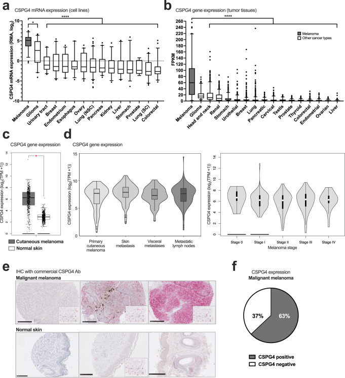
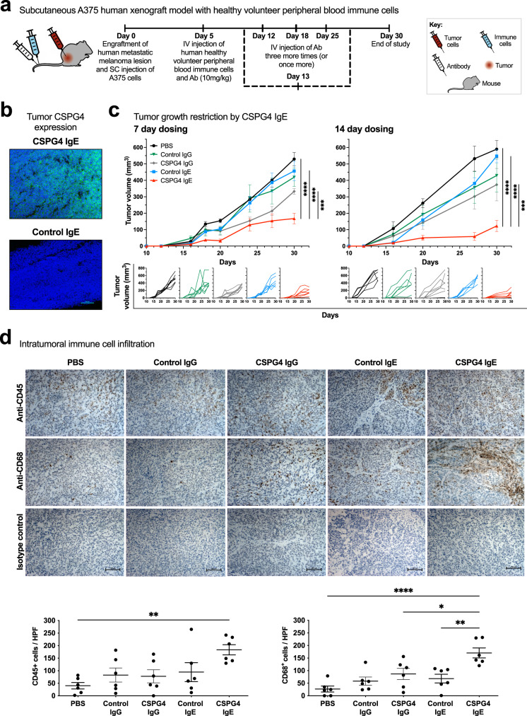
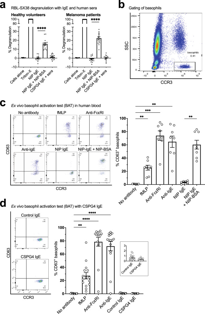
Outcomes for half of patients with melanoma remain poor despite standard-of-care checkpoint inhibitor therapies. The prevalence of the melanoma-associated antigen chondroitin sulfate proteoglycan 4 (CSPG4) expression is ~70%, therefore effective immunotherapies directed at CSPG4 could benefit many patients. Since IgE exerts potent immune-activating functions in tissues, we engineer a monoclonal IgE antibody with human constant domains recognizing CSPG4 to target melanoma. CSPG4 IgE binds to human melanomas including metastases, mediates tumoricidal antibody-dependent cellular cytotoxicity and stimulates human IgE Fc-receptor-expressing monocytes towards pro-inflammatory phenotypes. IgE demonstrates anti-tumor activity in human melanoma xenograft models engrafted with human effector cells and is associated with enhanced macrophage infiltration, enriched monocyte and macrophage gene signatures and pro-inflammatory signaling pathways in the tumor microenvironment. IgE prolongs the survival of patient-derived xenograft-bearing mice reconstituted with autologous immune cells. No ex vivo activation of basophils in patient blood is measured in the presence of CSPG4 IgE. Our findings support a promising IgE-based immunotherapy for melanoma.


Conflict of interest statement

J.S. and S.N.K. are founders and shareholders of Epsilogen Ltd. S.N.K., J.S., D.H.J., G.P., and H.J.B. declare patents on antibodies for cancer. J.C., M.G., J.L-A, H.S.S, and H.J.B have been employed through a fund provided by Epsilogen Ltd. P.K. has conducted advisory work for Janssen and is stockholder in Abbvie. J.D. has acted as a consultant for AstraZeneca, Jubilant, Theras, BridgeBio and Vividion, and has funded research agreements with BMS and Revolution Medicines. All other authors declare no competing interests.

Figures

Fig. 1
Fig. 1. CSPG4 expression in malignant melanoma and normal tissues.
a CSPG4 mRNA expression, derived from RNAseq data, across cell lines of different cancer cell types. n represents the number of cell lines (data from Cancer Cell Line Encyclopedia (CCLE), portals.broadinstitute.org/ccle, n = 56, n = 59, n = 26, n = 57, n = 28, n = 27, n = 48, n = 127, n = 41, n = 32, n = 26, n = 37, n = 8, n = 50, and n = 58, respectively) (p = 0.0156 and p ≤ 0.0001). b CSPG4 gene expression in tissues across cancer types (data and images from Human Protein Atlas, v20.proteinatlas.org, , n = 153, n = 499, n = 877, n = 354, n = 406, n = 1075, n = 994, n = 176, n = 291, n = 134, n = 494, n = 501, n = 597, n = 541, n = 373, and n = 365 samples, respectively per cancer type; p ≤ 0.0001). c Comparison of CPSG4 gene expression between cutaneous melanoma and normal skin tissues (from GEPIA) (n = 461 and n = 558, respectively). TPM = transcripts per million. d CPSG4 gene expression across primary cutaneous melanoma lesions, skin metastases, visceral metastases and metastatic lymph nodes (left; TCGA-SKCM data was obtained from xenabrowser.net, n = 103, n = 116, n = 36, n = 208, respectively), and across disease stages of melanoma (right; from GEPIA). e Representative immunohistochemical (IHC) images of malignant melanoma samples showing low, intermediate, and high CSPG4 expression (pink staining, left to right) respectively, and normal skin tissue (showing no/low CSPG4 expression). Samples were stained with a commercially sourced anti-human CSPG4 antibody and CSPG4 expression was detected by alkaline phosphatase (AP; pink) staining. Nuclei were stained with hematoxylin (blue). Scale bar = 250 μm. f Quantitative analyses of CSPG4 expression detected in human melanoma and non-malignant tissues by IHC: expression was detected in 63% of all melanoma tissues (n = 428). Boxes denote 25th to 75th percentile with median line. Whiskers mark the minima 5th percentile to the maxima 95th percentile. Data shown as mean ± SEM. Source data are provided as a Source Data file. Kruskal–Wallis (a, b, d left), One-way ANOVA (d right), Student’s t test (c): *p ≤ 0.05; ****p ≤ 0.0001.
Fig. 2
Fig. 2. Generation, biophysical characterization, and cancer specificity of CSPG4 IgE.
a Structure of CSPG4 IgE: CSPG4-specific variable domains (white), constant heavy chain domains (orange) and constant light chain domains (gray). b SDS-PAGE (non-reduced (left) and reduced (middle) conditions) and size-exclusion HPLC (right) analyses confirmed comparable size and purity of CSPG4 IgE in relation to a previously engineered anti-Folate Receptor alpha (FRα) IgE. c Flow cytometric analyses of CSPG4 IgE (black line) confirmed binding to CSPG4-expressing human melanoma cell lines (A2058, A375, and WM1366), but not to FRα- or Her2-expressing cancer cells (IGROV1 and SKBR3, respectively). Antibody Fc-binding to human FcεRI on RBL-SX38 rat basophilic leukemia cells was also demonstrated (representative data). d Representative immunohistochemical images of cutaneous melanoma and lymph node metastases specimens stained with the engineered CSPG4 IgE (detected by alkaline phosphatase (AP; pink)) staining; nuclei were stained with hematoxylin (blue). Scale bar = 1 mm. e Quantitation of CSPG4 expression detected by IHC using the engineered CSPG4 IgE: the clone detected CSPG4 low/intermediate expression in 50% of benign nevus samples (n = 18) and variable high to low expression levels in 72–73% of malignant specimens (n = 468; including further analysis of antibody binding to cutaneous lesions, lymph node metastases and distant metastases (n = 150, n = 75 and n = 77, respectively)) (right), and high to low CSPG4 expression in 58–93% of melanoma lesions across stages I–IV (left; n = 12, n = 102, n = 14 and n = 6, respectively). f CSPG4 IgE IHC staining indicated absent or low/intermediate expression of CSPG4 in non-malignant tissue specimens (n = 297). Source data are provided as a Source Data file.
Fig. 3
Fig. 3. Anti-tumor and Fc-mediated effector functions of CSPG4 IgE in vitro.
a Left: CSPG4 expression levels by human cancer cell lines and melanocytes (as control cells) normalized relative to the mean fluorescent intensity (MFI) of CSPG4 IgE binding to A375 melanoma cells. Inset: Analyses of CSPG4 mRNA expression data in cell lines extracted from the CCLE database (Cancer Cell Line Encyclopedia (CCLE), portals.broadinstitute.org/ccle) and cell-surface binding of CSPG4 IgE (r = 0.2680). Right: Representative histograms for the highest expressing A375 (blue) and A2058 (orange) cells (light gray, anti-IgE-FITC only; dark gray, CSPG4 IgE + anti-IgE-FITC). b Treatment of A375 melanoma cells with CSPG4 IgE resulted in moderate restrictions in cancer cell adhesion (n = 5), migration (n = 5) and invasion (n = 7) compared with an isotype IgE control (Control IgE) (p = 0.0263, p = 0.0022, and p = 0.0131, respectively). c CSPG4 IgE-mediated degranulation of FcεRI-expressing RBL-SX38 cells when cross-linked by polyclonal anti-IgE (left, n = 3) or with CSPG4-expressing cancer cells (A375, middle, n = 3; A2058, right, n = 5). dg Compared to cells alone and treatment with isotype control IgE, CSPG4 IgE-mediated significant levels of antibody-dependent cell-mediated cytotoxicity (ADCC; white bars; ADCP; gray bars) of CSPG4-expressing melanoma cell lines (A2058, orange; A375, blue). d ADCC/ADCP by healthy volunteer and melanoma patient-derived PBMCs (healthy volunteer: left, n = 17, p ≤ 0.0001; melanoma patients: right, n = 14, p ≤ 0.0001). e ADCC/ADCP by U937 monocytic cells (A375: left, n = 10, p = 0.0075 and p = 0.0038; A2058: right, n = 10, p = 0.0008 and p = 0.0004). f Left: Flow cytometric histograms show cell-surface detection of endogenous bound IgE (anti-IgE-FITC) and of CSPG4 IgE (CSPG4 IgE + anti-IgE-FITC) to primary human monocytes from two healthy volunteers; Right: ADCC/ADCP by healthy volunteer monocytes (A375: left, n = 8, p = 0.0004 and p = 0.0089; A2058: right, n = 4, p ≤ 0.0001). g ADCC/ADCP by patient-derived monocytes (A375: left, n = 9, p ≤ 0.0001; A2058: right, n = 3, p = 0.0010 and p = 0.0010). No phagocytosis (ADCP, gray bars) was triggered by CSPG4 IgE above controls. Data shown as mean ± SEM. Source data are provided as a Source Data file. Two-tailed Student’s t test (b), One-way ANOVA (ce right, f, g), Kruskal–Wallis test (e left): *p ≤ 0.05; **p ≤ 0.01; ***p ≤ 0.001; ****p ≤ 0.0001.
Fig. 4
Fig. 4. Cross-linking of CSPG4 IgE on the surface of human monocytes promotes secretion of pro-inflammatory cytokines and enhanced expression of co-stimulatory cell-surface markers.
a Serum analysis of total IgE levels in healthy volunteers (n = 38) and melanoma patients (n = 13). Flow cytometric analysis of % monocytes in total PMBCs (healthy volunteers, n = 25; melanoma patients, n = 44) and % of monocytes expressing FcɛRI (healthy volunteers, n = 25; melanoma patients, n = 46). Concentrations of cytokines and chemokines measured in the sera of sex-matched healthy volunteers (n = 13) and melanoma patients (n = 13) (p = 0.0387). b Quantitative PCR (qPCR) analysis of TNF expression following cross-linking of human IgE bound to human monocytic U937 cells (n = 4 independent experimental repeats). Anti-IgE vs. CSPG4 IgE + anti-IgE, p≤0.0001; Anti-IgE vs. NIP IgE + anti-IgE, p = 0.0039; NIP IgE vs. NIP IgE + anti-IgE, p = 0.0113; CSPG4 IgE vs. CSPG4 IgE + anti-IgE, p = 0.0014. c Cytokine and chemokine secretion in primary monocyte culture supernatants following cross-linking of IgE (n = 7 healthy volunteers; p = 0.0085, p = 0.0152, p = 0.0001, and p = 0.0156, respectively). d Flow cytometric analysis (MFI change) in the expression levels of cell-surface markers of healthy volunteer monocytes untreated or stimulated with CSPG4 IgE with and without cross-linking with anti-IgE (n = 7 healthy volunteers; CD80: p = 0.0004 and p = 0.0003, CD86: p = 0.0038, CD163: p = 0.0005, PD-L1: p = 0.0033 and p = 0.0021, CD40: p = 0.0227 and p = 0.0060, HLA-DR: p = 0.0046, CCR2: p-0.0015 and ≤0.0001). e Cytokine and chemokine secretion in supernatants from primary human monocyte and A2058 cancer cell co-cultures in the presence of CSPG4 IgE or control NIP IgE (n = 8 healthy volunteers; TNF: p = 0.0053 and p = 0.0121, CCL-2/MCP-1: p = 0.0103 and p = 0.0110, IL-10: p = 0.0093 and p = 0.0492, IL-6: p = 0.0081 and p = 0.0081). Data shown as mean ± SEM. Source data are provided as a Source Data file. Mann–Whitney (a left), Two-tailed Student’s t test (a right, c), One-way ANOVA (b, d, e): *p ≤ 0.05; **p ≤ 0.01; ***p ≤ 0.001; ****p ≤ 0.0001.
Fig. 5
Fig. 5. CSPG4 IgE treatment can restrict tumor growth and induce human immune cell infiltration in a subcutaneous A375 in vivo model engrafted with healthy volunteer immune cells.
a Design and dosing regimen for in vivo model. SC = subcutaneous, IV = intravenous, Ab = antibody. b Immunofluorescence images of CSPG4 expression (green) in established A375 melanoma xenografts grown subcutaneously in immunocompromised mice. Frozen tumor sections were labeled with CSPG4 IgE (top, green) or isotype control IgE antibody (bottom), followed by fluorescently conjugated anti-human IgE. DAPI (blue): nuclear staining; 10× magnification; scale bar = 150 μm. c CSPG4 IgE significantly inhibited the growth of subcutaneous A375 tumors in immunodeficient mice engrafted with human peripheral blood immune cells. Mice challenged with subcutaneous melanomas were treated every 7 days (left) or 14 days (right) with either vehicle alone (PBS, black), CSPG4 IgE (red), isotype control IgE (blue), CSPG4 IgG (gray), isotype control IgG (green) (7 day dosing: n = 7 mice per group; 14 day dosing: PBS, CSPG4 IgE and CSPG4 IgG; n = 7, MOv18 IgE and MOv18 IgG; n = 6 mice per group). Inset graphs below show tumor growth curves for individual animals. Full statistical analyses shown in Supplementary Tables 1, 2. d Compared to CSPG4 IgG and control-treated mice, immunohistochemical studies showed elevated levels of human CD45+ leukocytes and CD68+ macrophages observed in subcutaneous A375 tumors excised from animals treated with CSPG4 IgE; top: representative images, ×10 magnification, scale bar = 100 μm; bottom: number of positive cell infiltrates per animal (n = 6; CD45+: p = 0.0072, CD68+: p ≤ 0.0001, p = 0.0201, p = 0.0036) (each value was derived from 3 independent images per high-power field (HPF)). Data shown as mean ± SEM. Source data are provided as a Source Data file. Two-way ANOVA (c), One-way ANOVA (d): *p ≤ 0.05; **p ≤ 0.01; ***p ≤ 0.001; ****p ≤ 0.0001.
Fig. 6
Fig. 6. Monocyte and macrophage signatures, and activation of Fcɛ receptor and pro-inflammatory immune pathways, with CSPG4 IgE treatment in vivo.
Gene expression and enriched pathways were studied in A375 subcutaneous tumors from mice treated intravenously with CSPG4 IgE (n = 4) or PBS (n = 5). a Significantly differential expression of monocyte and macrophage gene signatures (signatures annotated as per Li et al.). b Differentially expressed genes were identified using the package limma, ranked according to fold change and calculated enrichment of gene sets were evaluated within Reactome. Selected example pathways are denoted with arrows: FcɛRI (black), TNF receptors (orange), Interleukin 1 (green), Interleukin 12 (blue), Interferon (red), Antigen presentation (purple), MHC class I/II presentation (cyan). c Differentially expressed (FDR corrected) genes are shown for each selected pathway (FcɛRI; n = 37, n = 49; TNF: n = 39, n = 13; Interleukin 1: n = 44; Interleukin 12: n = 17; Interferon: n = 67; Antigen presentation: n = 29, n = 44, n = 50, n = 45; and MHC class I/II presentation: n = 42, n = 125, differentially expressed genes for each example pathway, respectively). Source data are provided as a Source Data file. Full statistical analyses shown in Supplementary Table 3. *p ≤ 0.05.
Fig. 7
Fig. 7. Efficacy of CSPG4 IgE in A375 and PDX tumor models.
a Upper left: Design and dosing regimen for in vivo model. IV = intravenous, Ab = antibody. Upper right: In an A375 human melanoma model of lung metastases, engrafted with healthy volunteer peripheral blood immune cells, the number of metastases per lung (n = 8; p = 0.0107), and % occupancy by tumor metastases per lung (n = 8) were reduced in CSPG4 IgE-treated animals, compared to those treated with a non-specific isotype control IgE (Control IgE). Lower: Representative images of Indian ink-stained lungs showing tumor metastases in white. b Upper: Design and dosing regimen for in vivo model. SC = subcutaneous. Lower left: In an A375 subcutaneous model engrafted with melanoma patient-derived PBMCs, mice treated with CSPG4 IgE had significantly lower tumor weights at the end of the study, compared to those treated with Control IgE (n = 11, n = 12, n = 10) (p = 0.0243 and p = 0.0408). Lower right: In the same model, tumor volume was significantly lower in animals treated with CSPG4 IgE, compared to Control IgE. Inset graphs to the right show tumor growth curves for individual animals (PBS and Control IgE; n = 8, CSPG4 IgE; n = 10). Full statistical analyses shown in Supplementary Table 4 (growth curve). c Upper: Design and dosing regimen for in vivo model. PBLs = peripheral blood lymphocytes, PDX = patient-derived xenograft. Lower: In mice transplanted with patient-derived xenografts from two patients with stage III and IV melanoma alongside intravenous autologous patient PBLs, survival was significantly greater with CSPG4 IgE treatment compared to vehicle control (n = 8 and n = 9, respectively) (p = 0.0425). Data presented as mean ± SEM and Kaplan–Meier survival curves. Source data are provided as a Source Data file. Two-tailed unpaired Student’s t test (a, left), Mann–Whitney (a, right), One-way ANOVA (b, left), Two-way ANOVA (b, right), Log-rank Mantel–Cox test (c): *p ≤ 0.05; **p ≤ 0.01; ***p ≤ 0.001.
Fig. 8
Fig. 8. CSPG4 IgE does not mediate RBL-SX38 cell degranulation in the presence of human sera from patients or healthy participants and does not trigger basophil activation in cancer patient blood ex vivo.
a In the absence of cancer cells, FcεRI-expressing RBL-SX38 cells sensitized with CSPG4 IgE did not degranulate when incubated with sera from healthy volunteers (left, n = 16) or from melanoma patients (right, n = 31) (p≤0.0001). bd Basophil activation test (BAT) was performed to assess the potential risk of hypersensitivity to CSPG4 IgE treatment in human blood samples ex vivo. b Gating strategy to identify CCR3highSSClow basophils in unfractionated whole blood samples. c Incubation of whole blood from cancer patients with positive control stimuli (fMLP, anti-FcεRI and anti-IgE), or NIP IgE and its polyclonal antigen NIP-BSA, triggered basophil activation as measured by increased CD63 cell-surface expression (representative plots, left; and summary of data from n = 8 independent experiments, right) (p = 0.0018, p = 0.0003, p = 0.0030, p = 0.0028). d CSPG4 IgE and isotype control IgE did not trigger human basophil activation in whole blood samples from melanoma patients (Left: representative plots; Right: n = 15 patient samples) (p ≤ 0.0001, p ≤ 0.0001, p = 0.0016). Inset graph shows CSPG4 IgE and Control IgE on a smaller axis scale. Data shown as mean ± SEM. Source data are provided as a Source Data file. One-way ANOVA (a, c, d): **p ≤ 0.01; ***p ≤ 0.001, ****p ≤ 0.0001.
Fig. 9
Fig. 9. Efficacy and mechanism of action of an IgE antibody specific for the tumor-associated antigen CSPG4 to target melanoma support the development of IgE therapies for CSPG4-expressing tumors.
CSPG4 IgE bound to a high proportion of melanomas, including metastases. CSPG4 IgE-mediated antibody-dependent cellular cytotoxicity (ADCC) of CSPG4-expressing melanoma cells by immune effector cells from healthy volunteers or patients with melanoma and stimulated human FcɛRI-expressing effector monocytes towards pro-inflammatory states. CSPG4 IgE restricted melanoma growth in patient-relevant in vivo models using human-derived immune cells and was associated with macrophage infiltration into tumors and activation of pro-inflammatory pathways. The antibody also prolonged the survival of mice bearing patient-derived xenografts (PDX) reconstituted with autologous immune cells from the same patient. Ex vivo basophil activation test (BAT) was used to predict that CSPG4 IgE may not induce type I hypersensitivity in melanoma patients. Created with BioRender.com.

References

    1. Ilieva KM, et al. Chondroitin sulfate proteoglycan 4 and its potential as an antibody immunotherapy target across different tumor types. Front. Immunol. 2017;8:1911. doi: 10.3389/fimmu.2017.01911. - DOI - PMC - PubMed
    1. Kozanoglu I, et al. Human bone marrow mesenchymal cells express NG2: possible increase in discriminative ability of flow cytometry during mesenchymal stromal cell identification. Cytotherapy. 2009;11:527–533. doi: 10.1080/14653240902923153. - DOI - PubMed
    1. Rivera Z, et al. CSPG4 as a target of antibody-based immunotherapy for malignant mesothelioma. Clin. Cancer Res. 2012;18:5352–5363. doi: 10.1158/1078-0432.CCR-12-0628. - DOI - PMC - PubMed
    1. Williams IP, et al. In vivo safety profile of a CSPG4-directed IgE antibody in an immunocompetent rat model. MAbs. 2020;12:1685349. doi: 10.1080/19420862.2019.1685349. - DOI - PMC - PubMed
    1. Hoffmann, R. M. et al. A novel antibody-drug conjugate (ADC) delivering a dna mono-alkylating payload to chondroitin sulfate proteoglycan (CSPG4)-expressing melanoma. Cancers (Basel)12, 10.3390/cancers12041029 (2020). - PMC - PubMed

Publication types