Review
. 2023 Mar 29;30(4):3859-3870.
doi: 10.3390/curroncol30040292.

Current Concepts in the Resection of Bone Tumors Using a Patient-Specific Three-Dimensional Printed Cutting Guide

Affiliations
Review

Current Concepts in the Resection of Bone Tumors Using a Patient-Specific Three-Dimensional Printed Cutting Guide

Hisaki Aiba et al. Curr Oncol. .

Abstract

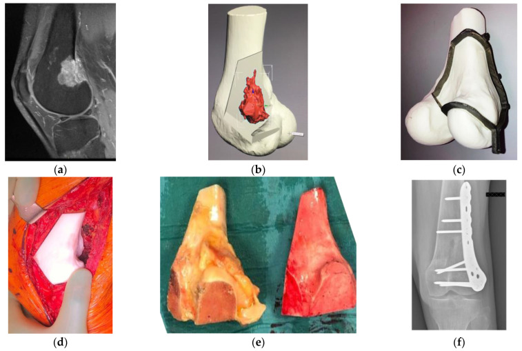
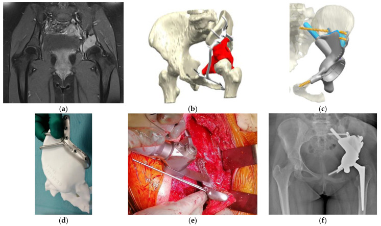
Orthopedic oncology has begun to use three-dimensional-printing technology, which is expected to improve the accuracy of osteotomies, ensure a safe margin, and facilitate precise surgery. However, several difficulties should be considered. Cadaver and clinical studies have reported more accurate osteotomies for bone-tumor resection using patient-specific cutting guides, especially in challenging areas such as the sacrum and pelvis, compared to manual osteotomies. Patient-specific cutting guides can help surgeons achieve resection with negative margins and reduce blood loss and operating time. Furthermore, this patient-specific cutting guide could be combined with more precise reconstruction using patient-specific implants or massive bone allografts. This review provides an overview of the basic technologies used in the production of patient-specific cutting guides and discusses their current status, advantages, and limitations. Moreover, we summarize cadaveric and clinical studies on the use of these guides in orthopedic oncology.

Keywords: bone tumor; limb-sparing surgery; orthopedic oncology; patient-specific implant; patients-specific guide; pelvic tumor; three-dimensional printed guide; tumor of the sacrum.


Conflict of interest statement

The authors declare no conflict of interest.

Figures

Figure 1
Figure 1
Osteosarcoma in the distal femur of a 56-year-old man. (a) High-intensity signal was seen in the sagittal MRI image (T1-fat suppression with contrast) at the posterior to the lateral side of the distal femur. (b) Resection planning in the computer-aided design phase. The location of the tumor was highlighted in red via an integrated image of CT and MRI and resection planes were determined with a discussion between surgeons and engineers. (c) Surgical planning with a PSG and an artificial bone. To preserve the surface of the knee joint and bone stock, hemi-cortical resection with the PSG, followed by reconstruction with a massive bone allograft was planned. The fitting of the PSG to the bone was confirmed. (d) Intraoperatively, after the resection with the PSG, the compatibility of the bone defect with the allograft, which was subsequently resected using the PSG, was confirmed by the artificial bone spacer. (e) Resected specimen (left) and resected massive bone allograft (right) via the PSG. We confirmed the identical fitting of a massive bone allograft to the large defect after the tumor resection. (f) Postoperative radiograph after the resection of the tumor and reconstruction with massive bone allograft. CT, computed tomography; PSG, patient-specific cutting guide; MRI, magnetic resonance imaging.
Figure 2
Figure 2
Osteosarcoma in the pelvis in a 14-year-old girl. (a) MRI image (T1-fat suppression with contrast) shows an osteolytic lesion in the left acetabulum with a high-intensity signal. (b) Preoperative planning for the P2–P3 resection of the pelvis using a PSG and reconstruction using a PSI. The tumor’s location was highlighted in red. The PSG was designed to have a sufficient margin with hooks for better attachment of the pelvic bone. The PSG was segmented for detachment into small parts, enabling a step-by-step surgical procedure. (c) In the computer-aided-design phase, the insertion of the PSI after the resection of the tumor and the points of fixation of the PSI were simulated. The actual cutting points were confirmed with a 3D-printed bone model. The points of fixation of cancellous screws and stems were also determined, and the appropriate length of drilling or the sizes of planned screws or stems were calculated. (d) The actual cutting points were preoperatively simulated with an artificial, 3D-printed bone model. (e) An intraoperative image of the resection along with the PSG. This procedure was supported by Waldemar Link GmbH & Co. KG (Norderstedt, Germany). (f) Postoperative radiograph after the resection of the tumor and reconstruction with a PSI. PSI, patient-specific implant; PSG, patient-specific cutting guide; MRI, magnetic resonance imaging.

References

    1. Gkagkalis G., Moerenhout K., Rüdiger H.A., Müller D.A., Letovanec I., Cherix S. Pelvic chondrosarcoma treated by en bloc resection with patient-specific osteotomy guides and reimplantation of the extracorporeally irradiated bone as an osseocartilaginous structural orthotopic autograft: A report of two cases with description of the surgical technique. Case. Rep. Orthop. 2021;2021:5512143. - PMC - PubMed
    1. Angelini A., Trovarelli G., Berizzi A., Pala E., Breda A., Ruggieri P. Three-dimension-printed custom-made prosthetic reconstructions: From revision surgery to oncologic reconstructions. Int. Orthop. 2019;43:123–132. doi: 10.1007/s00264-018-4232-0. - DOI - PubMed
    1. Vaishya R., Patralekh M.K., Vaish A., Agarwal A.K., Vijay V. Publication trends and knowledge mapping in 3D printing in orthopaedics. J. Clin. Orthop. Trauma. 2018;9:194–201. doi: 10.1016/j.jcot.2018.07.006. - DOI - PMC - PubMed
    1. Meng M., Wang J., Sun T., Zhang W., Zhang J., Shu L., Li Z. Clinical applications and prospects of 3D printing guide templates in orthopaedics. J. Orthop. Translat. 2022;34:22–41. doi: 10.1016/j.jot.2022.03.001. - DOI - PMC - PubMed
    1. Cartiaux O., Docquier P.L., Paul L., Francq B.G., Cornu O.H., Delloye C., Raucent B., Dehez B., Banse X. Surgical inaccuracy of tumor resection and reconstruction within the pelvis: An experimental study. Acta Orthop. 2008;79:695–702. doi: 10.1080/17453670810016731. - DOI - PubMed