. 2023 Apr 25;14(4):293.
doi: 10.1038/s41419-023-05802-3.

The long non-coding RNA keratin-7 antisense acts as a new tumor suppressor to inhibit tumorigenesis and enhance apoptosis in lung and breast cancers

Affiliations

The long non-coding RNA keratin-7 antisense acts as a new tumor suppressor to inhibit tumorigenesis and enhance apoptosis in lung and breast cancers

Zhe Zhao et al. Cell Death Dis. .

Abstract

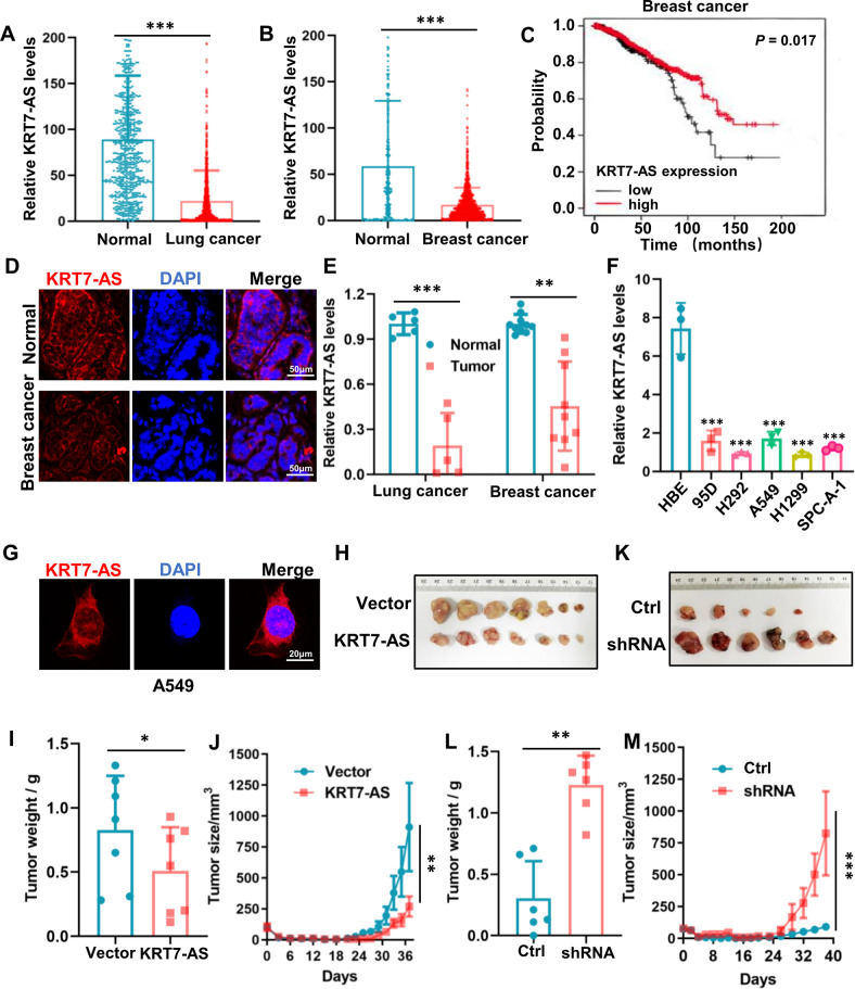
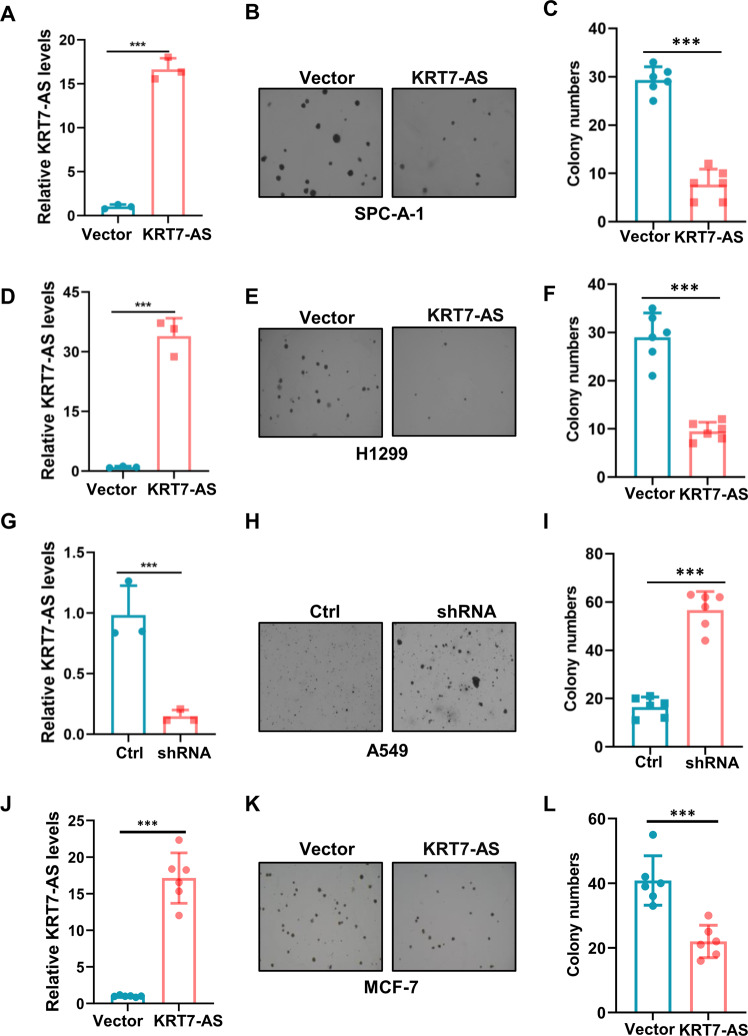
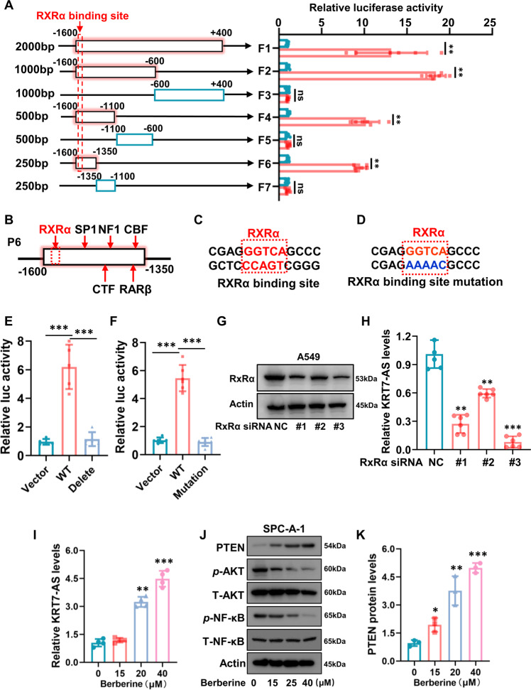
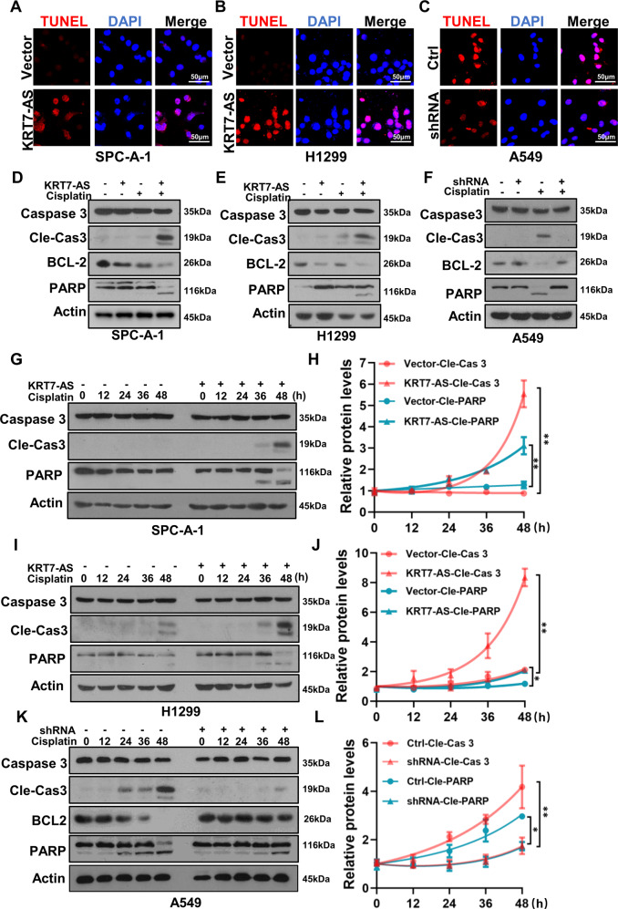
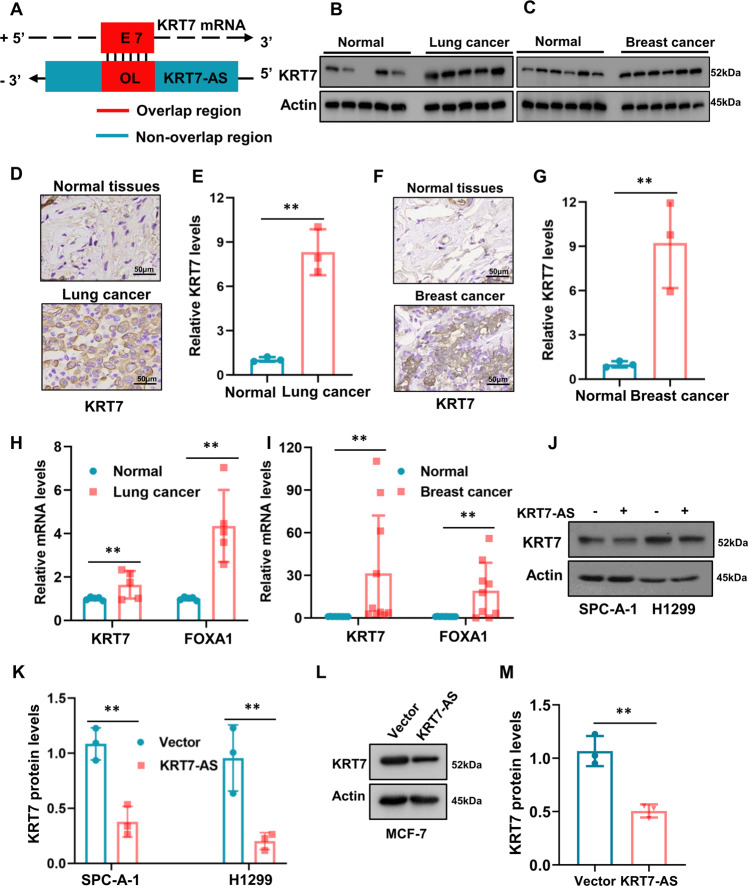
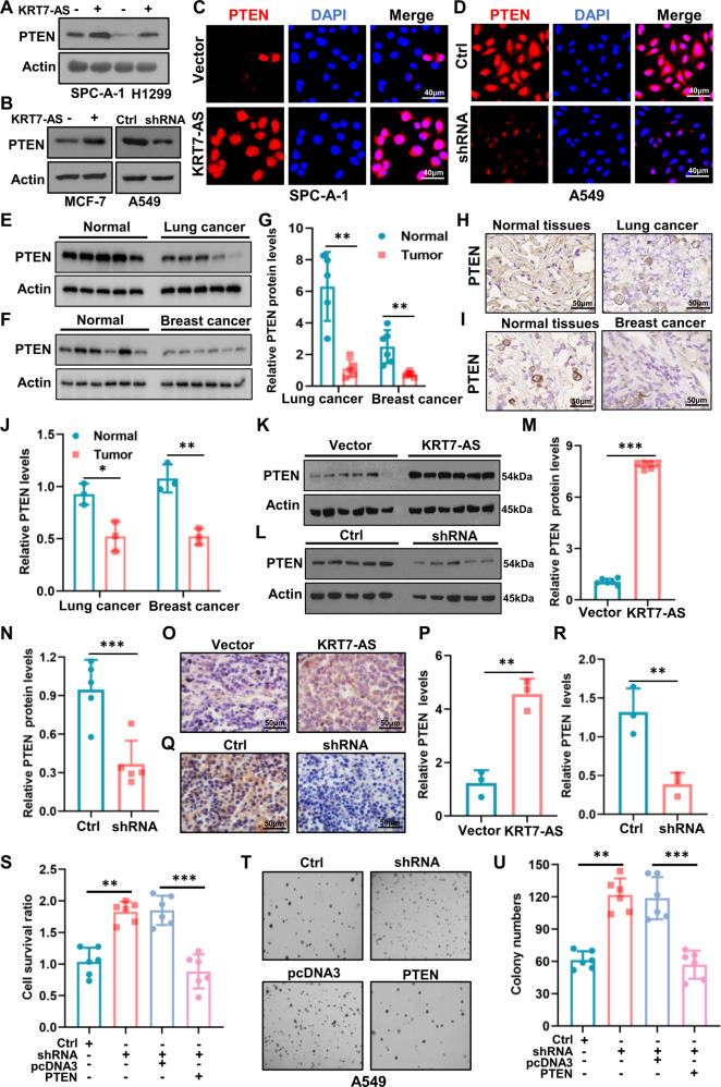
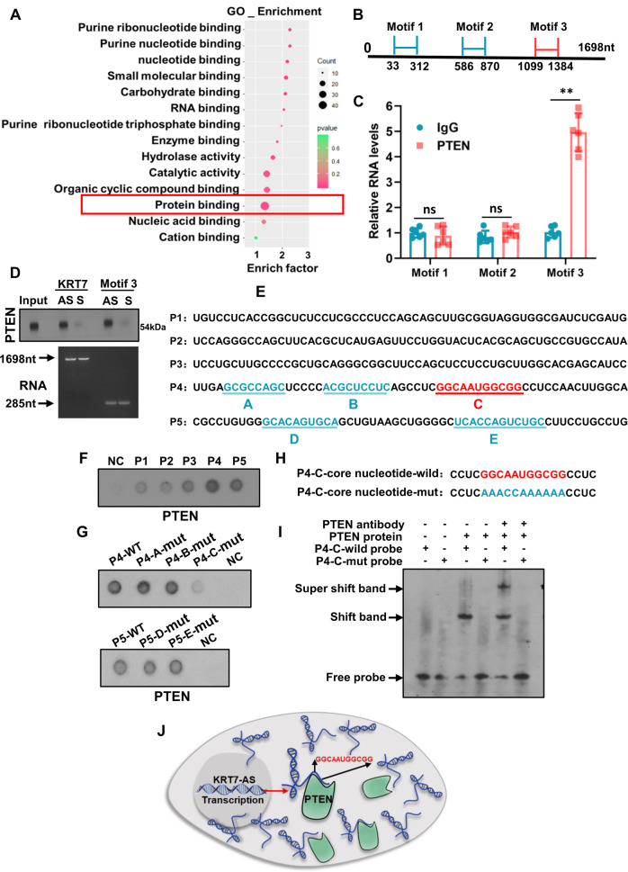
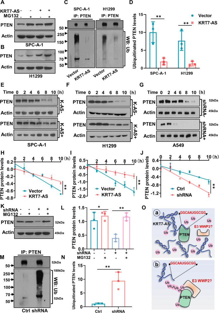
Expression of the long non-coding RNA (lncRNA) keratin-7 antisense (KRT7-AS) is downregulated in various types of cancer; however, the impact of KRT7-AS deficiency on tumorigenesis and apoptosis is enigmatic. We aim to explore the influence of KRT7-AS in carcinogenesis and apoptosis. We found that KRT7-AS was deficient in breast and lung cancers, and low levels of KRT7-AS were a poor prognostic factor in breast cancer. Cellular studies showed that silencing of KRT7-AS in lung cancer cells increased oncogenic Keratin-7 levels and enhanced tumorigenesis, but diminished cancer apoptosis of the cancer cells; by contrast, overexpression of KRT7-AS inhibited lung cancer cell tumorigenesis. Additionally, KRT7-AS sensitized cancer cells to the anti-cancer drug cisplatin, consequently enhancing cancer cell apoptosis. In vivo, KRT7-AS overexpression significantly suppressed tumor growth in xenograft mice, while silencing of KRT7-AS promoted tumor growth. Mechanistically, KRT7-AS reduced the levels of oncogenic Keratin-7 and significantly elevated amounts of the key tumor suppressor PTEN in cancer cells through directly binding to PTEN protein via its core nucleic acid motif GGCAAUGGCGG. This inhibited the ubiquitination-proteasomal degradation of PTEN protein, therefore elevating PTEN levels in cancer cells. We also found that KRT7-AS gene transcription was driven by the transcription factor RXRα; intriguingly, the small molecule berberine enhanced KRT7-AS expression, reduced tumorigenesis, and promoted apoptosis of cancer cells. Collectively, KRT7-AS functions as a new tumor suppressor and an apoptosis enhancer in lung and breast cancers, and we unraveled that the RXRα-KRT7-AS-PTEN signaling axis controls carcinogenesis and apoptosis. Our findings highlight a tumor suppressive role of endogenous KRT7-AS in cancers and an important effect the RXRα-KRT7-AS-PTEN axis on control of cancer cell tumorigenesis and apoptosis, and offer a new platform for developing novel therapeutics against cancers.


Conflict of interest statement

The authors declare no competing interest.

Figures

Fig. 1
Fig. 1. Expression and tumor-suppressive function of the lncRNA KRT7-AS in lung and breast cancers.
The levels of KRT7-AS in lung cancer (A), breast cancer (B), and the adjacent normal tissues were analyzed by integrative analysis using TCGA database. The influence of KRT7-AS levels on the survival of breast cancer patients was also analyzed using TCGA database (C). The expression of KRT7-AS (red) in breast cancer and adjacent normal tissues was examined by FISH (D), the nuclei were stained with DAPI (blue). The expression levels of KRT7-AS in paired human breast cancer tissues and lung cancer tissues (E), and in lung cancer cell lines (95D, NCI-H292, A549, H1299, SPC-A-1), and in the immortalized normal human bronchial epithelial cell line HBE were analyzed by real time qPCR (F). The sub-cellular localization of KRT7-AS (red) was examined by FISH (G), the nuclei were stained with DAPI (blue). The inhibitory effect of KRT7-AS on tumorigenesis was accessed in xenograft mice (HJ, n = 7). Additionally, the tumor size (J) and weight (I) of xenograft mice were measured and statistically analyzed, and silencing of KRT7-AS strongly enhanced tumor growth in xenograft mice (KM, n = 6). The tumor volume was also quantified using GraphPad (M). Above data are shown as mean ± SD of three independent replicates. *P < 0.05, **P < 0.01, ***P < 0.001.
Fig. 2
Fig. 2. KRT7-AS inhibits lung and breast cancer cell colony formation ability in vitro.
KRT7-AS expression levels in lung cancer SPC-A-1 cells were detected by real time qPCR (A). Colonies of KRT7-AS-overexpressed SPC-A-1 cells were imaged and quantified (B, C). The expression levels of KRT7-AS in H1299 were measured by real time qPCR (D). The colony formation ability of H1299 cells was accessed (E, F). The KRT7-AS expression levels in lung cancer A549 cells were detected by real time qPCR (G). Colonies of KRT7-AS-silenced A549 cells were imaged and quantified (H, I). The colony formation ability of breast cancer MCF-7 cells overexpressed KRT7-AS or vector control was accessed (JL). Data are shown as mean ± SD of three independent replicates. *P < 0.05, **P < 0.01, ***P < 0.001.
Fig. 3
Fig. 3. RXRα drives the transcription of KRT7-AS and the RXRα agonist berberine promotes KRT7-AS expression.
The KRT7-AS genomic DNA fragment (−1600 to +400) from the transcription initial site was separated into seven fragments (A, left panel). Luciferase assay showed that the fragments which contain the transcription factor RXRα-binding site had promoter activity (A, right panel), and indicated that the promoter DNA from −1600 to −1350 from the transcription initial site was the shortest fragment with strong promoter activity (A, B). LASAGNA method indicated that the DNA fragment from -1600 to -1350 has six putative transcription factor binding sites (B), The core nucleic acids GGTCA in wild type of RXRα-binding site in the sub-fragment P6 (C) was mutated to AAAAC (D). Luciferase assay showed that both deletion and mutation the core nucleic acids CGAGGGTCAGCCC in the RXRα-binding site lost the promoter activity (E, F). RXRα levels were detected in RXRα-silenced A549 cells (G). KRT7-AS levels were detected in RXRα-silenced A549 cells (H). The RXRα agonist berberine significantly increased KRT7-AS transcription levels (I), and concurrently raised PTEN protein levels (J, K). Data are shown as mean ± SD of three independent replicates. *P < 0.05, **P < 0.01, ***P < 0.001.
Fig. 4
Fig. 4. Upregulation of KRT7-AS enhances cisplatin-induced DNA damage and increases lung cancer cell sensibility to the drug.
Cell Titer-Glo luminescent (CTG) assay showed that KRT7-AS overexpression enhanced the sensibility of SPC-A-1 (A) and H1299 (C) lung cancer cells to the anti-cancer drug cisplatin. Flow cytometry analysis showed that KRT7-AS overexpression significantly enhanced cisplatin-induced apoptosis of SPC-A-1 (B, E) and H1299 (D, E) lung cancer cells. CTG and flow cytometry analysis showed that in KRT7-AS- silenced A549 lung cancer cells, reduction of KRT7-AS levels diminished cisplatin-induced apoptosis (F, G, H). The effect of KRT7-AS on the DNA damage marker γ-H2AX in KRT7-AS-overexpressed SPC-A-1 and KRT7-AS-silenced A549 cells in the presence of cisplatin was accessed by IF staining (I, J) and Western blotting (K, L). Time course study showed that KRT7-AS-induced elevation of γ-H2AX levels in SPC-A-1 (M, P), H1299 (N, Q), and A549 (O, R) lung cancer cells was in a time-dependent manner (M-R). Data are shown as mean ± SD of three independent replicates. **P < 0.01, ***P < 0.001.
Fig. 5
Fig. 5. KRT7-AS enhances cisplatin-induced apoptosis and activates apoptotic signaling pathways in cancer cells.
TUNEL assay showed that KRT7-AS enhanced cisplatin-induced apoptosis in SPC-A-1 (A) and H1299 (B) lung cancer cells, whereas, silencing of KRT7-AS in A549 (C) lung cancer cells reduced apoptosis. The levels of Caspase 3, cleaved-Caspase 3, BCL-2, and PARP in KRT7-AS-overexpressed SPC-A-1 (D) and H1299 (E) lung cancer cells, or KRT7-AS-silenced A549 (F) lung cancer cells were measured by Western blotting. Time course study showed that KRT7-AS elevated the levels of cleaved-Caspase3 and cleaved PARP in cisplatin concentration- and time-dependent manner (GL). Data are shown as mean ± SD of three independent replicates. *P < 0.05, **P < 0.01.
Fig. 6
Fig. 6. KRT7-AS increases the levels of oncogenic KRT7 in lung cancer and breast cancer.
The relationship between KRT7 and KRT7-AS in genomic DNA is depictured in (A). KRT7 protein levels in lung cancer (B) and breast cancer (C) were detected by Western blotting. The levels of KRT7 protein in the lung cancer (D, E) and breast cancer (F, G) tissues and in the paired normal tissues were examined by IHC. Expression levels of KRT7 downstream FOXA1 gene in lung cancer (H) and breast cancer (I) were detected using real time qPCR. Western blotting showed that KRT7 protein was decreased in KRT7-AS-overexpressed SPC-A-1 and H1299 lung cancer cells (J, K). In KRT7-ASoverexpressed MCF-7 cells, KRT7 protein levels were reduced (L, M). Data are shown as mean ± SD of three independent replicates. **P < 0.01.
Fig. 7
Fig. 7. KRT7-AS elevates amounts of the key tumor suppressor PTEN in cancer cells and inhibits tumorigenesis.
Western blotting showed that overexpression of KRT7-AS elevated PTEN protein levels in SPC-A-1 and H1299 lung cancer cells (A), and in MCF-7 breast cancer cells (B), whereas silencing of KRT7-AS reduced the levels of PTEN in A549 lung cancer cells (B). The effect of either overexpression of KRT7-AS or silencing of KRT7-AS on PTEN levels in SPC-A-1 cells (C) and A549 lung cancer cells (D) was accessed by IF staining. Expression of PTEN in lung cancer (E, G) and breast cancer tissues (F, G), and their paired normal tissues (EG) was detected by Western blotting. PTEN protein levels in lung cancer (H, J) and breast cancer (I, J) tissues were measured by IHC staining. The effect of KRT7-AS on PTEN levels in the tumor tissues from the xenograft mice was accessed by western blotting (KN) and IHC staining (OR), respectively. Cell viability and colony formation were detected in KRT7-AS-silenced or PTEN re-overexpressed A549 cells, and control cells (SU). Data are shown as mean ± SD of three independent replicates. **P < 0.01, ***P < 0.001.
Fig. 8
Fig. 8. KRT7-AS directly binds to PTEN and stabilizes the protein in lung cancer cells.
GO analysis indicated that KRT7-AS possesses putative protein-binding motifs with the highest count as shown in a red frame (A). CatRAPID analysis showed that KRT7-AS has three putative motifs that potentially bind to PTEN protein (B). RIP assay showed that the motif 3 of KRT7-AS bound to PTEN protein (C), RNA pulldown assay indicated that the anti-sense KRT7-AS (full length or motif 3), but not the sense KRT7-AS (full length or motif 3), bound to PTEN protein directly (D). CatRAPID further predicted that there are five sub-fragments (AE) in the motif 3 (E). Dot blot showed that the binding of fragments P4 and P5 with PTEN protein was stronger than the other 3 fragments (F). Dot blot showed that the P4 mutation (P4-C-mut) lost its ability for binding to PTEN protein (G, H). EMSA assay showed that wild type of the P4-C sub-fragment GGCAAUGGCGG bound to PTEN protein directly, whereas mutant P4-C lost the binding capability (I). Schematic figure showed the binding of KRT7-AS to PTEN protein (J). Data are shown as mean ± SD of three independent replicates. **P < 0.01.
Fig. 9
Fig. 9. KRT7-AS protects PTEN protein from degradation via inhibiting the ubiquitination-proteasome system in lung cancer cells.
The ubiquitination inhibitor MG132 raised PTEN levels in SPC-A-1 (A) and H1299 (B) lung cancer cells. KRT7-AS overexpression significantly reversed the ubiquitination of PTEN protein in SPC-A-1 and H1299 cells (C, D). The half-life of PTEN protein was prolonged by KRT7-AS overexpression in the presence of the protein synthesis inhibitor CHX in SPC-A-1 (E, H) and H1299 cells (F, I). Silencing of KRT7-AS shortened the half-life of PTEN protein in A549 lung cancer cells (G, J). KRT7-AS-mediated degradation of PTEN protein in A549 cells was significantly reversed by the ubiquitination inhibitor MG132 (K, L). Silencing of KRT7-AS significantly increased ubiquitination of PTEN protein in A549 cells (M, N). Schematic figure showed that KRT7-AS binds to PTEN and protects the protein from degradation by ubiquitination system (O). Data are shown as mean ± SD of three independent replicates. *P < 0.05, **P < 0.01.

Similar articles

Cited by

References

    1. Nagano T, Fraser P. No-nonsense functions for long noncoding RNAs. Cell. 2011;145:178–81. doi: 10.1016/j.cell.2011.03.014. - DOI - PubMed
    1. Prensner JR, Chinnaiyan AM. The emergence of lncRNAs in cancer biology. Cancer Discov. 2011;1:391–407. doi: 10.1158/2159-8290.CD-11-0209. - DOI - PMC - PubMed
    1. Statello L, Guo CJ, Chen LL, Huarte M. Gene regulation by long non-coding RNAs and its biological functions. Nat Rev Mol Cell Biol. 2021;22:96–118. doi: 10.1038/s41580-020-00315-9. - DOI - PMC - PubMed
    1. Evan GI, Vousden KH. Proliferation, cell cycle and apoptosis in cancer. Nature. 2001;411:342–8. doi: 10.1038/35077213. - DOI - PubMed
    1. Wu J, Minikes AM, Gao M, Bian H, Li Y, Stockwell BR, et al. Intercellular interaction dictates cancer cell ferroptosis via NF2-YAP signalling. Nature. 2019;572:402–6. doi: 10.1038/s41586-019-1426-6. - DOI - PMC - PubMed

Publication types