Review
. 2023 Mar 30;13(4):439.
doi: 10.3390/bios13040439.

Optical Detection of Cancer Cells Using Lab-on-a-Chip

Affiliations
Review

Optical Detection of Cancer Cells Using Lab-on-a-Chip

Luis Abraham García-Hernández et al. Biosensors (Basel). .

Abstract

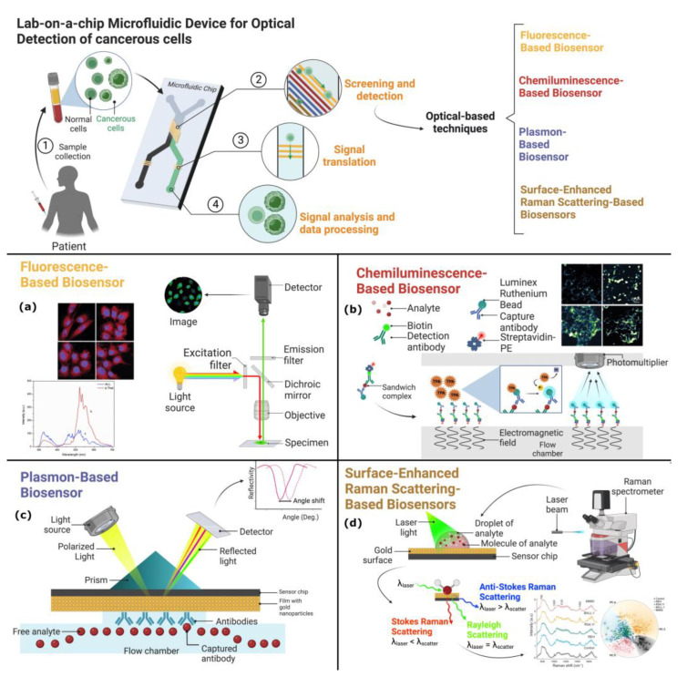
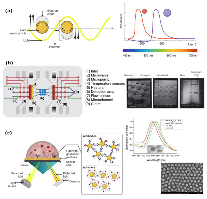
The global need for accurate and efficient cancer cell detection in biomedicine and clinical diagnosis has driven extensive research and technological development in the field. Precision, high-throughput, non-invasive separation, detection, and classification of individual cells are critical requirements for successful technology. Lab-on-a-chip devices offer enormous potential for solving biological and medical problems and have become a priority research area for microanalysis and manipulating cells. This paper reviews recent developments in the detection of cancer cells using the microfluidics-based lab-on-a-chip method, focusing on describing and explaining techniques that use optical phenomena and a plethora of probes for sensing, amplification, and immobilization. The paper describes how optics are applied in each experimental method, highlighting their advantages and disadvantages. The discussion includes a summary of current challenges and prospects for cancer diagnosis.

Keywords: cancer; chemiluminescence; fluorescence; lab-on-a-chip; optical detection; surface plasmon resonance SPR; surface-enhanced Raman scattering SERS.


Conflict of interest statement

The authors declare no conflict of interest.

Figures

Figure 5
Figure 5
(a) Energetic levels in SERS: understanding the interaction between molecules and plasmonic nanoparticles. (b) Bright field accompanying with Raman spectra of chronic lymphocytic leukemia cells stained with Giemsa and labeled with anti-CD19-SERS nanoparticles. Reproduced with the permission of [99]. (c) Integrated microfluidic and biosensing SERS system for high sensitivity detection in biomedical applications. (Created with Biorender.com).
Figure 1
Figure 1
The schematic diagram displays lab-on-a-chip microfluidic devices utilizing optical-based detection techniques. At the top, a standard microfluidic channel is shown, where a human sample containing normal and cancerous cells has been introduced. The cells pass through microchannels, and a screening and detection step is performed using an optical-based detection technique. The four types of sensor chips depicted are (a) fluorescence-based biosensor, (b) chemiluminescence-based biosensor, (c) localized surface plasmon resonance, and (d) surface-enhanced Raman scattering sensor chips. Insets of micrographs adapted with permission from Refs. [20,21] a and b, respectively and [22] for d. Copyright 2019 WILEY and 2019 and 2021 Elsevier. (Created with Biorender.com).
Figure 2
Figure 2
A lab-on-a-chip fluorescence device for biosensing. The device consists of microchannels and sensing regions that allow for the detection and analysis of specific biomolecules in biological samples. Fluorescent probes are used to detect the presence of these biomolecules, and the fluorescence signal is read by a detector integrated into the chip. Created with Biorender.com.
Figure 3
Figure 3
Schematic illustration of the microfluidic chemiluminescence biosensor. Created with Biorender.com.
Figure 4
Figure 4
(a) Schematic configuration of a surface plasmon resonance biosensor. A visual representation of the electron cloud’s behavior in a thin gold film when stimulated by light, resulting in the surface plasmon effect, indicated by an absorption peak at a specific frequency that varies based on the size and shape of the nanoparticles. (b) This micrograph illustrates the different zones of Lee’s microfluidic channels (reproduced with permission) used for the operation of a lab-on-a-chip for immunoassays and detection of plasmonic signals [74]. (c) The presented scheme depicts the process occurring in the SPR sensor. Resonance produces an amplified increase in the extinction signal when gold nanoparticles bind to antibodies, enabling the detection of the immune signal that identifies the presence of cancerous cells in the sample. Adapted with permission from Ref. [75]. Copyright 2019 American Chemical Society. (Created with Biorender.com).

Similar articles

Cited by

References

    1. De Martel C., Georges D., Bray F., Ferlay J., Clifford G.M. Global burden of cancer attributable to infections in 2018: A worldwide incidence analysis. Lancet Glob. Health. 2020;8:e180–e190. doi: 10.1016/S2214-109X(19)30488-7. - DOI - PubMed
    1. Yu Z., Gao L., Chen K., Zhang W., Zhang Q., Li Q., Hu K. Nanoparticles: A New Approach to Upgrade Cancer Diagnosis and Treatment. Nanoscale Res. Lett. 2021;16:88. doi: 10.1186/s11671-021-03489-z. - DOI - PMC - PubMed
    1. Sackmann E.K., Fulton A.L., Beebe D.J. The present and future role of microfluidics in biomedical research. Nature. 2014;507:181–189. doi: 10.1038/nature13118. - DOI - PubMed
    1. Bargahi N., Ghasemali S., Jahandar-Lashaki S., Nazari A. Recent advances for cancer detection and treatment by microfluidic technology, review and update. Biol. Proced. Online. 2022;24:5. doi: 10.1186/s12575-022-00166-y. - DOI - PMC - PubMed
    1. Pereira S.P., Oldfield L., Ney A., Hart P.A., Keane M.G., Pandol S.J., Li D., Greenhalf W., Jeon C.Y., Koay E.J., et al. Early detection of pancreatic cancer. Lancet Gastroenterol. Hepatol. 2020;5:698–710. doi: 10.1016/S2468-1253(19)30416-9. - DOI - PMC - PubMed