. 2023 Mar 31;13(4):444.
doi: 10.3390/bios13040444.

Advanced Urea Precursors Driven NiCo2O4 Nanostructures Based Non-Enzymatic Urea Sensor for Milk and Urine Real Sample Applications

Affiliations

Advanced Urea Precursors Driven NiCo2O4 Nanostructures Based Non-Enzymatic Urea Sensor for Milk and Urine Real Sample Applications

Sanjha Mangrio et al. Biosensors (Basel). .

Abstract

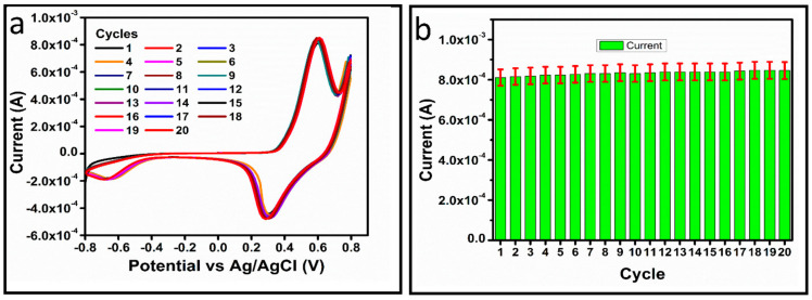
The electrochemical performance of NiCo2O4 with urea precursors was evaluated in order to develop a non-enzymatic urea sensor. In this study, NiCo2O4 nanostructures were synthesized hydrothermally at different concentrations of urea and characterized using scanning electron microscopy and X-ray diffraction. Nanostructures of NiCo2O4 exhibit a nanorod-like morphology and a cubic phase crystal structure. Urea can be detected with high sensitivity through NiCo2O4 nanostructures driven by urea precursors under alkaline conditions. A low limit of detection of 0.05 and an analytical range of 0.1 mM to 10 mM urea are provided. The concentration of 006 mM was determined by cyclic voltammetry. Chronoamperometry was used to determine the linear range in the range of 0.1 mM to 8 mM. Several analytical parameters were assessed, including selectivity, stability, and repeatability. NiCo2O4 nanostructures can also be used to detect urea in various biological samples in a practical manner.

Keywords: NiCo2O4 nanostructures; alkaline conditions; non-enzymatic sensor; urea precursors.


Conflict of interest statement

Authors declare no competing interest in the presented research.

Figures

Scheme 1
Scheme 1
Synthesis of NiCo2O4 nanostructured materials by hydrothermal method followed by calcination in air.
Figure 1
Figure 1
(a) XRD patterns of NiCo2O4 samples prepared with various concentrations of urea (Samples 1–3), (bd) effect of various urea concentrations (Samples 1–3) on the morphology of NiCo2O4 nanomaterial.
Figure 2
Figure 2
(a) CV curves of bare GCE (BGCE) and modified glassy carbon electrode (MGCE) with Sample 3 NiCo2O4 nanomaterial, Samples 1 and 2, 50 mV/s in the presence of 0.1 mM urea and only in 0.1 M NaOH, (b) CV curves of Samples 1 and 2 based NiCo2O4 nanomaterial, at 50 mV/s in the presence of 0.1 mM urea prepared in 0.1 M NaOH.
Figure 3
Figure 3
(a) CV curves of Sample 2 based on NiCo2O4 nanomaterial, at various scan rates in the presence of 0.1 mM urea prepared in 0.1 M NaOH, (b) Linear plots of anodic and cathodic peak currents.
Figure 4
Figure 4
(a) CV curves of Sample 2 based on NiCo2O4 nanomaterial, at 50 mV/s in the presence of various urea concentrations prepared in 0.1 M NaOH, (b) Linear plot of oxidation peak current versus different urea concentrations.
Figure 5
Figure 5
(a) chronoamperometry response of Sample 2 based on NiCo2O4 nanomaterial, at 0.6 V in the presence of various urea concentrations prepared in 0.1 M NaOH, (b) Linear plot of amperometeric generated current against various urea concentrations.
Figure 6
Figure 6
(a) CV curves of Sample 2 based on NiCo2O4 nanomaterial for the monitoring of stability and repeatability, at 50 mV/s in the presence of 0.1 mM urea concentration prepared in 0.1 M NaOH, (b) illustration of peak current in bar graph for the statistical error for the demonstration of stability and repeatability of material.
Figure 7
Figure 7
(ac) Cyclic voltammetry measured for Samples 2 and 3 and Sample 1 in 0.5 mM urea solution at different scan rates. (d) Linear plots of anodic and cathodic current density difference for each scan rate for the quantification of (ECSA).

References

    1. Jonker J., Kohn R., Erdman R. Using milk urea nitrogen to predict nitrogen excretion and utilization efficiency in lactating dairy cows. J. Dairy Sci. 1998;81:2681–2692. doi: 10.3168/jds.S0022-0302(98)75825-4. - DOI - PubMed
    1. Roy B., Brahma B., Ghosh S., Pankaj P., Mandal G. Evaluation of milk urea concentration as useful indicator for dairy herd management: A review. Asian J. Anim. Vet. Adv. 2011;6:1–19. doi: 10.3923/ajava.2011.1.19. - DOI
    1. Francis P.S., Lewis S.W., Lim K.F. Analytical methodology for the determination of urea: Current practice and future trends. TrAC Trends Anal. Chem. 2002;21:389–400. doi: 10.1016/S0165-9936(02)00507-1. - DOI
    1. Bisht V., Takashima W., Kaneto K. An amperometric urea biosensor based on covalent immobilization of urease onto an electrochemically prepared copolymer poly (N-3-aminopropyl pyrrole-co-pyrrole) film. Biomaterials. 2005;26:3683–3690. - PubMed
    1. Deng H.-H., Hong G.-L., Lin F.-L., Liu A.-L., Xia X.-H., Chen W. Colorimetric detection of urea, urease, and urease inhibitor based on the peroxidase-like activity of gold nanoparticles. Anal. Chim. Acta. 2016;915:74–80. doi: 10.1016/j.aca.2016.02.008. - DOI - PubMed

LinkOut - more resources