Review
. 2023 Sep;21(9):573-589.
doi: 10.1038/s41579-023-00889-z. Epub 2023 Apr 25.

Cell entry and release of quasi-enveloped human hepatitis viruses

Affiliations
Review

Cell entry and release of quasi-enveloped human hepatitis viruses

Anshuman Das et al. Nat Rev Microbiol. 2023 Sep.

Abstract

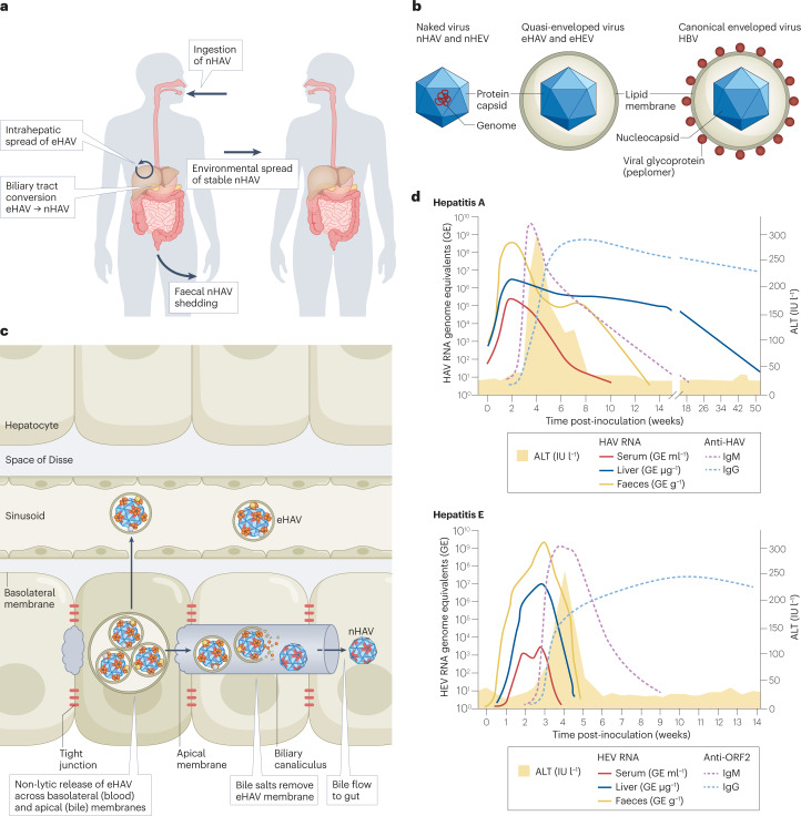
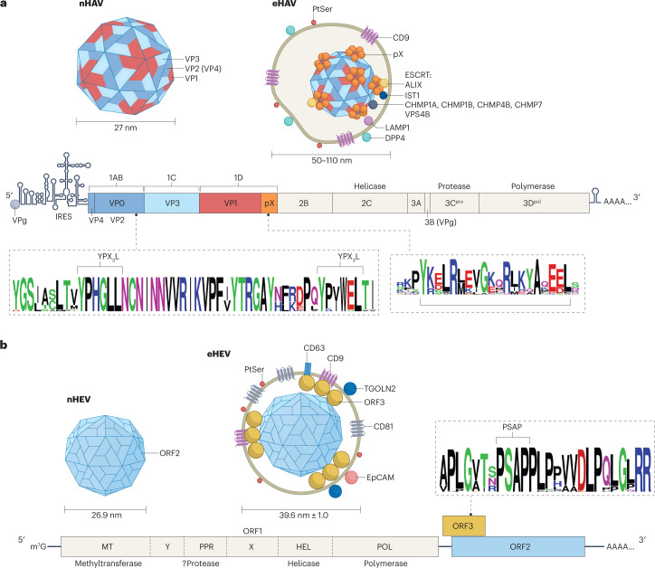
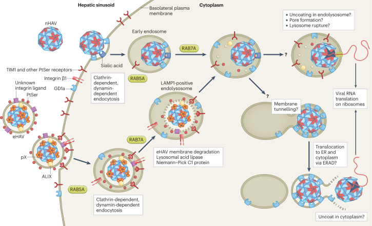
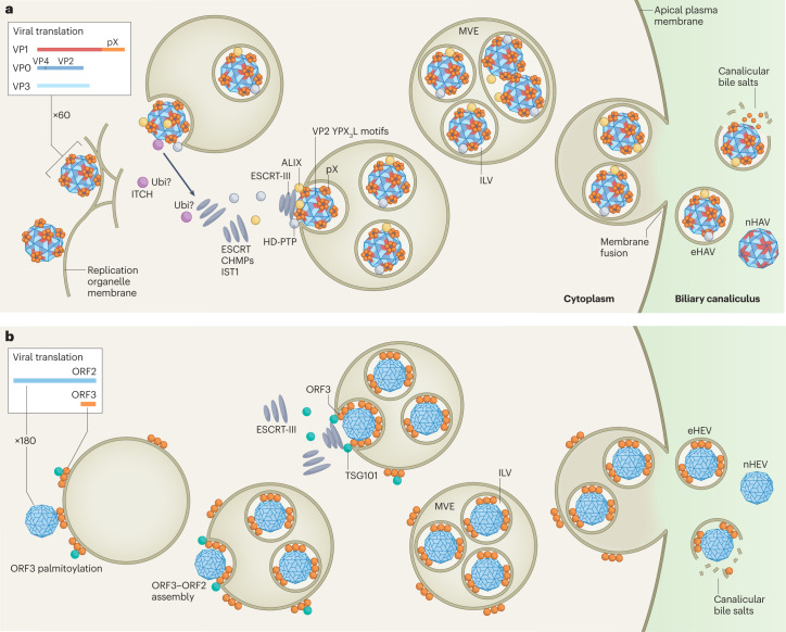
Infectious hepatitis type A and type E are caused by phylogenetically distinct single-stranded, positive-sense RNA viruses that were once considered to be non-enveloped. However, studies show that both are released nonlytically from hepatocytes as 'quasi-enveloped' virions cloaked in host membranes. These virion types predominate in the blood of infected individuals and mediate virus spread within the liver. They lack virally encoded proteins on their surface and are resistant to neutralizing anti-capsid antibodies induced by infection, yet they efficiently enter cells and initiate new rounds of virus replication. In this Review, we discuss the mechanisms by which specific peptide sequences in the capsids of these quasi-enveloped virions mediate their endosomal sorting complexes required for transport (ESCRT)-dependent release from hepatocytes through multivesicular endosomes, what is known about how they enter cells, and the impact of capsid quasi-envelopment on host immunity and pathogenesis.


Conflict of interest statement

SML is co-inventor on a pending patent application related to antiviral compounds with activity against hepatitis A virus. The other authors have no competing interests to declare.

Figures

Fig. 1
Fig. 1. Pathogenesis of enterically transmitted hepatitis A and hepatitis E virus.
a, Hepatitis A virus (HAV) life cycle, showing per-oral infection with naked HAV (nHAV), in-host spread of quasi-enveloped HAV (eHAV) and faecal shedding of nHAV resulting in environmental transmission to naive hosts. nHAV particles shed in faeces are produced by bile acid conversion of eHAV released from hepatocytes. The hepatitis E virus (HEV) life cycle is similar (not shown). b, Basic structures of naked versus quasi-enveloped hepatitis viruses and a canonical enveloped virus (hepatitis B virus (HBV)), showing the absence of virus-encoded proteins on the surface of quasi-enveloped virions,. c, Liver architecture, showing basolateral release of eHAV from polarized hepatocytes into blood flowing through hepatic sinusoids and apical release into a bile canaliculus. The quasi-envelope is stripped from eHAV by bile acids, resulting in faecal shedding of nHAV. Events are similar in hepatitis E (not shown). d, Virological and serological markers in acute hepatitis A (top) and hepatitis E (bottom). ALT, alanine aminotransferase; IgG, immunoglobulin G; IgM, immunoglobulin M; IU l−1, international units per litre.
Fig. 2
Fig. 2. Genome organization and structures of naked and quasi-enveloped hepatitis A and hepatitis E virions.
a, Hepatitis A (HAV) RNA (7.5 kb) contains a lengthy 5′ untranslated region with secondary structure essential for genome replication and an internal ribosomal entry site (IRES) that initiates cap-independent translation of a single long open reading frame (ORF) encoding a large polyprotein. The polyprotein is processed by the HAV protease 3Cpro into three proteins that form the capsid, VP0 (also known as 1AB; subsequently processed into VP4 (1A) and VP2 (1B)), VP3 (1C) and VP1pX (1D), and six nonstructural proteins that mediate genome replication, 2B, 2C, 3A, 3B, 3Cpro and 3Dpol. The 8-kDa pX segment is present in quasi-enveloped HAV (eHAV), but cleaved from VP1 upon loss of the membrane, and it is not present in naked HAV (nHAV). Proteomics studies show that programmed cell death 6-interacting protein (ALIX), vacuolar protein sorting-associated protein IST1 homologue (IST1, a component of the endosomal complexes required for transport (ESCRT)-III complex), and multiple charged multivesicular body proteins (CHMPs) such as CHMP1A, CHMP1B, CHMP4B and CHMP7 (also components of ESCRT-III) are physically associated with eHAV. b, Hepatitis E virus (HEV) RNA (7.2 kb) has a 5′ 7-methylguanosine (m7G) cap and three ORFs that are translated in a cap-dependent manner. ORF1 encodes a multifunctional polyprotein; whether this is processed into smaller proteins in infected cells is uncertain. ORF2 and ORF3 are translated from a subgenomic RNA. ORF2 encodes the capsid protein, but also produces a secreted protein from an in-frame start codon that is thought to decoy neutralizing antibody. ORF3 protein associates with membranes and recruits TSG101, a component of ESCRT-I; it is not present in nHEV,,. An additional ORF4 exists within the ORF1 coding region of genotype 1 virus (not shown). WebLogos show conserved domains in VP2 and pX, proteins of HAV and in ORF3 protein of HEV (sequences from 40 paslahepeviruses) that recruit ESCRT during capsid quasi-envelopment. Naked and quasi-enveloped virions are shown at the top of each panel at the left and right, respectively, with associated host proteins and lipids,,. EpCAM, epithelial cell adhesion molecule (or CD326 antigen); LAMP1, lysosome-associated membrane protein 1; PtSer, phosphatidylserine; TGOLN2, trans-Golgi network integral membrane protein 2.
Fig. 3
Fig. 3. Cellular entry of naked and quasi-enveloped hepatitis A virus.
Both virion types undergo clathrin-dependent endocytosis driven by interactions between distinct ligands, including phosphatidylserine (PtSer) on the quasi-enveloped hepatitis A virus (eHAV) surface and cellular PtSer receptors such as T cell immunoglobulin mucin receptor 1 (TIM1). Endocytosis of naked HAV (nHAV) is inhibited by either sialidase or trypsin treatment of the cell, suggesting that gangliosides and proteins (possibly sialylated) are involved. Integrin β1, presumably in association with different α-integrins, is required for endocytosis of both virion types. Both virion types traffic through early (RAB5A-positive) and late (RAB7A-positive) endosomal compartments. Subsequent steps that involve uncoating and genome release into the cytoplasm require endolysosomal gangliosides, preferably GD1a (ref. ). These late steps are delayed for eHAV, which must traffic first to lysosomal-associated membrane protein 1 (LAMP1)-positive endolysosomes, where the quasi-envelope is degraded by lysosomal enzymes and cholesterol transporters, such as lysosomal acid lipase and Niemann–Pick C1 protein. Like nHAV entry, eHAV entry is dependent upon endosomal gangliosides. Subsequent steps in entry are not well understood, but progressive binding of the now-naked capsid to membrane-bound gangliosides may result in tunnelling of the capsid into the membrane. The trigger for capsid disassembly is not known, nor is it known whether uncoating occurs in the endolysosomal lumen or following transport of the capsid across the endolysosomal membrane to the cytoplasm. Entry of naked hepatitis E virus is not dependent upon RAB5A or RAB7A, but entry of quasi-enveloped hepatitis E virus is similar to eHAV entry and also involves trafficking to endolysosomes for degradation of the quasi-envelope. Receptors have not been identified for the hepatitis E virus capsid. Steps in entry that are not understood are indicated by a question mark. ER, endoplasmic reticulum; ERAD, endoplasmic reticulum-associated degradation pathway.
Fig. 4
Fig. 4. Multivesicular endosome-dependent biogenesis of quasi-enveloped virions.
a, Hepatitis A virus (HAV) capsids assemble from 60 copies of three major capsid proteins in close association with the limiting membrane of endosomes into which they bud. An 8-kDa C-terminal extension of the VP1 protein, pX, recruits two endosomal complexes required for transport (ESCRT)-associated proteins — programmed cell death 6-interacting protein (ALIX) and tyrosine-protein phosphatase non-receptor type 23 (HD-PTP) — as well as the ubiquitin ligase ITCH. This induces assembly of ESCRT-III complexes containing multiple charged multivesicular body proteins (CHMPs) and vacuolar protein sorting-associated protein IST1 homologue (IST1) that pinch off the membrane, creating an intraluminal vesicle (ILV) that contains single or multiple HAV capsids. Ubiquitin (Ubi) tags conjugated to viral or ESCRT-associated proteins by ITCH may facilitate this process. ALIX-interacting late domains also exist within the VP2 capsid protein (YPX3L motifs). Multivesicular endosomes (MVEs) containing multiple HAV-laden ILVs traffic to the apical (shown) and basolateral (not shown) membranes, where fusion leads to the release of quasi-enveloped HAV (eHAV) into the biliary tract and sinusoidal blood, respectively. High concentrations of bile acids strip the membrane from eHAV in the bile canaliculus, resulting in faecal shedding of naked virus (nHAV). b, The biogenesis of quasi-enveloped hepatitis E virus (eHEV) is similar to eHAV, with capsids assembling from 180 copies of ORF2 protein interacting with ORF3 protein associated with the cytosolic leaflet of endosomal membranes via its palmitoylated N terminus. ORF3 protein recruits the ESCRT-I protein TSG101 to initiate ESCRT-dependent membrane scission and ILV formation,. Additional interactions of ORF2 or ORF3 proteins with other ESCRT-associated proteins seem likely to occur but have not been identified. eHEV ILVs generally contain only a single capsid, possibly reflecting a more integrated process of capsid assembly and quasi-envelopment. Fusion of the MVE and plasma membranes and release of HEV virions is similar to HAV.

References

    1. Balayan MS, et al. Evidence for a virus in non-A, non-B hepatitis transmitted via the fecal-oral route. Intervirology. 1983;20:23–31. doi: 10.1159/000149370. - DOI - PubMed
    1. Feinstone SM, Kapikian AZ, Purcell RH. Hepatitis A: detection by immune electron microscopy of a viruslike antigen associated with acute illness. Science. 1973;182:1026–1028. doi: 10.1126/science.182.4116.1026. - DOI - PubMed
    1. Lanford RE, et al. Acute hepatitis A virus infection is associated with a limited type I interferon response and persistence of intrahepatic viral RNA. Proc. Natl Acad. Sci. USA. 2011;108:11223–11228. doi: 10.1073/pnas.1101939108. - DOI - PMC - PubMed
    1. Yu C, et al. Pathogenesis of hepatitis E virus and hepatitis C virus in chimpanzees: similarities and differences. J. Virol. 2010;84:11264–11278. doi: 10.1128/JVI.01205-10. - DOI - PMC - PubMed
    1. Purcell RH, et al. Pathobiology of hepatitis E: lessons learned from primate models. Emerg. Microbes Infect. 2013;2:e9. doi: 10.1038/emi.2013.9. - DOI - PMC - PubMed

Publication types

Substances