Review
. 2023 Jul;20(7):417-432.
doi: 10.1038/s41575-023-00766-3. Epub 2023 Apr 25.

Paracellular permeability and tight junction regulation in gut health and disease

Affiliations
Review

Paracellular permeability and tight junction regulation in gut health and disease

Arie Horowitz et al. Nat Rev Gastroenterol Hepatol. 2023 Jul.

Abstract

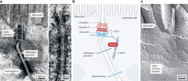
Epithelial tight junctions define the paracellular permeability of the intestinal barrier. Molecules can cross the tight junctions via two distinct size-selective and charge-selective paracellular pathways: the pore pathway and the leak pathway. These can be distinguished by their selectivities and differential regulation by immune cells. However, permeability increases measured in most studies are secondary to epithelial damage, which allows non-selective flux via the unrestricted pathway. Restoration of increased unrestricted pathway permeability requires mucosal healing. By contrast, tight junction barrier loss can be reversed by targeted interventions. Specific approaches are needed to restore pore pathway or leak pathway permeability increases. Recent studies have used preclinical disease models to demonstrate the potential of pore pathway or leak pathway barrier restoration in disease. In this Review, we focus on the two paracellular flux pathways that are dependent on the tight junction. We discuss the latest evidence that highlights tight junction components, structures and regulatory mechanisms, their impact on gut health and disease, and opportunities for therapeutic intervention.


Conflict of interest statement

J.R.T. is a consultant for Entrinsic and Kallyope. The other authors declare no competing interests.

Figures

Fig. 1
Fig. 1. Tight junction structure and morphology.
a, Transmission electron micrograph showing the tight junction, adherens junction and desmosome, which, together, comprise the apical junctional complex. The tight junction is located just below the base of the microvilli. The magnification shows the transition from the luminal space, between the microvilli, into the tight junction, where morphologically detectable paracellular space is obliterated. b, Schematic of the apical junctional complex shown in part a, showing the location of the tight junction proteins zonula occludens 1 (ZO-1), occludin, claudin-2 and other claudin family members. Long myosin light chain kinase 1 (MLCK1) is associated with perijunctional F-actin and is a key regulator of tight junction permeability. c, Freeze–fracture electron micrograph showing tight junction strands at the base of apical microvilli.
Fig. 2
Fig. 2. Coordination of transcellular and paracellular transport.
The gradient of Na+ between the gut lumen and the cytoplasm of epithelial cells provides the driving force for nutrient absorption across the apical brush border membrane, such as glucose absorption via the intestinal epithelial Na+–glucose cotransporter SGLT1. Nutrients then exit the cell via facilitated exchange proteins, such as the glucose transporter GLUT2, and Na+ exits via the Na+–K+ ATPase. Na+–glucose cotransport also triggers signal transduction pathways that activate long myosin light chain kinase 1 (MLCK1) and increase tight junction permeability. The osmotic gradient generated by transcellular nutrient and Na+ transport draws water across the tight junction and, owing to the high concentration of nutrient monomers in the unstirred layer, nutrients are carried along with this fluid in a mechanism known as solvent drag. This process would quickly exhaust luminal Na+ if not for claudin-2 and claudin-15, which form paracellular Na+ channels that enable efflux of absorbed Na+ in order to provide the driving force for continued transcellular nutrient absorption,.
Fig. 3
Fig. 3. Promotion of pathogen clearance by paracellular fluid efflux.
Citrobacter rodentium infection triggers an immune response that leads to IL-22 release within the lamina propria within 2 days of infection. IL-22 signalling activates claudin-2 transcription and increases claudin-2 channel-mediated Na+ and water efflux via the tight junction pore pathway, resulting in diarrhoea that promotes clearance of the infection. Adapted with permission from ref. , Elsevier.
Fig. 4
Fig. 4. Epithelial and smooth muscle myosin light chain kinase.
a, The human MYLK gene encodes long (non-muscle) and short (smooth muscle) isoforms of myosin light chain kinase (MLCK) protein. Two long MLCK transcriptional start sites that result in expression of the same protein have been identified. However, extensive alternative splicing within the 5′ half of the transcript occurs, which, in intestinal epithelial cells, results in expression of two long MLCK splice variants, MLCK1 and MLCK2. These variants differ by a single exon (black), removal of which causes the third of the nine immunoglobulin-cell adhesion molecule (IgCAM) domains to be incomplete in long MLCK2. The short MLCK promoter is located within a long MLCK intron and drives transcription of smooth muscle MLCK, which lacks the six amino-terminal IgCAM domains that are present in long MLCK1. The kinase and calmodulin (CaM)-binding domains are encoded by sequences within the 3′ half of MYLK and are identical in long and short MLCK proteins. b, Inflammatory signals, such as tumour necrosis factor (TNF), trigger MLCK1 binding to FKBP8. This binding facilitates MLCK1 recruitment to the perijunctional actomyosin ring, where it phosphorylates MLC. This phosphorylation causes occludin internalization to increase leak pathway permeability. In contrast to MLCK1, MLCK2 distribution is not affected by TNF. c, MLCK1 expression and recruitment to the perijunctional actomyosin ring (arrows) are increased in Crohn’s disease. The insets show the boxed areas. MLCK1 and total MLCK are shown, as the absence of unique MLCK2 sequences prevents generation of MLCK2-specific antibodies. Nuclei appear yellow. Part a adapted from ref. , Springer Nature Limited.

References

    1. Simon DB, et al. Paracellin-1, a renal tight junction protein required for paracellular Mg2+ resorption. Science. 1999;285:103–106. doi: 10.1126/science.285.5424.103. - DOI - PubMed
    1. Kausalya PJ, et al. Disease-associated mutations affect intracellular traffic and paracellular Mg2+ transport function of claudin-16. J. Clin. Invest. 2006;116:878–891. doi: 10.1172/JCI26323. - DOI - PMC - PubMed
    1. Wilcox ER, et al. Mutations in the gene encoding tight junction claudin-14 cause autosomal recessive deafness DFNB29. Cell. 2001;104:165–172. doi: 10.1016/S0092-8674(01)00200-8. - DOI - PubMed
    1. Ben-Yosef T, et al. Claudin 14 knockout mice, a model for autosomal recessive deafness DFNB29, are deaf due to cochlear hair cell degeneration. Hum. Mol. Genet. 2003;12:2049–2061. doi: 10.1093/hmg/ddg210. - DOI - PubMed
    1. Zhao J, et al. Multiple claudin–claudin cis interfaces are required for tight junction strand formation and inherent flexibility. Commun. Biol. 2018;1:50. doi: 10.1038/s42003-018-0051-5. - DOI - PMC - PubMed

Publication types