. 2023 Sep;44(9):1777-1789.
doi: 10.1038/s41401-023-01086-7. Epub 2023 Apr 25.

JMJD6 protects against isoproterenol-induced cardiac hypertrophy via inhibition of NF-κB activation by demethylating R149 of the p65 subunit

Affiliations

JMJD6 protects against isoproterenol-induced cardiac hypertrophy via inhibition of NF-κB activation by demethylating R149 of the p65 subunit

Zhen Guo et al. Acta Pharmacol Sin. 2023 Sep.

Abstract

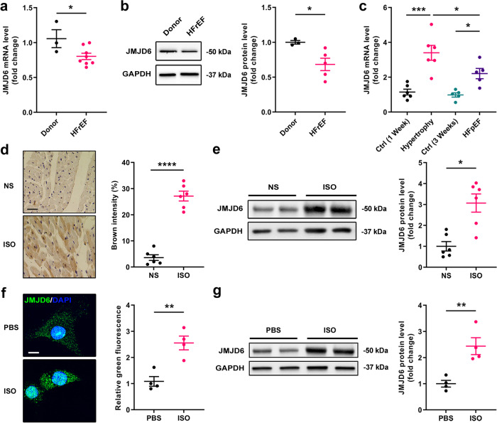
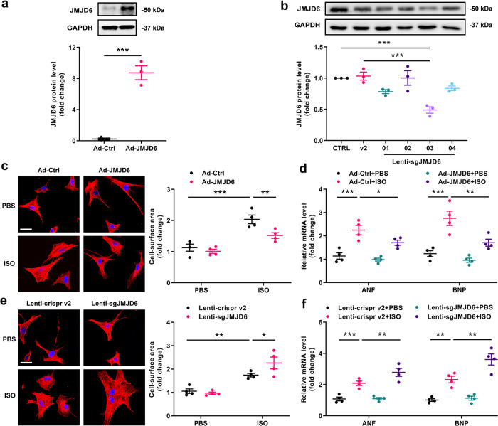
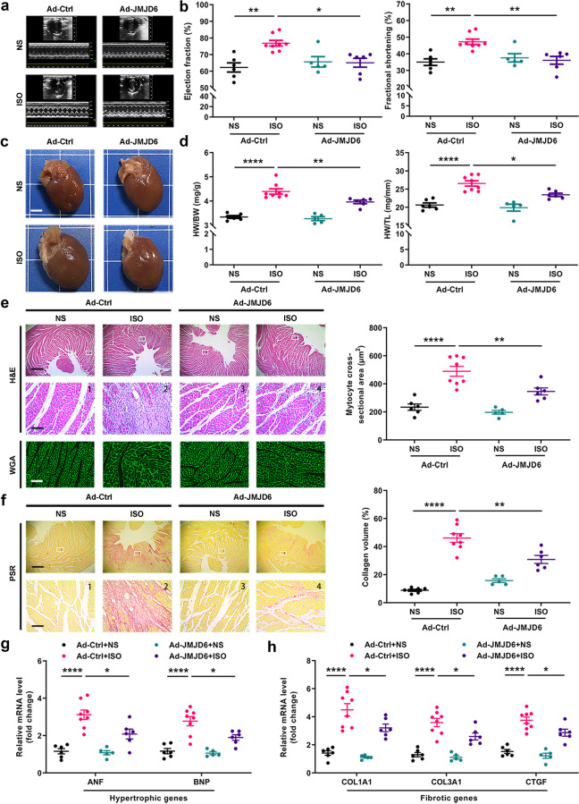
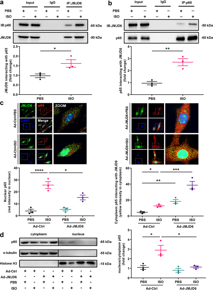
Histone modification plays an important role in pathological cardiac hypertrophy and heart failure. In this study we investigated the role of a histone arginine demethylase, Jumonji C domain-containing protein 6 (JMJD6) in pathological cardiac hypertrophy. Cardiac hypertrophy was induced in rats by subcutaneous injection of isoproterenol (ISO, 1.2 mg·kg-1·d-1) for a week. At the end of the experiment, the rats underwent echocardiography, followed by euthanasia and heart collection. We found that JMJD6 levels were compensatorily increased in ISO-induced hypertrophic cardiac tissues, but reduced in patients with heart failure with reduced ejection fraction (HFrEF). Furthermore, we demonstrated that JMJD6 overexpression significantly attenuated ISO-induced hypertrophy in neonatal rat cardiomyocytes (NRCMs) evidenced by the decreased cardiomyocyte surface area and hypertrophic genes expression. Cardiac-specific JMJD6 overexpression in rats protected the hearts against ISO-induced cardiac hypertrophy and fibrosis, and rescued cardiac function. Conversely, depletion of JMJD6 by single-guide RNA (sgRNA) exacerbated ISO-induced hypertrophic responses in NRCMs. We revealed that JMJD6 interacted with NF-κB p65 in cytoplasm and reduced nuclear levels of p65 under hypertrophic stimulation in vivo and in vitro. Mechanistically, JMJD6 bound to p65 and demethylated p65 at the R149 residue to inhibit the nuclear translocation of p65, thus inactivating NF-κB signaling and protecting against pathological cardiac hypertrophy. In addition, we found that JMJD6 demethylated histone H3R8, which might be a new histone substrate of JMJD6. These results suggest that JMJD6 may be a potential target for therapeutic interventions in cardiac hypertrophy and heart failure.

Keywords: JMJD6; NF-κB; arginine demethylation; cardiac hypertrophy; heart failure; isoproterenol.


Conflict of interest statement

The authors declare no competing interests.

Figures

Fig. 1
Fig. 1. JMJD6 expression was changed in HFrEF patients and hypertrophic rat hearts.
a The mRNA level of JMJD6 in non-failing controls (Donor) or heart failure with reduced ejection fraction (HFrEF) patients (n = 3 in Donor group, n = 8 in HFrEF group). b The protein level of JMJD6 in Donor control or HFrEF patients (n = 3 in Donor group, n = 5 in HFrEF group). c The mRNA level of JMJD6 in ISO-induced hypertrophic cardiac tissues (1.2 mg·kg−1·d−1 × 7 days, SQ, n = 6) and heart failure with preserved ejection fraction (HFpEF) cardiac tissues (2.5 mg·kg−1·d-1 × 21 days, IP, n = 5). d The protein expression of JMJD6 in ISO-induced hypertrophic cardiac tissues was shown as brown intensity by immunohistochemistry staining, scale bar = 30 μm (n = 6). e The protein level of JMJD6 in ISO-induced hypertrophic cardiac tissues was shown by Western blot analysis (n = 6). f The protein expression of JMJD6 in ISO-induced NRCMs was identified by confocal immunofluorescence microscopy, scale bar = 10 μm (n = 4). g The protein expression of JMJD6 in ISO-induced NRCMs was identified by Western blot analysis (n = 4). Student’s t test was used in (a), (b) and (dg), One-way ANOVA with Tukey’s correction for multiple comparisons was used in (c). *P < 0.05, **P < 0.01, ***P < 0.001, ****P < 0.0001. NS normal saline, PBS phosphate-buffered saline.
Fig. 2
Fig. 2. JMJD6 negatively regulates ISO-induced cardiac hypertrophy in NRCMs.
a The adenovirus infection efficiency was confirmed by Western blot in NRCMs infected with Ad-Ctrl (empty vector) or Ad-JMJD6 (n = 3). b JMJD6 protein level was detected by Western blot in NRCMs infected with Lenti-crispr v2 (empty vector) or Lenti-sgJMJD6-01, 02, 03 or 04 (n = 3). c Cell surface area was measured by rhodamine-phalloidin staining in NRCMs infected with Ad-Ctrl or Ad-JMJD6 for 48 h in the presence of ISO (10 μM, 12 h) or PBS, scale bar = 20 μm (n = 4). d The mRNA level of hypertrophic genes (ANF and BNP) in NRCMs infected with Ad-Ctrl or Ad-JMJD6 (n = 4). e Cell surface area was measured in NRCMs infected with Lenti-crispr v2 or Lenti-sgJMJD6, scale bar = 20 μm (n = 4). f The mRNA level of hypertrophic genes in NRCMs infected with Lenti-crispr v2 or Lenti-sgJMJD6 (n = 4). Student’s t test was used in (a), One-way ANOVA with Tukey’s correction for multiple comparisons was used in (bf). *P < 0.05, **P < 0.01, ***P < 0.001. Ad-Ctrl control adenovirus, Ad-JMJD6 adenovirus overexpressing JMJD6, ANF atrial natriuretic factor, BNP brain natriuretic polypeptide. Lenti-sgJMJD6 means Lenti-sgJMJD6-03.
Fig. 3
Fig. 3. JMJD6 overexpression represses ISO-induced cardiac hypertrophy in rats.
a Representative echocardiography of SD rats infected with Ad-Ctrl or Ad-JMJD6 for 14 days in the presence of ISO or NS. b Ejection fraction (EF) and fractional shortening (FS) of Ad-Ctrl or Ad-JMJD6 infected rats treated with ISO or NS (n = 5–8). c Representative heart morphology of SD rats infected with Ad-Ctrl or Ad-JMJD6 for 14 days in the presence of ISO or NS, scale bar = 2 mm. d Heart weight-to-body weight (HW/BW) ratios and HW to-tibia length (HW/TL) ratios of Ad-Ctrl or Ad-JMJD6 infected rats treated with ISO or NS (n = 5–8). e Hematoxylin-Eosin (H&E, scale bar = 1 mm or 50 μm) and wheat germ agglutinin (WGA, scale bar = 50 μm) staining were performed to determine the cardiomyoctye size of Ad-Ctrl or Ad-JMJD6 infected rats treated with ISO or NS (n = 5–8). f Picrosirius red (PSR, scale bar = 1 mm or 50 μm) staining was performed to determine cardiac fibrosis of the hearts from Ad-Ctrl or Ad-JMJD6 infected rats treated with ISO or NS (n = 5–8). g The mRNA levels of hypertrophic genes in Ad-Ctrl or Ad-JMJD6 infected rats treated with ISO or NS (n = 5–8). h The mRNA levels of fibrotic genes (COL1A1, COL3A1 and CTGF) in Ad-Ctrl or Ad-JMJD6 infected rats treated with ISO or NS (n = 5–8). Kruskal-Wallis test with Dunn’s post-hoc for multiple comparisons was used in panel (b) for ejection fraction, One-way ANOVA with Tukey’s correction for multiple comparisons was used in (b) for fractional shortening and (dh). *P < 0.05, **P < 0.01, ****P < 0.0001. COL1A1 collagen1a1, COL3A1 collagen3a1, CTGF connective tissue growth factor.
Fig. 4
Fig. 4. JMJD6 regulates NF-κB signaling in ISO-induced cardiac hypertrophy in vitro.
a Immunoprecipitation analysis showed that interaction of p65 and JMJD6 in NRCMs using JMJD6 antibody in presence of ISO or PBS (n = 3). b Immunoprecipitation analysis showed that interaction of JMJD6 and p65 in NRCMs using p65 antibody in presence of ISO or PBS (n = 3). c Subcellular location of p65 in NRCMs. NRCMs were infected with Ad-JMJD6 or Ad-Ctrl for 24 h in presence of ISO or PBS, followed by immunofluorescence to detect the JMJD6 and p65 protein. Upper: Representative immunofluorescence showing the location of JMJD6 (green) and p65 (red), scale bar = 25 μm or 10 μm. Lower: Blinded quantification of nuclear p65 and cytoplasmic p65 interacting with JMJD6 (n = 3). d The protein expression of p65 was measured by Western blot analysis in the cytoplasm and nucleus in NRCMs infected with Ad-Ctrl or Ad-JMJD6 for 24 h in the presence of ISO or PBS. Left: Representative Western blot images showing the level of p65. Right: The quantification of nucleus/cytoplasm p65 (n = 3). Student’s t test was used in (a) and (b), One-way ANOVA with Tukey’s correction for multiple comparisons was used in (c) and (d). *P < 0.05, **P < 0.01, ***P < 0.001, ****P < 0.0001.
Fig. 5
Fig. 5. JMJD6 regulates NF-κB signaling in ISO-induced cardiac hypertrophy in vivo.
a Immunofluorescence analysis showed that the expression of p65 and JMJD6 in SD rats infected with Ad-Ctrl or Ad-JMJD6 for 14 days in the presence of ISO or NS. Left: Representative immunofluorescence showing the location of JMJD6 (green) and p65 (red), scale bar = 50 μm. Right: Blinded quantification of nuclear p65 and cytoplasmic p65 interacting with JMJD6 (n = 5). b Representative Western blot images and quantitative results showing p65 phosphorylation at Ser536 and relative IκBα expression in cardiac tissues of Ad-Ctrl or Ad-JMJD6 infection for 14 days in the presence of ISO or NS (n = 5). c Immunofluorescence analysis showed that the expression of IκBα (green) and p65 (red) in NRCMs infected with Ad-Ctrl or Ad-JMJD6 for 24 h, scale bar = 25 μm (n = 4). One-way ANOVA with Tukey’s correction for multiple comparisons was used in (a) and (b), Student’s t test was used in (c). *P < 0.05, **P < 0.01, ****P < 0.0001, ns not significant.
Fig. 6
Fig. 6. JMJD6 decreased the methylation level in ISO-induced cardiac hypertrophy.
a The histone H4R3 methylation level was detected by Western blot in 293 T cells treated with PRMT5, WT-JMJD6 and mutant-JMJD6 (H187A, D189A or Double) transfection for 48 h. Left: Representative Western blot images showing the expression of H4R3me1 and H4R3me2s. Middle: The quantification of H4R3me1 level. Right: The quantification of H4R3me2s level (n = 6). b The histone H3R8 methylation level was detected by Western blot in 293 T cells treated with PRMT5, WT-JMJD6 and mutant-JMJD6 (H187A, D189A or Double) transfection for 48 h. Left: Representative Western blot images showing the expression of H3R8me1 and H3R8me2s. Middle: The quantification of H3R8me1 level. Right: The quantification of H3R8me2s level (n = 3). c Representative Western blot images and quantitative result showing the monomethylation level of arginine (R) in NRCMs infected with Ad-JMJD6 or Ad-Ctrl for 48 h in presence of ISO or PBS (n = 4). d Representative Western blot images and quantitative result showing the symmertrical dimethylation level of R in NRCMs infected with Ad-JMJD6 or Ad-Ctrl for 48 h in presence of ISO or PBS (n = 4). One-way ANOVA with Tukey’s correction for multiple comparisons was used in (ad). *P < 0.05, **P < 0.01, ***P < 0.001, ns not significant.
Fig. 7
Fig. 7. JMJD6 demethylated R149 of p65 to inactivate NF-κB.
a Immunoprecipitation analysis showed the monomethylation and symmertrical dimethylation level of arginine in p65 in NRCMs using p65 antibody. b The potential arginine methylation sites in amino acid sequence of Rattus norvegicus JMJD6 using a methylation site prediction platform. c Representative Western blot images and quantitative result showing p65 phosphorylation at Ser536 in 293 T cells co-transfected with WT-JMJD6 and p65 (WT, R30A, R149A, R236A or R336A) (n = 4). d Representative Western blot images and quantitative result showing p65 phosphorylation at Ser536 in 293 T cells co-transfected with WT-PRMT5 and p65 (WT, R30A, R149A, R236A or R336A) (n = 3). One-way ANOVA with Tukey’s correction for multiple comparisons was used in (c) and (d). *P < 0.05, **P < 0.01.

References

    1. Virani SS, Alonso A, Benjamin EJ, Bittencourt MS, Callaway CW, Carson AP, et al. Heart disease and stroke statistics-2020 update: a report from the American Heart Association. Circulation. 2020;141:e139–e596. doi: 10.1161/CIR.0000000000000757. - DOI - PubMed
    1. Frey N, Katus HA, Olson EN, Hill JA. Hypertrophy of the heart: a new therapeutic target? Circulation. 2004;109:1580–9. doi: 10.1161/01.CIR.0000120390.68287.BB. - DOI - PubMed
    1. Maillet M, van Berlo JH, Molkentin JD. Molecular basis of physiological heart growth: fundamental concepts and new players. Nat Rev Mol Cell Biol. 2013;14:38–48. doi: 10.1038/nrm3495. - DOI - PMC - PubMed
    1. MacDonald MR, Petrie MC, Hawkins NM, Petrie JR, Fisher M, McKelvie R, et al. Diabetes, left ventricular systolic dysfunction, and chronic heart failure. Eur Heart J. 2008;29:1224–40. doi: 10.1093/eurheartj/ehn156. - DOI - PubMed
    1. Mano H. Epigenetic abnormalities in cardiac hypertrophy and heart failure. Environ Health Prev Med. 2008;13:25–29. doi: 10.1007/s12199-007-0007-8. - DOI - PMC - PubMed

LinkOut - more resources