. 2023 Jul:63:102739.
doi: 10.1016/j.redox.2023.102739. Epub 2023 May 9.

ROS-Drp1-mediated mitochondria fission contributes to hippocampal HT22 cell apoptosis induced by silver nanoparticles

Affiliations

ROS-Drp1-mediated mitochondria fission contributes to hippocampal HT22 cell apoptosis induced by silver nanoparticles

Xiaoru Chang et al. Redox Biol. 2023 Jul.

Abstract

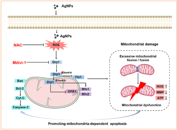
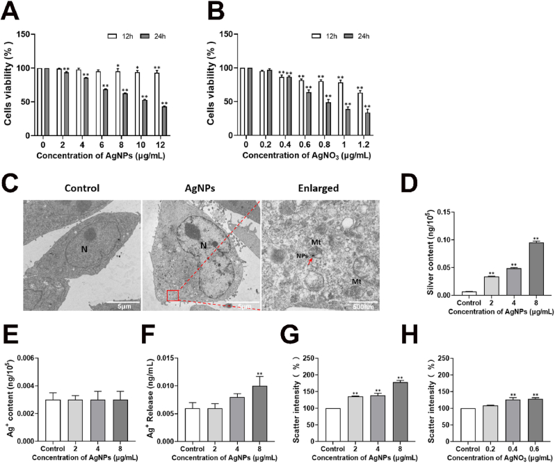
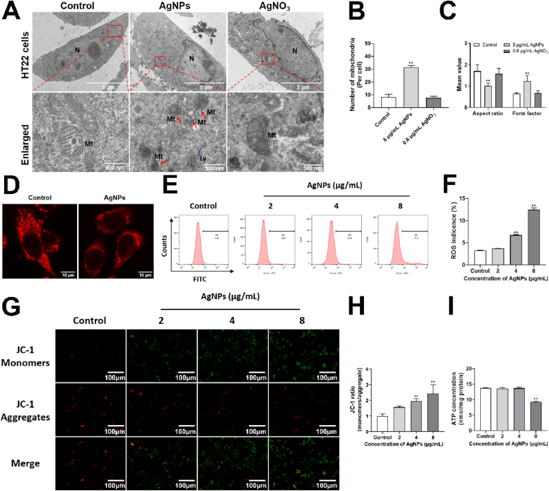
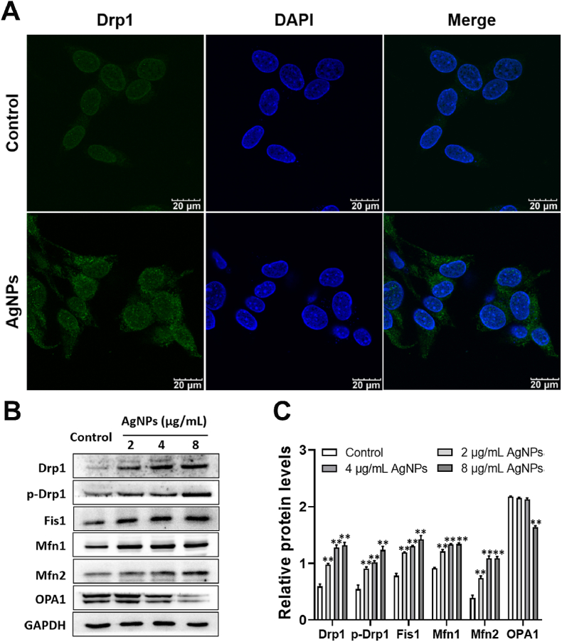
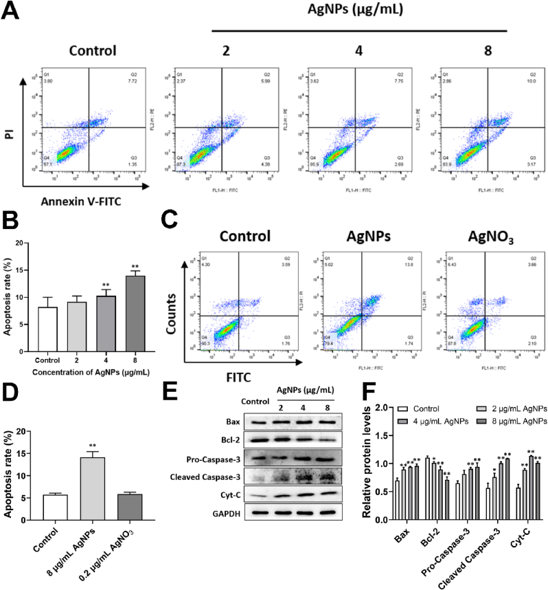
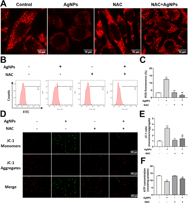
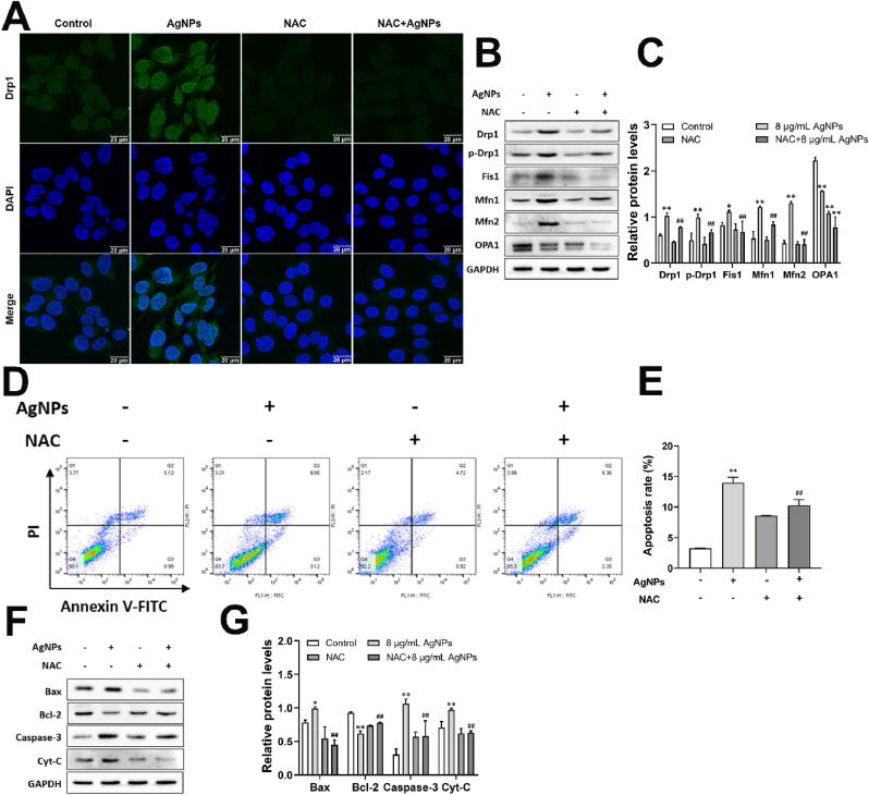
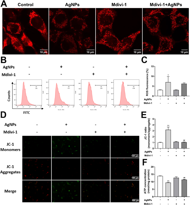
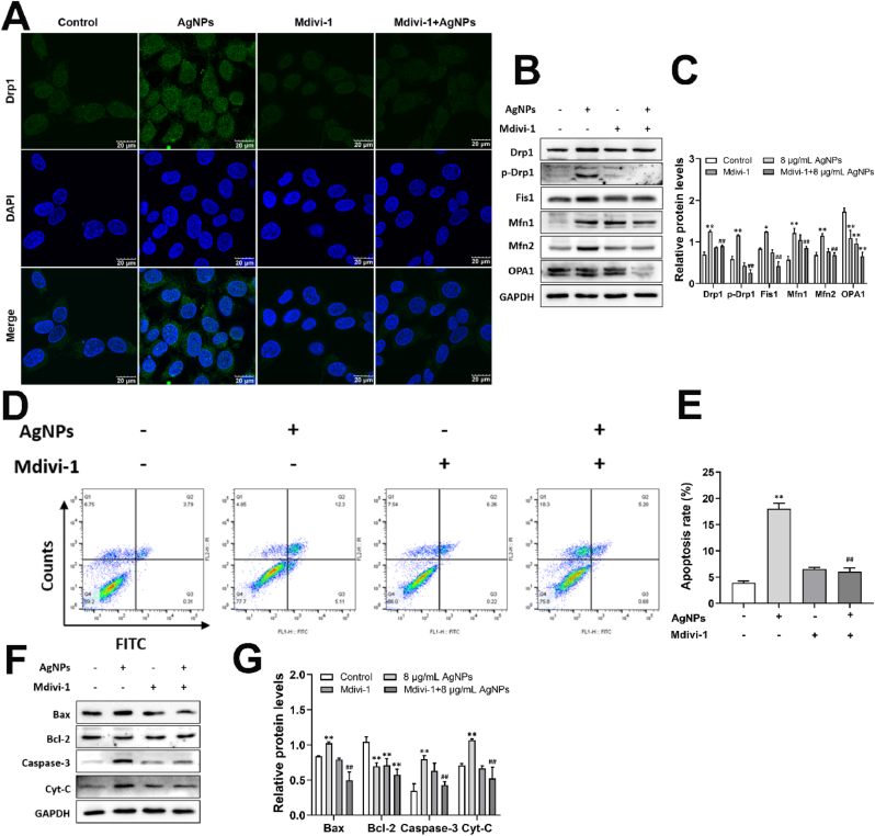
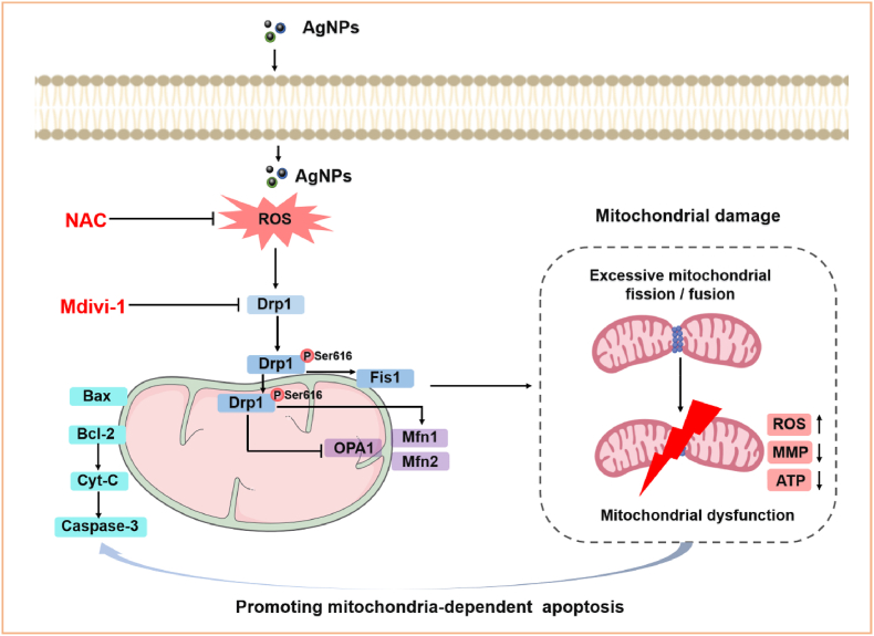
Silver nanoparticles (AgNPs) have widely used in industrial and medical applications for their excellent antibacterial activities. AgNPs can penetrate into the brain and cause neuronal death, but limited evidence focused on toxic effects and mechanic study in hippocampal neuron. This study aimed to investigate the molecular mechanisms of mitochondrial damage and apoptosis in mouse hippocampal HT22 cells and further to explore role of reactive oxygen species (ROS) and GTPase dynamin-related protein 1 (Drp1) in AgNPs-induced neurotoxicity. Our results showed that acute exposure to AgNPs at low doses (2-8 μg/mL) increased ROS generation, decreased mitochondrial membrane potential (MMP) and ATP synthesis in HT22 cells. In addition, AgNPs promoted mitochondrial fragmentation and mitochondria-dependent apoptosis via excessive mitochondrial fission/fusion by 8 μg/mL AgNPs treatment for 24 h. The mechanism was involved in increased protein expression of Drp1, mitochondrial fission protein 1 (Fis1), mitofusin 1/2 (Mfn1/2) and inhibited optic atrophy 1 (OPA1), and mainly mediated by phosphorylation of Drp1 Ser616. The AgNPs-induced mitochondrial impairment and apoptosis was mainly due to their particle-specific effect rather than silver ions release. Furthermore Drp1-mediated mitochondrial fission contributed to mitochondria-dependent apoptosis induced by AgNPs, all aforementioned changes were significantly rescued by N-acetyl-l-cysteine (NAC) and Mdivi-1 except for OPA1 protein expression. Hence, our results provide a novel neurotoxic mechanism to AgNPs-induced neurotoxicity and revealed that the mechanism of mitochondria-dependent apoptosis in HT22 cells was mediated by excessive activation of ROS-Drp1-mitochondrial fission axis. These findings can deepen current evidences on neurotoxicological evaluation of AgNPs and aid in guiding their proper applications in different areas, especially in biomedical use.

Keywords: Apoptosis; Drp1; Mitochondrial fission; Neurotoxicity; Silver nanoparticles.


Conflict of interest statement

Declaration of competing interest The authors declare that they have no known competing financial interests or personal relationships that could have appeared to influence the work reported in this paper.

Figures

Image 1
Graphical abstract
Fig. 1
Fig. 1
Characterizations of AgNPs. (A) The hydrodynamic sizes of the AgNPs in cell culture medium (DMEM containing 10% FBS). (B) UV–vis spectrum of AgNPs in cell culture medium.
Fig. 2
Fig. 2
Cytotoxicity, distribution and uptake of AgNPs in HT22 cells. (A–B) Cytotoxicity of HT22 cells, after AgNPs (0–12 μg/mL) or AgNO3 (0–1.2 μg/mL) exposure for 24 h; (C) Cellular distribution of AgNPs in HT22 cells, after AgNPs (8 μg/mL) exposure for 24 h, the red arrow indicates engulfed AgNPs; (D–F) Silver content (D), Intracellular Ag+ content (E) and released Ag+ content in cell culture medium (F) in HT22 cells, after AgNPs (0–8 μg/mL) exposure for 24 h; (G–H) Dose-dependent scattering intensities as a percentage of control for HT22 cells treated with AgNPs (0–8 μg/mL) or Ag+ (0–0.6 μg/mL) for 24 h. NPs: nanoparticles; Mt: mitochondria. Data are expressed as mean ± SD from three independent experiments. *p < 0.05, **p < 0.01 when compared with the control. (For interpretation of the references to color in this figure legend, the reader is referred to the Web version of this article.)
Fig. 3
Fig. 3
AgNPs disrupted mitochondrial morphology and function in HT22 cells. (A–C) Alterations of mitochondrial morphology (A), mitochondrial number (B), mitochondrial AR and FF parameter (C) in HT22 cells, after AgNPs (8 μg/mL) or AgNO3 (0.6 μg/mL) exposure for 24 h, the red arrow indicates mitochondrial fission and fragmentation, the yellow arrow indicates mitochondrial fusion; (D) Alterations of mitochondrial structure and Mitotracker fluorescence in HT22 cells, after AgNPs (8 μg/mL) exposure for 24 h; (E–F) Intracellular ROS levels, (G–H) MMP changes, (I) ATP content in HT22 cells, after AgNPs (0–8 μg/mL) exposure for 24 h. AR: aspect ratio (ratio between major and minor axes of an ellipse); FF: form factor (perimeter2/4π∙area; degree of branching). Data are expressed as mean ± SD from three independent experiments. *p < 0.05, **p < 0.01 when compared with the control. (For interpretation of the references to color in this figure legend, the reader is referred to the Web version of this article.)
Fig. 4
Fig. 4
AgNPs induced disruption of the mitochondrial dynamics in HT22 cells. (A) Immunofluorescence of Drp1in HT22 cells exposed to AgNPs (8 μg/mL) for 24 h; (B–C) Western blot for protein expression of Drp1, p-Drp1, Fis1, Mfn1, Mfn2, OPA1 in HT22 cells, after AgNPs (0–8 μg/mL) exposure for 24 h. Data are expressed as mean ± SD from three independent experiments. *p < 0.05, **p < 0.01 when compared with the control.
Fig. 5
Fig. 5
AgNPs induced mitochondria-dependent apoptosis in HT22 cells. (A–D) Apoptosis rate of HT22 cells, after AgNPs (0–8 μg/mL) or AgNO3 (0.2 μg/mL) exposure for 24 h; (E–F) Western blot for protein expression of Bax, Bcl-2, Caspase-3, Cyt-C in HT22 cells, after AgNPs (0–8 μg/mL) exposure for 24 h. Data are expressed as mean ± SD from three independent experiments. *p < 0.05, **p < 0.01 when compared with the control.
Fig. 6
Fig. 6
ROS promoted mitochondrial fragmentation and mitochondrial dysfunction induced by AgNPs in HT22 cells. HT22 cells were pretreated with/without NAC (5 mM) 2 h before exposure to 8 μg/mL AgNPs for 24 h. (A) Alteration of Mitotracker fluorescence in HT22 cells; (B–C) Intracellular ROS levels in HT22 cells; (D–E) MMP changes in HT22 cells; (F) ATP content in HT22 cells. Data are expressed as mean ± SD from three independent experiments. *p < 0.05, **p < 0.01 when compared with the control. #p < 0.05, ##p < 0.01 when compared with the AgNPs (8 μg/mL) group.
Fig. 7
Fig. 7
ROS promoted mitochondrial fission/fusion and enhance mitochondria-dependent apoptosis induced by AgNPs in HT22 cells. HT22 cells were pretreated with/without NAC (5 mM) 2 h before exposure to 8 μg/mL AgNPs for 24 h. (A) Immunofluorescence of Drp1in HT22 cells; (B–C) Western blot for protein expression of Drp1, p-Drp1, Fis1, Mfn1, Mfn2, OPA1 in HT22 cells; (D–E) Apoptosis rate of HT22 cells; (F–G) Western blot for protein expression of Bax, Bcl-2, Caspase-3, Cyt-C in HT22 cells. Data are expressed as mean ± SD from three independent experiments. *p < 0.05, **p < 0.01 when compared with the control. #p < 0.05, ##p < 0.01 when compared with the AgNPs (8 μg/mL) group.
Fig. 8
Fig. 8
Role of Drp1 on mitochondrial fragmentation and mitochondrial dysfunction induced by AgNPs in HT22 cells. HT22 cells were pretreated with/without Mdivi-1 (5 μM) 2 h before exposure to 8 μg/mL AgNPs for 24 h. (A) Alteration of Mitotracker fluorescence in HT22 cells; (B–C) Intracellular ROS levels in HT22 cells; (D–E) MMP changes in HT22 cells; (F) ATP content in HT22 cells. Data are expressed as mean ± SD from three independent experiments. *p < 0.05, **p < 0.01 when compared with the control. #p < 0.05, ##p < 0.01 when compared with the AgNPs (8 μg/mL) group.
Fig. 9
Fig. 9
Role of Drp1-mediated mitochondrial fission on apoptosis induced by AgNPs in HT22 cells. HT22 cells were pretreated with/without Mdivi-1 (5 μM) 2 h before exposure to 8 μg/mL AgNPs for 24 h. (A) Immunofluorescence of Drp1in HT22 cells; (B–C) Western blot for protein expression of Drp1, p-Drp1, Fis1, Mfn1, Mfn2, OPA1 in HT22 cells; (D–E) Apoptosis rate of HT22 cells; (F–G) Western blot for protein expression of Bax, Bcl-2, Caspase-3, Cyt-C in HT22 cells. Data are expressed as mean ± SD from three independent experiments. *p < 0.05, **p < 0.01 when compared with the control. #p < 0.05, ##p < 0.01 when compared with the AgNPs (8 μg/mL) group.
Fig. 10
Fig. 10
Schematic diagram of signaling pathways involved in AgNPs-induced mitochondrial damage and apoptosis in hippocampal HT22 cells. AgNPs promoted mitochondrial fragmentation via excessive mitochondrial fission/fusion, increased ROS generation, decreased MMP and ATP synthesis, eventually induced mitochondria-dependent apoptosis in HT22 cells. The mechanism was involved in increased Drp1, Fis1, Mfn1, Mfn2 and decreased OPA1 expression, mainly mediated by phosphorylation and mitochondrial translocation of Drp1 Ser616. Furthermore, all aforementioned changes were significantly rescued by NAC and Mdivi-1 except for OPA1 expression, indicating that mechanism of mitochondria-dependent apoptosis in HT22 cells was mediated by excessive activation of ROS-Drp1-mitochondrial fission axis.

References

    1. Yin I.X., Zhang J., Zhao I.S., Mei M.L., Li Q., Chu C.H. The antibacterial mechanism of silver nanoparticles and its application in dentistry. Int. J. Nanomed. 2020;15:2555–2562. doi: 10.2147/IJN.S246764. - DOI - PMC - PubMed
    1. Li Y., Cummins E. Hazard characterization of silver nanoparticles for human exposure routes. J. Environ. Sci. Health. A Tox. Hazard. Subst. Environ. Eng. 2020;55:704–725. doi: 10.1080/10934529.2020.1735852. - DOI - PubMed
    1. Chang X., Li J., Niu S., Xue Y., Tang M. Neurotoxicity of metal-containing nanoparticles and implications in glial cells. J. Appl. Toxicol. 2021;41:65–81. doi: 10.1002/jat.4037. - DOI - PubMed
    1. Li J., Zhang B., Chang X., Gan J., Li W., Niu S., Kong L., Wu T., Zhang T., Tang M., Xue Y. Silver nanoparticles modulate mitochondrial dynamics and biogenesis in HepG2 cells. Environ. Pollut. 2020;256 doi: 10.1016/j.envpol.2019.113430. - DOI - PubMed
    1. Li L., Cui J., Liu Z., Zhou X., Li Z., Yu Y., Jia Y., Zuo D., Wu Y. Silver nanoparticles induce sh-sy5y cell apoptosis via endoplasmic reticulum- and mitochondrial pathways that lengthen endoplasmic reticulum-mitochondria contact sites and alter inositol-3-phosphate receptor function. Toxicol. Lett. 2018;285:156–167. doi: 10.1016/j.toxlet.2018.01.004. - DOI - PubMed

Publication types

Substances

LinkOut - more resources