. 2023 Jun;299(6):104819.
doi: 10.1016/j.jbc.2023.104819. Epub 2023 May 13.

The NSs protein of severe fever with thrombocytopenia syndrome virus differentially inhibits the type 1 interferon response among animal species

Affiliations

The NSs protein of severe fever with thrombocytopenia syndrome virus differentially inhibits the type 1 interferon response among animal species

Rokusuke Yoshikawa et al. J Biol Chem. 2023 Jun.

Abstract

Severe fever with thrombocytopenia syndrome virus (SFTSV), which has been reported in China, Korea, Japan, Vietnam, and Taiwan, is a causative agent of severe fever thrombocytopenia syndrome. This virus has a high mortality and induces thrombocytopenia and leukocytopenia in humans, cats, and aged ferrets, whereas immunocompetent adult mice infected with SFTSV never show symptoms. Anti-SFTSV antibodies have been detected in several animals-including goats, sheep, cattle, and pigs. However, there are no reports of severe fever thrombocytopenia syndrome in these animals. Previous studies have reported that the nonstructural protein NSs of SFTSV inhibits the type I interferon (IFN-I) response through the sequestration of human signal transducer and activator of transcription (STAT) proteins. In this study, comparative analysis of the function of NSs as IFN antagonists in human, cat, dog, ferret, mouse, and pig cells revealed a correlation between pathogenicity of SFTSV and the function of NSs in each animal. Furthermore, we found that the inhibition of IFN-I signaling and phosphorylation of STAT1 and STAT2 by NSs depended on the binding ability of NSs to STAT1 and STAT2. Our results imply that the function of NSs in antagonizing STAT2 determines the species-specific pathogenicity of SFTSV.

Keywords: NSs; SFTSV; STAT2; animal; animal virus; innate immunity; interferon; negative-strand RNA virus; signal transducers and activators of transcription 1.


Conflict of interest statement

Conflict of interest The authors declare that they have no conflicts of interest with the contents of this article.

Figures

Figure 1
Figure 1
Growth of SFTSV WT and SFTSV ΔNSs in cells derived from different animal species.A, HEK293T (human), (B) NIH3T3 (mouse), (C) MDTF (mouse), (D) CRFK (cat), (E) FEA (cat), (F) A72 (dog), (G) Cf2Th (dog), (H) Mpf (ferret), and (I) PK15 (pig) cells infected with either SFTSV WT or SFTSV ΔNSs at an MOI of 0.1. Each virus released in the culture supernatants was monitored by focus-forming assay. The black dots (●) and blue triangles (formula image) represent SFTSV WT and SFTSV ΔNSs, respectively. Values are the averages with SDs of data from three independent experiments (n = 3). ∗p < 0.05 and ∗∗p < 0.01, SFTSV WT infected versus SFTSV ΔNSs infected. Each exact p value, average, and SD is shown in Table S1. CRFK, Crandell–Rees feline kidney; HEK293T, human embryonic kidney 293T cell line; MOI, multiplicity of infection; ND, not detected; NSs, nonstructural protein; SFTSV, severe fever with thrombocytopenia syndrome virus.
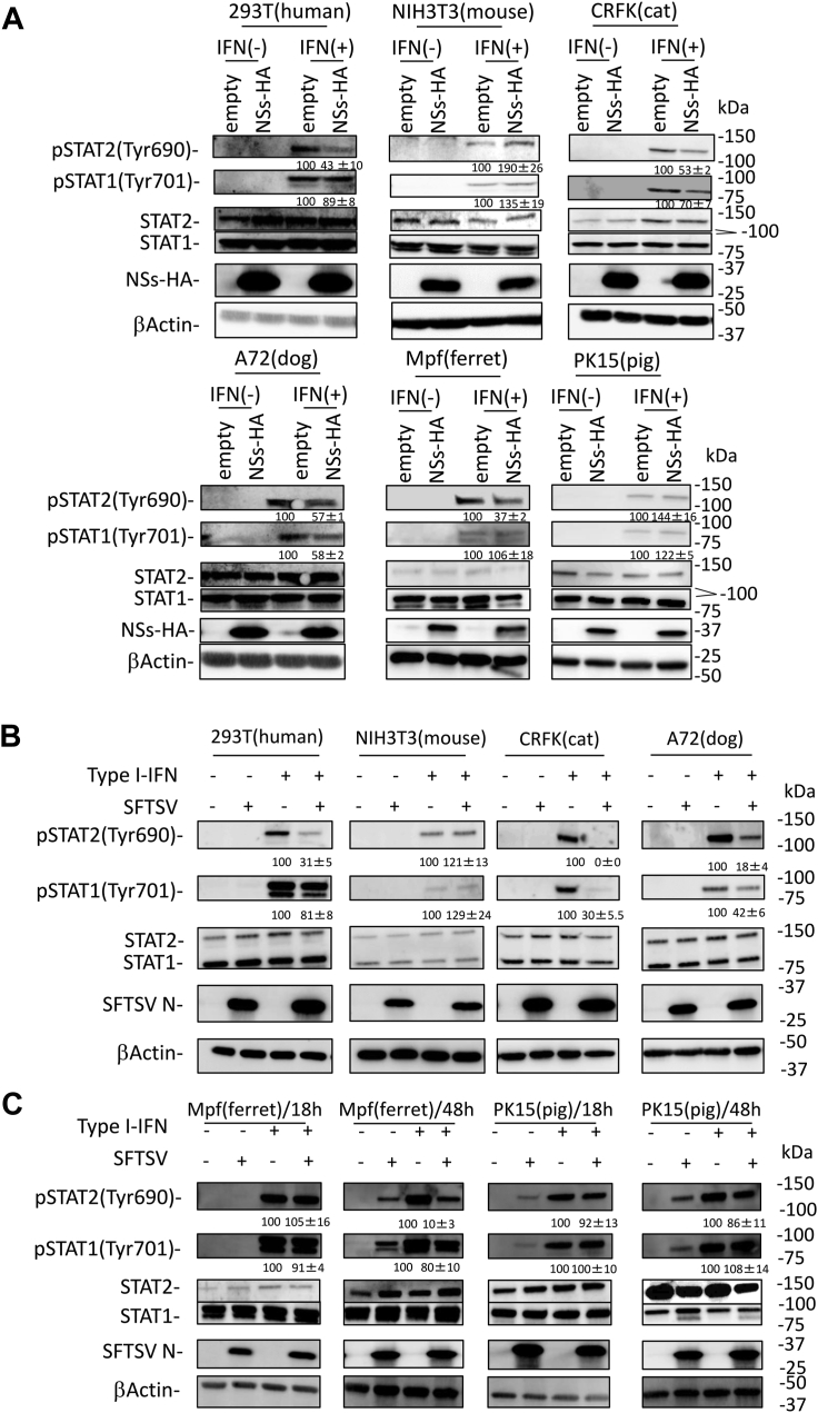
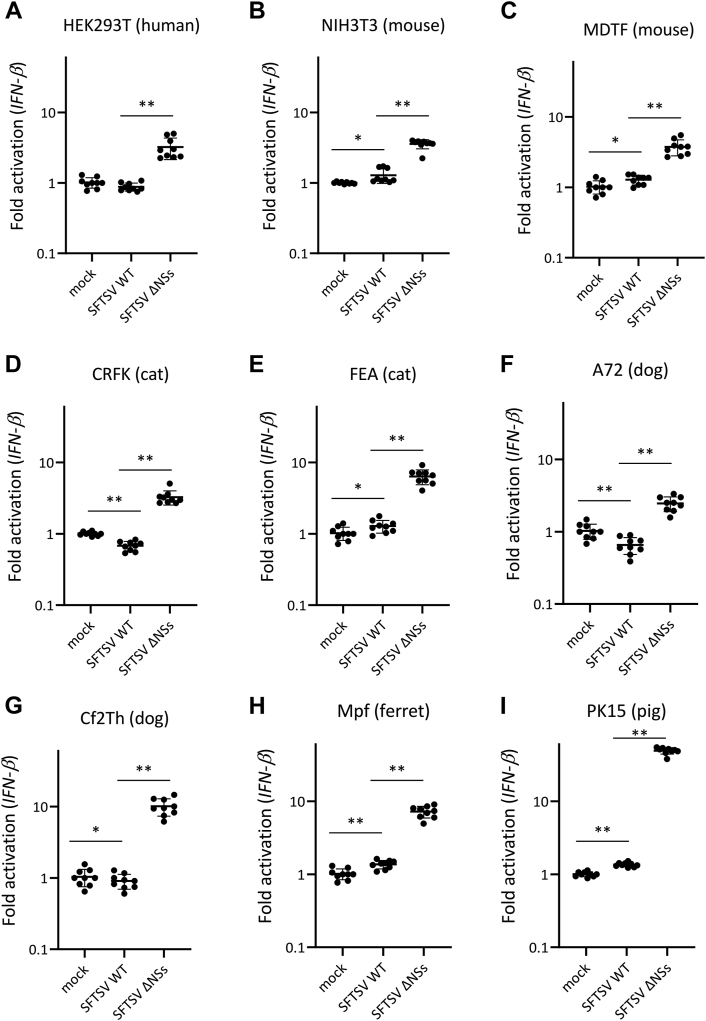
Figure 2
Figure 2
Ability of NSs as an antagonist to IFN-I induction during infection in cells derived from different animal species. (A) HEK293T (human), (B) NIH3T3 (mouse), (C) MDTF (mouse), (D) CRFK (cat), (E) FEA (cat), (F) A72 (dog), (G) Cf2Th (dog), (H) Mpf (ferret), and (I) PK15 (pig) cells were mock infected or infected with SFTSV WT or SFTSV ΔNSs at an MOI of 10. After 18 h, total RNA was extracted from these cells, and an mRNA expression level of IFN-β was quantified by real-time RT–PCR. The mRNA expression levels of IFN-β in mock-infected cells were set as 1. These assays were independently performed in triplicate. Values are the averages with SDs of data from nine results obtained from three experiments (n = 9). ∗p < 0.05, ∗∗p < 0.01, SFTSV WT infected versus SFTSV ΔNSs infected. Each exact p value, average, and SD is shown in Table S2. CRFK, Crandell–Rees feline kidney; HEK293T, human embryonic kidney 293T cell line; IFN-I, type I interferon; MDTF, Mus dunni tail fibroblast; MOI, multiplicity of infection; NSs, nonstructural protein; SFTSV, severe fever with thrombocytopenia syndrome virus.
Figure 3
Figure 3
Binding of NSs with TBK1.A, HEK293T (human) cells were transfected with the expression plasmid for HA-tagged NSs and each of the FLAG-tagged TBK1, and then co-IP assays were performed. B, colocalization of NSs with TBK1. HEK293T (human) cells were transfected with the expression plasmid for HA-tagged NSs and each of the FLAG-tagged TBK1. IFA was also performed with NSs, TBK1, and the nuclei, shown in red, green, and blue, respectively. Scale bar represents 20 μm. Representative results of Western blotting assays (A) and IFA (B) are shown. co-IP, coimmunoprecipitation; HA, hemagglutinin; HEK293T, human embryonic kidney 293T cell line; IFA, immunofluorescence assay; NSs, nonstructural protein; TBK1, TANK-binding kinase 1.
Figure 4
Figure 4
Function of NSs as an antagonist to IFN-I signaling in cells derived from different animal species.A, HEK293T (human), (B) NIH3T3 (mouse), (C) MDTF (mouse), (D) CRFK (cat), (E) FEA (cat), (F) A72 (dog), (G) Cf2Th (dog), (H) Mpf (ferret), and (I) PK15 (pig) cells were mock infected or infected with SFTSV WT or SFTSV ΔNSs at an MOI of 10. After 24 or 48 h, the cells were treated with IFN-αA/D (500 U/ml) or left untreated for 18 h. Expression levels of ISG56 mRNAs in each cell line were measured by real-time RT–PCR. The mRNA expression levels of ISG56 in IFN-αA/D-untreated cells were set as 1. These assays were independently performed in triplicate. Values are the averages with SDs of data from nine results obtained from three experiments (n = 9). The black and blue dots show the result at 42 and 66 h after infection, respectively. ∗p < 0.05, ∗∗p < 0.01, versus SFTSV WT infected. Each exact p value, average, and SD is shown in Table S3. CRFK, Crandell–Rees feline kidney; HEK293T, human embryonic kidney 293T cell line; IFN-I, type I interferon; MDTF, Mus dunni tail fibroblast; MOI, multiplicity of infection; NSs, nonstructural protein; SFTSV, severe fever with thrombocytopenia syndrome virus.
Figure 5
Figure 5
Suppression of STAT1 and STAT2 phosphorylation by NSs.A, HEK293T (human), NIH3T3 (mouse), CRFK (cat), A72 (dog), Mpf (ferret), or PK15 (pig) cells transfected with expression plasmid for HA-tagged NSs were treated with IFN-αA/D (2000 U/ml) or left untreated for 30 min and were then lysed for detection of each protein expression by immunoblotting. B and C, HEK293T (human), NIH3T3 (mouse), CRFK (cat), A72 (dog), Mpf (ferret), or PK15 (pig) cells were infected with SFTSV at an MOI of 10. C, after 18 h or 48 h, these cells were treated with IFN-αA/D (2000 U/ml) or left untreated for 30 min and were then lysed for detection of each protein expression by immunoblotting. The relative expression rates of pSTAT1 and pSTAT2 in mock-transfected cells and uninfected cells treated with IFN-I are set at 100%. The assays were independently performed in triplicate. The data represent averages with SDs. CRFK, Crandell–Rees feline kidney; HA, hemagglutinin; HEK293T, human embryonic kidney 293T cell line; IFN, interferon; MOI, multiplicity of infection; NSs, nonstructural protein; SFTSV, severe fever with thrombocytopenia syndrome virus; STAT, signal transducer and activator of transcription.
Figure 6
Figure 6
Influence of NSs to STAT1 and STAT2 activation in cells derived from different animal species. HEK293T (human), NIH3T3 (mouse), CRFK (cat), A72 (dog), Mpf (ferret), or PK15 (pig) cells were transfected with the expression plasmid for HA-tagged NSs and treated with IFN-αA/D (2000 U/ml) for 30 min at 48 h post-transfection. IFA was performed to detect NSs, the nuclei, and pSTAT1 or pSTAT2, shown in red, blue, and green, respectively. Each figure in the red frame and blue frame indicates the result of indirect IFA with anti-pSTAT1 and anti-pSTAT2, respectively. The arrow and arrowhead indicate NSs-expressing and nonexpressing cells, respectively. Scale bar represents 50 μm. Representative results of IFA are shown. CRFK, Crandell–Rees feline kidney; HA, hemagglutinin; HEK293T, human embryonic kidney 293T cell line; IFA, immunofluorescence assay; IFN, interferon; NSs, nonstructural protein; STAT, signal transducer and activator of transcription.
Figure 7
Figure 7
Interaction of NSs with STAT2.A, NIH3T3 (mouse) cells were transfected with the expression plasmid for HA-tagged NSs and each of the His-tagged STAT2, and then co-IP assays were performed. B, colocalization of NSs with STAT2. NIH3T3 (mouse) cells were transfected with the expression plasmid for HA-tagged NSs and each of the His-tagged STAT2. IFA was also performed with NSs, STAT2, and the nuclei, shown in green, red, and blue, respectively. Scale bar represents 20 μm. C, identification of binding regions in porcine STAT2 to NSs. Schematic representation of the chimeric mutants of human, mouse, and pig STAT2 (upper). NIH3T3 (mouse) cells were transfected with the HA-tagged NSs and each of the His-tagged STAT2 chimeras, following which co-IP assays were performed (lower). D, the phylogenetic tree of the 101 to 315 region of each STAT2. This phylogenetic tree was constructed using the neighbor-joining method. The bootstrap values are indicated on each node. Representative results of Western blotting assays (A and C) and IFA (B) are shown. co-IP, coimmunoprecipitation; HA, hemagglutinin; IFA, immunofluorescence assay; NSs, nonstructural protein; STAT, signal transducer and activator of transcription.
Figure 8
Figure 8
Function of human, murine, porcine, and chimeric STAT2s in SFTSV infection. SFTSV was inoculated into HEK293T (human) cells transfected with expression plasmid for each STAT2 at MOI 1. After 24 hpi, cells were treated with IFN-αA/D (500 U/ml) for 48 h and then collected culture supernatants. The SFTSV titer in culture supernatants was determined by focus-forming assay (upper). The protein expression was detected by Western blotting (lower). The assays were independently performed in triplicate. Values are the averages with SDs of data from nine results obtained from three experiments (n = 9). ∗∗p < 0.01, versus human STAT2. Each exact p value, average, and SD is shown in Table S4. HEK293T, human embryonic kidney 293T cell line; hpi, hours postinfection; IFN, interferon; MOI, multiplicity of infection; SFTSV, severe fever with thrombocytopenia syndrome virus; STAT, signal transducer and activator of transcription.
Figure 9
Figure 9
Interaction of NSs with STAT1.A, HEK293T (human), NIH3T3 (mouse), CRFK (cat), A72 (dog), Mpf (ferret), and PK15 (pig) cells were transfected with the expression plasmid for HA-tagged NSs, and then co-IP assays were performed. The relative binding rates of NSs to human STAT1 are set at 100%. The assays were independently performed in triplicate. The data represent averages with SDs. The asterisks indicate the nonspecific bands. B, colocalization of NSs with STAT2. The expression plasmid for HA-tagged NSs was transfected to HEK293T (human), NIH3T3 (mouse), CRFK (cat), A72 (dog), Mpf (ferret), and PK15 (pig) cells. IFA was also performed with NSs, STAT1, and the nuclei, shown in green, red, and blue, respectively. Scale bar represents 20 μm. C, interaction of NSs with exogenous STAT1. HEK293T (human), NIH3T3 (mouse), and CRFK (cat) cells were transfected with the expression plasmid for HA-tagged NSs, His-tagged human STAT1 (hSTAT1), murine STAT1 (mSTAT1), feline STAT1 (fSTAT1), and human STAT2 (hSTAT2), and then co-IP assays were performed. D, Interaction activity of NSs to STAT1 and STAT2. The expression plasmid for HA-tagged NSs was transfected into NIH3T3 (mouse) cells with the expression plasmid for His-tagged human STAT1(H) or feline STAT1(F) and FLAG-tagged human STAT2 or feline STAT2, and then co-IP assays were performed. Representative results of Western blotting assays (A, C, and D) and IFA (B) are shown. CRFK, Crandell–Rees feline kidney; HA, hemagglutinin; HEK293T, human embryonic kidney 293T cell line; IFA, immunofluorescence assay; NSs, nonstructural protein; STAT, signal transducer and activator of transcription.
Figure 10
Figure 10
Schematic diagram of NSs functionality in each animal. Schematic summary of the differential mechanisms by which SFTSV NSs inhibit the (A) IFN-I induction and (B) signaling pathways among cells derived from various animal species. Dashed red lines indicate week interactions. Solid red lines indicate strong interactions. P indicates the phosphorylation. IFNAR, interferon-α/β receptor; IKKε, inhibitor of kappa B kinase-ε; JAK1, Janus kinase; MAVS, mitochondrial antiviral signaling protein; NSs, nonstructural protein; SFTSV, severe fever with thrombocytopenia syndrome virus.

References

    1. Yu X.J., Liang M.F., Zhang S.Y., Liu Y., Li J.D., Sun Y.L., et al. Fever with thrombocytopenia associated with a novel bunyavirus in China. N. Engl. J. Med. 2011;364:1523–1532. - PMC - PubMed
    1. Denic S., Janbeih J., Nair S., Conca W., Tariq W.U.Z., Al-Salam S. Acute thrombocytopenia, leucopenia, and multiorgan dysfunction: the first case of SFTS bunyavirus outside China? Case Rep. Infect. Dis. 2011;2011 - PMC - PubMed
    1. Kim K.H., Yi J., Kim G., Choi S.J., Jun K.I., Kim N.H., et al. Severe fever with thrombocytopenia syndrome, South Korea, 2012. Emerg. Infect. Dis. 2013;19:1892–1894. - PMC - PubMed
    1. Takahashi T., Maeda K., Suzuki T., Ishido A., Shigeoka T., Tominaga T., et al. The first identification and retrospective study of severe fever with thrombocytopenia syndrome in Japan. J. Infect. Dis. 2014;209:816–827. - PMC - PubMed
    1. Tran X.C., Yun Y., Van An.L., Kim S.H., Thao N.T.P., Man P.K.C., et al. Endemic severe fever with thrombocytopenia syndrome, Vietnam. Emerg. Infect. Dis. 2019;25:1029–1031. - PMC - PubMed

Publication types

Substances