Review
. 2023 Apr 28:11:1173311.
doi: 10.3389/fped.2023.1173311. eCollection 2023.

Neonatologist-performed point-of-care abdominal ultrasound: What have we learned so far?

Affiliations
Review

Neonatologist-performed point-of-care abdominal ultrasound: What have we learned so far?

Archana Priyadarshi et al. Front Pediatr. .

Abstract

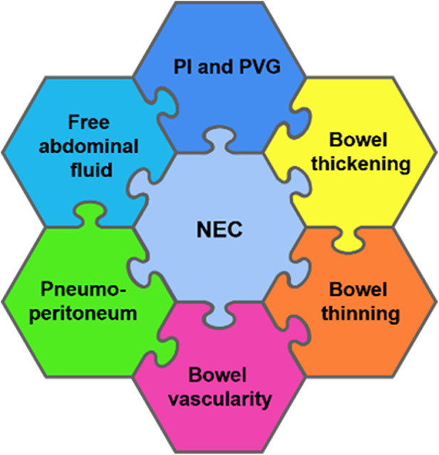
This review describes the sonographic appearances of the neonatal bowel in Necrotising enterocolitis. It compares these findings to those seen in midgut-Volvulus, obstructive intestinal conditions such as milk-curd obstruction, and slow gut motility in preterm infants on continuous positive airway pressure (CPAP)-CPAP belly syndrome. Point-of-care bowel ultrasound is also helpful in ruling out severe and active intestinal conditions, reassuring clinicians when the diagnosis is unclear in a non-specific clinical presentation where NEC cannot be excluded. As NEC is a severe disease, it is often over-diagnosed, mainly due to a lack of reliable biomarkers and clinical presentation similar to sepsis in neonates. Thus, the assessment of the bowel in real-time would allow clinicians to determine the timing of re-initiation of feeds and would also be reassuring based on specific typical bowel characteristics visualised on the ultrasound.

Keywords: NEC=necrotizing enterocolitis; bowel; neonates; ultrasound; vovulus.


Conflict of interest statement

The authors declare that the research was conducted in the absence of any commercial or financial relationships that could be construed as a potential conflict of interest.

Figures

Figure 1
Figure 1
A schematic approach to perform a structured point-of-care abdominal ultrasound for bowel assessment in neonates.
Figure 2
Figure 2
The sonographic appearance of normal bowel. (A) 2-D Grayscale image of normal bowel with normal intraluminal air seen as echogenic dots. (B) Color Doppler imaging of bowel showing normal bowel vascularity.
Figure 3
Figure 3
A schematic diagram showing a detailed radiological assessment of the neonatal bowel for the diagnosis of necrotizing enterocolitis (NEC). PI, pneumatosis intestinalis and PVG, portal venous gas.
Figure 4
Figure 4
Stages of necrotizing enterocolitis (NEC). Laparotomy findings of (A) Localized NEC. (B) Severe extensive NEC with micro-perforations.
Figure 5
Figure 5
The sonographic appearance of the bowel wall during various stages of necrotizing enterocolitis on point-of-care abdominal ultrasound. (A) Bowel wall thickening. (B) Bowel wall thinning.
Figure 6
Figure 6
The sonographic appearance of edema and thickening of small intestinal mucosal folds as seen in necrotizing enterocolitis on point-of-care abdominal ultrasound. Grayscale imaging showing the echogenic stripping of the small intestinal folds due to edema of the valve conniventes. Color Doppler imaging of these folds showing hyperemia.
Figure 7
Figure 7
The sonographic appearance of intramural gas trapping—pneumatosis intestinalis, which is seen as white echogenic dots.
Figure 8
Figure 8
The sonographic appearance demonstrating real-time visualization of portal venous gas release seen as bright echogenic dots (streaming of gas bubbles through the portal vein). The entrapment of these bubbles within the porto-hepatic venous architecture results in abnormal hepatic echotexture seen as interspersed bright echogenic dots as opposed to the normal homogenous hepatic echotexture of a normal Liver.
Figure 9
Figure 9
The sonographic appearance on the color Doppler showing the specific vascular patterns seen in necrotizing enterocolitis: the Y, ring, and the Zebra pattern.
Figure 10
Figure 10
The sonographic appearance of the intra-abdominal complex fluid collections seen in necrotizing enterocolitis. (A) Thickened bowel loops floating within the fluid with echogenic foci (complex ascites). (B) Thickening of the omentum as seen within this complex fluid. * Thickened omentum.
Figure 11
Figure 11
The sonographic appearance of neonatal midgut volvulus—the whirlpool sign. Color Doppler. Grayscale imaging.
Figure 12
Figure 12
The sonographic appearance of superior mesenteric vessels—superior mesenteric artery (SMA) and the superior mesenteric vein (SMV) anatomical configuration. (A) Normal SMV and SMA relationship (SMV seen at 11 O’clock position to the SMA). (B) Arterial Doppler through the SMA confirming its anatomical location. (C) Sagittal image showing the relationship of the superior mesenteric artery to the aorta and the coeliac trunk. (D) Comparison of the normal (SMV seen at 11 O’clock position to the SMA) and abnormal positions (SMV seen at 01 O’clock position to the SMA).
Figure 13
Figure 13
Images showing the clinical presentation of the abdomen in milk curd obstruction and the intra-operative findings in the same patient.
Figure 14
Figure 14
Milk-curd obstruction concurrent x-ray and point-of-care abdominal ultrasound findings. (A) Plain abdominal x-ray showing paucity of bowel gas with a soap-bubbly appearance on the right. (B) The sonographic appearance of the bowel showing islets of echogenic homogenous material within the lumen maintaining the normal bowel-wall sonographic appearance.

References

    1. Cuna AC, Lee JC, Robinson AL, Allen NH, Foley JE, Chan SS. Bowel ultrasound for the diagnosis of necrotizing enterocolitis: a meta-analysis. Ultrasound Q. (2018) 34(3):113–8. 10.1097/RUQ.0000000000000342 - DOI - PubMed
    1. Epelman M, Daneman A, Navarro OM, Morag I, Moore AM, Kim JH, et al. Necrotizing enterocolitis: review of state-of-the-art imaging findings with pathologic correlation. RadioGraphics. (2007) 27(2):285–305. 10.1148/rg.272055098 - DOI - PubMed
    1. Dinakara Prithviraj BS, Suresh A, Balaraju V, Swagata Mondal. Comparison between x-ray and abdominal ultrasound findings of necrotizing enterocolitis, its usefulness in early diagnosis, prognosis, and to assess, is this is the time to change our view of surgeon’s intervention according to the bell’s criteria. Int J Sci Study. (2015) 3(4):119–30. 10.17354/ijss/2015/319 - DOI
    1. Choi G, Je BK, Kim YJ. Gastrointestinal emergency in neonates and infants: a pictorial essay. Korean J Radiol. (2022) 23(1):124–38. 10.3348/kjr.2021.0111 - DOI - PMC - PubMed
    1. Priyadarshi A, Rogerson S, Hinder M, Tracy M. Neonatologist performed point-of-care bowel ultrasound: is the time right? Australas J Ultrasound Med. (2019) 22(1):15–25. 10.1002/ajum.12114 - DOI - PMC - PubMed