

Diagnostic and Therapeutic Challenges of Concurrent Intracranial Hemorrhage and Cerebral Venous Thrombosis in a Patient With Acute Lymphoblastic Leukemia: A Case Report and Literature Review
- PMID: 37187664
- PMCID: PMC10177010
- DOI: 10.7759/cureus.37482
Diagnostic and Therapeutic Challenges of Concurrent Intracranial Hemorrhage and Cerebral Venous Thrombosis in a Patient With Acute Lymphoblastic Leukemia: A Case Report and Literature Review
Abstract
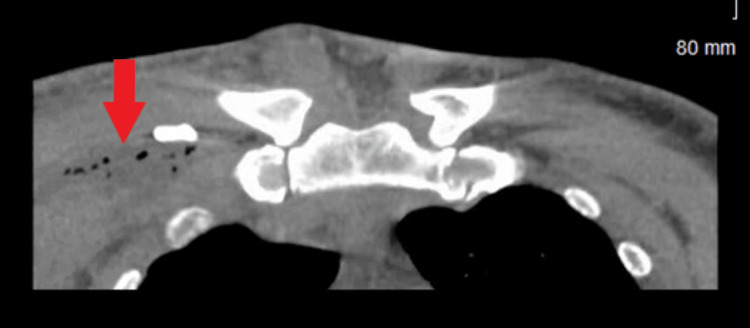
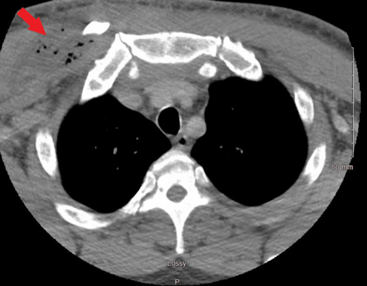
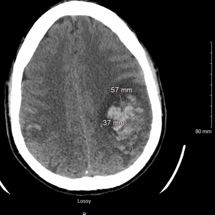
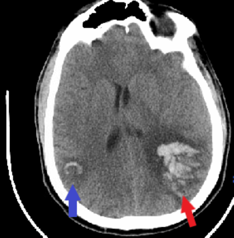
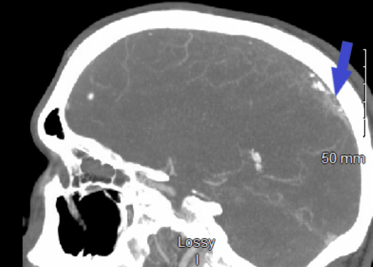
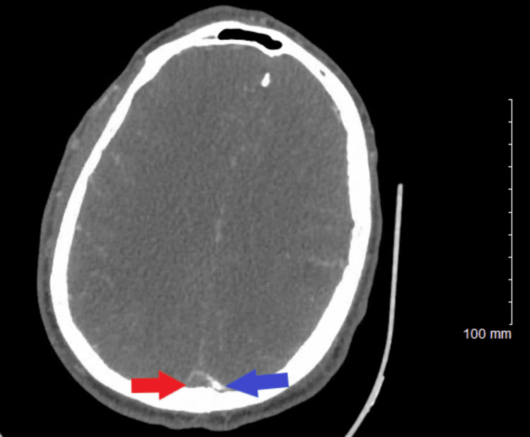
Cerebral venous sinus thrombosis (CVST) is a cerebrovascular condition due to the thrombosis of cerebral venous sinuses, leading to intracranial hemorrhage, increased intracranial pressure, focal deficit, seizure, toxic edema, encephalopathy, and death. The diagnosis and therapeutic approach of CVST remain challenging because of its highly nonspecific clinical presentation including headaches, seizures, focal neurologic deficits, and altered mental status, etc. Anticoagulation is the mainstay of CVST treatment and should be started as soon as the diagnosis is confirmed. Here, we present the case of a 34-year-old male construction worker who presented to the emergency department with a complaint of right chest wall pain and swelling. He was admitted to the hospital following a diagnosis of anterior chest wall abscess and mediastinitis. During hospitalization, his complete blood count revealed pancytopenia with blast cells, and bone marrow biopsy revealed 78.5% lymphoid blasts by aspirate differential count and hypercellular marrow (100%) with decreased hematopoiesis. He developed concurrent CVST and intracranial hemorrhage while receiving CALGB10403 (vincristine, daunorubicin, pegaspargase, prednisone) with intrathecal cytarabine induction chemotherapy for acute lymphoblastic leukemia (ALL). The patient failed two standard chemotherapy for ALL and achieved remission while on third-line chemotherapy with an anti-CD19 monoclonal antibody, blinatumomab. Although this patient had an MRI scan of the brain with multiple follow-up non-contrast CT scans, it was CT angiography that revealed CVST. This showed the diagnostic challenge in CVST, with CT and MRI venography having excellent sensitivity in diagnosing CVST. Risk factors for CVST in our patient were ALL and its intensive induction chemotherapy with pegaspargase.
Keywords: acute lymphoblastic leukemia (all); cerebral venous sinus thrombosis (cvst); cerebral venous thrombosis; concurrent intracranial hemorrhage; intracranial hemorrhage.
Copyright © 2023, Torere et al.
Conflict of interest statement
The authors have declared that no competing interests exist.
Figures
References
-
- Diagnosis and management of cerebral venous thrombosis: a statement for healthcare professionals from the American Heart Association/American Stroke Association. Saposnik G, Barinagarrementeria F, Brown RD Jr, et al. Stroke. 2011;42:1158–1192. - PubMed
-
- Prognosis of cerebral vein and dural sinus thrombosis: results of the International Study on Cerebral Vein and Dural Sinus Thrombosis (ISCVT) Ferro JM, Canhão P, Stam J, Bousser MG, Barinagarrementeria F. Stroke. 2004;35:664–670. - PubMed
-
- Cerebral venous and sinus thrombosis in women. Coutinho JM, Ferro JM, Canhão P, Barinagarrementeria F, Cantú C, Bousser MG, Stam J. Stroke. 2009;40:2356–2361. - PubMed
Publication types
LinkOut - more resources
Full Text Sources
Research Materials