. 2023 Apr 28:16:1161086.
doi: 10.3389/fnmol.2023.1161086. eCollection 2023.

Neonatal subarachnoid hemorrhage disrupts multiple aspects of cerebellar development

Affiliations

Neonatal subarachnoid hemorrhage disrupts multiple aspects of cerebellar development

David F Butler et al. Front Mol Neurosci. .

Abstract

Over the past decade, survival rates for extremely low gestational age neonates (ELGANs; <28 weeks gestation) has markedly improved. Unfortunately, a significant proportion of ELGANs will suffer from neurodevelopmental dysfunction. Cerebellar hemorrhagic injury (CHI) has been increasingly recognized in the ELGANs population and may contribute to neurologic dysfunction; however, the underlying mechanisms are poorly understood. To address this gap in knowledge, we developed a novel model of early isolated posterior fossa subarachnoid hemorrhage (SAH) in neonatal mice and investigated both acute and long-term effects. Following SAH on postnatal day 6 (P6), we found significant decreased levels of proliferation with the external granular layer (EGL), thinning of the EGL, decreased Purkinje cell (PC) density, and increased Bergmann glial (BG) fiber crossings at P8. At P42, CHI resulted in decreased PC density, decreased molecular layer interneuron (MLI) density, and increased BG fiber crossings. Results from both Rotarod and inverted screen assays did not demonstrate significant effects on motor strength or learning at P35-38. Treatment with the anti-inflammatory drug Ketoprofen did not significantly alter our findings after CHI, suggesting that treatment of neuro-inflammation does not provide significant neuroprotection post CHI. Further studies are required to fully elucidate the mechanisms through which CHI disrupts cerebellar developmental programming in order to develop therapeutic strategies for neuroprotection in ELGANs.

Keywords: Purkinje cells; cerebellar development; cerebellar granule cells; cerebellar hemorrhage; preterm brain injury.


Conflict of interest statement

The authors declare that the research was conducted in the absence of any commercial or financial relationships that could be construed as a potential conflict of interest.

Figures

Figure 1
Figure 1
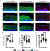
CHI results in decreased BrDU and Ki67 labeling indices, thinning of the EGL, decreased PC density, and increased BG fiber crossings at P8. (A) Schematic illustrating isolated posterior fossa hemorrhage model with typical P6 cerebellar architecture. (B) Representative comparative images of gross findings between aCSF and CHI groups; note large hematoma formation overlying left cerebellar hemisphere in the CHI specimen. (C) Representative confocal images at 40X of BrDU and Ki67 staining within the EGL. Only cells within the EGL were included in analysis. Scale bars are 25 μm. (D) Quantification of labeling indices for BrDU (left) and Ki67 (right); data obtained from 200 μm segment from lobule VI and VII (N = 2, n = 12 for Bup_Blood; N = 3, n = 16 for Bup_aCSF and Keto_Blood). (E) Representative confocal images at 20X magnification of lobule VII demonstrating differences in local findings including presence of large RBC collections (yellow dashed outline), PC density (white arrows), thickness of EGL, and architecture of BG fibers. Scale bars are 100 μm. (F) Quantification of EGL thickness for all treatment groups (N = 4, n = 16). (G) Quantification of PC density in lobule VI and VII for all treatment groups (N = 3, n = 24). (H) Quantification of BG fiber crossings in lobule I, VII-IX for all treatment groups (N = 3, n = 27). Significance defined as p < 0.05 by one-way ANOVA with Tukey post-test. Data are presented as SEM with minimum and maximum values. N = number of animals per group included for analysis; n = number of section segments per group included for analysis. **p-value ≤ 0.01; ****p-value ≤ 0.0001.
Figure 2
Figure 2
CHI results in decreased PC density, decreased MLI density, and increased BG fiber crossings within the ML at P42. (A) Representative confocal images at 20X demonstrating difference in PC (white arrowhead) density (left), MLI (white arrowhead) density (middle), and BG fiber density and morphology (right). Scale bars are 100 μm. (B) Quantification of PC density in lobule VI-X for all treatment groups (N = 3, n = 9). (C) Quantification of MLI density in lobule VI-X for all treatment groups (N = 3, n = 9). (D) Quantification of BG fiber crossings within the ML for all treatment groups (N = 3, n = 27). Significance defined as p < 0.05 by one-way ANOVA with Tukey's post-test. Data are presented as SEM with minimum and maximum values. N = number of animals per group included for analysis, n = number of section segments per group included for analysis.
Figure 3
Figure 3
CHI did not result in significant motor learning/coordination deficits. (A) Schematic of motor testing assays and timeline. (B) Quantification of time to fall for the inverted screen test for all groups (N = 10*). (C) Quantification of the time to fall for Day 1 of Rotarod training for all groups (N = 10*). (D) Quantification of the time to fall for Days 1-3 of Rotarod training for all groups (N = 10*). Data are presented as mean ± SEM (B) or SEM with minimum and maximum values (C, D). N = number of animals per group included for analysis. *Keto_Blood group with N = 8. Significance defined as p < 0.05 by one-way ANOVA with Tukey's post-test. Significance defined as p < 0.05 by two-way ANOVA with Tukey's post-test.

Update of

References

    1. Abraham H., Tornoczky T., Kosztolanyi G., Seress L. (2001). Cell formation in the cortical layers of the developing human cerebellum. Int. J. Dev. Neurosci. 19, 53–62. 10.1016/S0736-5748(00)00065-4 - DOI - PubMed
    1. Aldinger K. A., Timms A. E., Thomson Z., Mirzaa G. M., Bennett J. T., Rosenberg A. B., et al. . (2019). Redefining the etiologic landscape of cerebellar malformations. Am. J. Hum. Genet. 105, 606–615. 10.1016/j.ajhg.2019.07.019 - DOI - PMC - PubMed
    1. Barenberg P., Strahlendorf H., Strahlendorf J. (2001). Hypoxia induces an excitotoxic-type of dark cell degeneration in cerebellar Purkinje neurons. Neurosci. Res. 40, 245–254. 10.1016/S0168-0102(01)00234-6 - DOI - PubMed
    1. Barron T., Kim J. H. (2020). Preterm birth impedes structural and functional development of cerebellar purkinje cells in the developing baboon cerebellum. Brain Sci. 10:897. 10.3390/brainsci10120897 - DOI - PMC - PubMed
    1. Bartschat S., Fieguth A., Könemann J., Schmidt A., Bode-Jänisch S. (2012). Indicators for acute hypoxia–an immunohistochemical investigation in cerebellar Purkinje-cells. Forensic Sci. Int. 223, 165–170. 10.1016/j.forsciint.2012.08.023 - DOI - PubMed