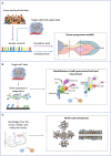
Review
. 2023 Apr 28:13:1164535.
doi: 10.3389/fonc.2023.1164535. eCollection 2023.

Tumor heterogeneity: preclinical models, emerging technologies, and future applications

Affiliations
Review

Tumor heterogeneity: preclinical models, emerging technologies, and future applications

Marco Proietto et al. Front Oncol. .

Abstract

Heterogeneity describes the differences among cancer cells within and between tumors. It refers to cancer cells describing variations in morphology, transcriptional profiles, metabolism, and metastatic potential. More recently, the field has included the characterization of the tumor immune microenvironment and the depiction of the dynamics underlying the cellular interactions promoting the tumor ecosystem evolution. Heterogeneity has been found in most tumors representing one of the most challenging behaviors in cancer ecosystems. As one of the critical factors impairing the long-term efficacy of solid tumor therapy, heterogeneity leads to tumor resistance, more aggressive metastasizing, and recurrence. We review the role of the main models and the emerging single-cell and spatial genomic technologies in our understanding of tumor heterogeneity, its contribution to lethal cancer outcomes, and the physiological challenges to consider in designing cancer therapies. We highlight how tumor cells dynamically evolve because of the interactions within the tumor immune microenvironment and how to leverage this to unleash immune recognition through immunotherapy. A multidisciplinary approach grounded in novel bioinformatic and computational tools will allow reaching the integrated, multilayered knowledge of tumor heterogeneity required to implement personalized, more efficient therapies urgently required for cancer patients.

Keywords: heterogeneity models; human in vitro models; tumor heterogeneity; tumor immune microenvironment; tumor microenvironment (TME).


Conflict of interest statement

The authors declare that the research was conducted in the absence of any commercial or financial relationships that could be construed as a potential conflict of interest.

Figures
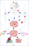
Figure 1
Figure 1
Stroma (A), immune cells (B), nutrients present in the microenvironment (C), and intrinsic factors such as DNA damage and epigenome (D) work together to produce the primary tumor heterogeneity (E). Intratumor heterogeneity can arise from the primary tumor (F) or from its metastasis (G). Both processes collaborate in the establishment of intertumoral heterogeneity in the population (H).
Figure 2
Figure 2
Models to study tumor heterogeneity: (A) non-murine models (Drosophila melanogaster, Danio renio, Saccharomyces cerevisiae), (B) murine models (syngeneic models, GEMMs), (C) human in vitro models (organoids, organ-on-chip), and (D) humanized murine models.
Figure 3
Figure 3
Brief description of the major spatial technologies. (A) CODEX is based on a panel of antibodies that binds to specific fluorescent reporters that reveal their position during the imaging phase. At the end of the first cycle of image acquisition, the reporters are detached, and another cycle with new reporters starts. (B) 10X Visium is based on slides of barcoded capture probes that bind to the polyA tail of RNAs released from the tissue. RNA is retrotranscribed into cDNA and sequenced. (C) GeoMX DSP is based on panels of antibodies or photocleavable barcoded probes. Once an area of interest is selected, a stream of light releases the probes that are lately sequenced. (D) MERFISH is based on fluorescently tagged probes that label RNA of interest. Sequential rounds of imaging enable spatial resolution. All the pictures have been adapted from the providers’ web pages.
Figure 4
Figure 4
Schematic representation of the inputs (left) and outputs (right) of the main families of computational approaches to tackle intratumor heterogeneity. (A) Cancer progression models. (B) Clustering of single cells. (C) Multiscale modeling and simulation.
Figure 5
Figure 5
The different components of the tumor microenvironment: (A) the immune system, (B) stroma, and (C) external factors.
Figure 6
Figure 6
Key properties (cost, time, scalability, ease of manipulation, direct translation to TME heterogeneity) of the different models highlighted in this review. From left to right: murine models, non-murine models, and human models. + = low, ++ = medium, +++ = good / high, ++++ = excellent / very high.

References

    1. Vendramin R, Litchfield K, Swanton C. Cancer evolution: Darwin and beyond. EMBO J (2021) 40(18). doi: 10.15252/embj.2021108389 - DOI - PMC - PubMed
    1. Kashyap A, Rapsomaniki MA, Barros V, Fomitcheva-Khartchenko A, Martinelli AL, Rodriguez AF, et al. . Quantification of tumor heterogeneity: from data acquisition to metric generation. Trends Biotechnol (2022) 40(6):647–76. http://www.cell.com/article/S0167779921002675/fulltext. doi: 10.1016/j.tibtech.2021.11.006 - DOI - PubMed
    1. McGranahan N, Swanton C. Clonal heterogeneity and tumor evolution: past, present, and the future. Cell (2017) 168(4):613–28. doi: 10.1016/j.cell.2017.01.018 - DOI - PubMed
    1. Gascoigne KE, Taylor SS. Cancer cells display profound intra- and interline variation following prolonged exposure to antimitotic drugs. Cancer Cell (2008) 14(2):111–22. doi: 10.1016/j.ccr.2008.07.002 - DOI - PubMed
    1. Jänicke RU, Sohn D, Schulze-Osthoff K. The dark side of a tumor suppressor: anti-apoptotic p53. Cell Death Differ (2008) 15(6):959–76. doi: 10.1038/cdd.2008.33 - DOI - PubMed