Case Reports
. 2023 Feb 17;7(3):249-253.
doi: 10.1177/24741264231152675. eCollection 2023 May-Jun.

Management of Atypical Central Serous Chorioretinopathy Mimicking Vogt-Koyanagi-Harada Disease

Affiliations
Case Reports

Management of Atypical Central Serous Chorioretinopathy Mimicking Vogt-Koyanagi-Harada Disease

Nam V Nguyen et al. J Vitreoretin Dis. .

Abstract

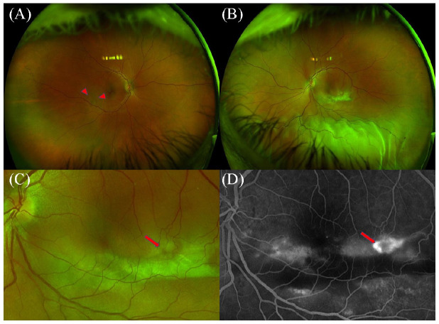
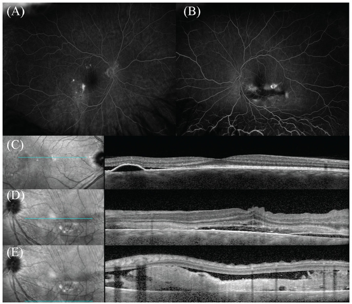
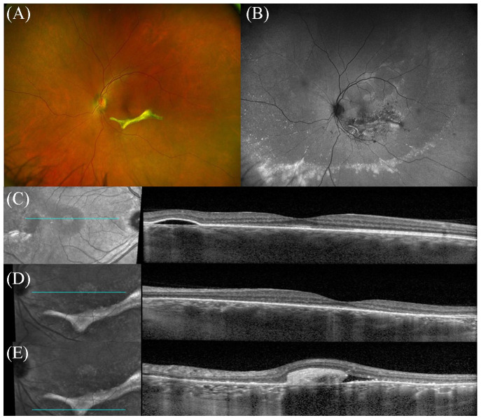
Purpose: To report a case of multifocal central serous chorioretinopathy (CSCR) mimicking Vogt-Koyanagi-Harada (VKH) disease. Methods: A 42-year-old man was evaluated for an exudative retinal detachment (RD) with a presumptive diagnosis of VKH while being treated with corticosteroids. The examination showed subretinal fibrin deposition with a bullous, exudative, macula-involved RD in the left eye and a progressive decline in visual acuity (VA) to hand motions. Multimodal imaging showed multifocal hyperfluorescent leaks bilaterally by angiography, highly suggestive of CSCR exacerbated by corticosteroids. Results: After the multifocal CSCR diagnosis, the systemic corticosteroids were tapered and eventually discontinued. The patient was then managed with focal laser photocoagulation, photodynamic therapy, and acetazolamide. The VA improved to 20/30 with complete resolution of the bullous RD at the 12-month follow-up. Conclusions: Extensive bullous RD with subretinal fibrin deposition is an infrequent manifestation of CSCR commonly associated with corticosteroid use that can mimic VKH. Thus, it is important to distinguish CSCR from VKH and the potential of combination therapy in managing chronic multifocal CSCR with a bullous RD.

Keywords: Vogt-Koyanagi-Harada; central serous chorioretinopathy; corticosteroids; retinal detachment.


Conflict of interest statement

The author(s) declared no potential conflicts of interest with respect to the research, authorship, and/or publication of this article.

Figures

Figure 1.
Figure 1.
Ultra-widefield fundus photography of the right eye shows retinal pigment epithelium detachment with pigmentary changes (arrowheads) descending inferiorly (A). A large subretinal fibrinous lesion is noted inferior to the macula along with inferior bullous retinal detachment in the left eye (B). Arrow points to the dark spot within the subretinal lesion (C), which corresponds to a focal area of hyperfluorescence on fluorescein angiography (arrow) (D).
Figure 2.
Figure 2.
(A) Fluorescein angiography (FA) of the right eye shows multiple late staining lesions with surrounding leakage temporal and superior to the macula. (B) FA of the left eye shows multiple pinpoint leakage with pooling inferiorly. (C) Optical coherence tomography (OCT) of the right eye shows serous retinal pigment epithelium detachment (PED) with associated subretinal fluid. (D) Left eye shows internal limiting membrane folds and serous PED with significant subretinal fluid. (E) Inferior B-scan of the left eye OCT shows significant fibrin deposit associated with significant subretinal fluid.
Figure 3.
Figure 3.
(A) Ultra-widefield fundus photography of the left eye still shows a well-demarcated area of subretinal fibrosis inferior the fovea with resolution of the exudative retinal. (B) Fundus autofluorescence of the left eye shows significant pigmentary changes around the fibrinous lesion as well as inferior hyperfluorescent deposits. (D and E) OCT of the left eye shows resolution of central subretinal fluid, although mild subretinal fluid associated with the subretinal fibrosis can be observed. (C) OCT of the right eye shows pigment epithelium detachment without associated subretinal fluid.

References

    1. Wang M, Munch IC, Hasler PW, Prunte C, Larsen M.Central serous chorioretinopathy. Acta Ophthalmol. 2008;86(2):126-145. doi:10.1111/j.1600-0420.2007.00889.x - DOI - PubMed
    1. Parchand S, Gupta V, Gupta A, Dogra MR, Singh R, Sharma A.Dark spot in fibrinous central serous chorioretinopathy masquerading choroiditis. Ocul Immunol Inflamm. 2013;21(3):201-206. doi:10.3109/09273948.2013.765015 - DOI - PubMed
    1. Aggarwal K, Agarwal A, Deokar A, et al.. Distinguishing features of acute Vogt-Koyanagi-Harada disease and acute central serous chorioretinopathy on optical coherence tomography angiography and en face optical coherence tomography imaging. J Ophthalmic Inflamm Infect. 2017;7(1):3. doi:10.1186/s12348-016-0122-z - DOI - PMC - PubMed
    1. Cebeci Z, Oray M, Bayraktar S, Tugal-Tutkun I, Kir N.Atypical central serous chorioretinopathy. Turk J Ophthalmol. 2017;47(4):238-242. doi:10.4274/tjo.38039 - DOI - PMC - PubMed
    1. Papadia M, Herbort CP.Central serous chorioretinopathy mistaken for tuberculous choroiditis. J Ophthalmic Vis Res. 2011;6(4):334-337. - PMC - PubMed

Publication types