. 2023 May 15;6(7):e202201740.
doi: 10.26508/lsa.202201740. Print 2023 Jul.

Functional characterization of C21ORF2 association with the NEK1 kinase mutated in human in diseases

Affiliations

Functional characterization of C21ORF2 association with the NEK1 kinase mutated in human in diseases

Mateusz Gregorczyk et al. Life Sci Alliance. .

Abstract

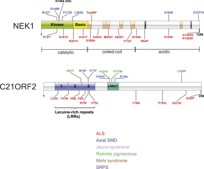
The NEK1 kinase controls ciliogenesis, mitosis, and DNA repair, and NEK1 mutations cause human diseases including axial spondylometaphyseal dysplasia and amyotrophic lateral sclerosis. C21ORF2 mutations cause a similar pattern of human diseases, suggesting close functional links with NEK1 Here, we report that endogenous NEK1 and C21ORF2 form a tight complex in human cells. A C21ORF2 interaction domain "CID" at the C-terminus of NEK1 is necessary for its association with C21ORF2 in cells, and pathogenic mutations in this region disrupt the complex. AlphaFold modelling predicts an extended binding interface between a leucine-rich repeat domain in C21ORF2 and the NEK1-CID, and our model may explain why pathogenic mutations perturb the complex. We show that NEK1 mutations that inhibit kinase activity or weaken its association with C21ORF2 severely compromise ciliogenesis, and that C21ORF2, like NEK1 is required for homologous recombination. These data enhance our understanding of how the NEK1 kinase is regulated, and they shed light on NEK1-C21ORF2-associated diseases.


Conflict of interest statement

D Durocher is a shareholder and advisor of Repare Therapeutics.

Figures

Figure S1.
Figure S1.. Schematic diagrams of NEK1 and C21ORF2 depicting domain organization.
Schematic diagram denoting amino acid substitutions encoded by pathogenic mutations in NEK1 or C21ORF2.  Asterisk denotes truncation after the amino acid specified. The amino acid substitutions encoded by missense mutations are colour-coded according to disease as indicated.
Figure 1.
Figure 1.. Characterization of the endogenous NEK1–C21ORF2 complex.
(A) ARPE-19 cells were transfected with siRNA targeting either C21ORF2 (siRNA C21ORF2-2) or NEK1. Cell lysates were subjected to immunoblotting using in-house sheep polyclonal antibodies against C21ORF2 or NEK1 antibodies or GAPDH antibodies. Cell lysates treated with siRNA targeting luciferase served as a negative control. Asterisk denotes non-specific band; arrow denotes the specific bands. One of at least three independent experiments is shown. (B) ARPE-19 cells extracts were subjected to immunoprecipitation with in-house sheep antibodies against NEK1 or C21ORF2, or sheep IgG. Precipitates were subjected to SDS–PAGE and immunoblotting with the antibodies indicated, and input cell extracts were also included. Arrow denotes the C21ORF2 band. One of at least three independent experiments is shown. (C) Extracts from parental ARPE-19, NEK1–KO (clone NC51), and C21ORF2–KO (clone CB1) cells were subjected to size exclusion chromatography using a Superose 6 Increase 10/300 column. Alternate fractions were subjected to SDS–PAGE gel followed by immunoblotting using antibodies against NEK1 (Bethyl Laboratories) and C21ORF2. The elution positions of molecular mass markers are indicated with black arrows. Red arrows indicate bands corresponding to NEK1 or C21ORF2. One of two independent experiments is shown. (D) The fractions indicated in C were pooled and subjected to three rounds of immunodepletion with either in-house antibodies against NEK1 or C21ORF2 or sheep IgG antibodies. Beads and supernatants were analysed by SDS–PAGE and Western blotting the antibodies indicated. Molecular weight markers “kD” are indicated. One of two independent experiments is shown. Source data are available for this figure.
Figure S2.
Figure S2.. NEK1 regulates C21ORF2 protein levels.
(A, B) U2-O-S or HeLa cells were transfected with siRNAs indicated. Cell extracts were subjected to SDS–PAGE and immunoblotting with polyclonal sheep antibodies against NEK1 or C21ORF2 or monoclonal GAPDH antibodies. IRDye secondary antibodies were used for detection in an Odyssey CLx imager (LI-COR Biosciences). Band intensities were quantified using the analysis tool provided in Image Studio Lite software. NEK1 or GAPDH band intensities were normalised using the intensity of the corresponding GAPDH band and divided by the intensity of the first siRNA control–treated band. Two independent experiments are shown for each cell line; two technical replicates are included for each independent experiment. (C, D) NEK1–KO ARPE-19 cells were treated with MG-132 (C) or MLN-4924 (D) for the times and concentrations indicated. Extracts were subjected to SDS–PAGE and immunoblotting with indicated antibodies. Immunostaining for HIF1α served as positive control. (E, F) Same as A, except that cells were treated with bafilomycin A1 or MRT68921. LC3B staining was used as positive control for bafilomycin treatment, and phosphorylation of ATG14 (pSer29; ULK1 site) was used as positive control for MRT68921 treatment. Molecular weight markers “kD” are indicated. Source data are available for this figure.
Figure S3.
Figure S3.. Generation of NEK1–KO ARPE-19 cells.
(A) ARPE-19 cells were transfected individually with plasmids encoding three different pairs of guide RNA sequences (NA, NB, NC) targeting exon 3 or exon 7 in NEK1. NEK1 gene disruption was tested by extract immunoblotting using the indicated antibodies. (B) Extracts of cells from clones NB32 and NC51 were subjected to immunoprecipitation with the in-house antibodies against NEK1 or C21ORF2. Precipitates (and input cell extracts) were subjected to SDS–PAGE and immunoblotting with the indicated antibodies. Molecular weight markers “kD” are indicated. (C) Genomic DNA from NC51 clone was extracted and subjected to PCR using primers flanking the site of genome editing within the NEK1 gene. The sequences of both alleles in clone NC51 are aligned with the sequence from parental ARPE-19 cells. The alteration in genomic DNA as a result of CRISPR/Cas9 activity in the form of insertion or deletion is highlighted in red and blue, respectively. Green highlighting is to aid visualisation of where each alteration occurred. The predicted protein sequence before and after genome editing is shown below the DNA sequence. Source data are available for this figure.
Figure S4.
Figure S4.. Generation of C21ORF2–KO ARPE-19 cells.
(A) ARPE-19 cells were transfected individually with plasmids encoding two pairs of guide RNA sequences (CA and CB) targeting exon 4 in C21ORF2. Extracts from the clones indicated were subjected to immunoblotting with the antibodies shown. (B) Extracts of cells clones CA2, CA5, CA40, CA61, and CB1 were subjected to immunoprecipitation with antibodies against C21ORF2 or NEK1. Precipitates (and input cell extracts) were subjected to SDS–PAGE and immunoblotting with the indicated antibodies. Molecular weight markers “kD” are indicated. (C) Genomic DNA from CB1 clone was extracted and subjected to PCR using primers flanking the site of genome editing within the C21ORF2 gene. The sequences of both alleles in clone CB1 are aligned with the sequence of parental ARPE-19 cells. Intronic DNA was removed from the figure as it was not subjected to genome modification. The alteration in genomic DNA as a result of CRISPR/Cas9 activity in the form of insertion or deletion is highlighted in red and blue, respectively. Green highlighting is to aid visualisation of where each alteration occurred. The predicted protein sequence before and after genome editing is shown below the DNA sequence. Source data are available for this figure.
Figure S5.
Figure S5.. C21ORF2 interacts with NEK1, HeLa, HEK293, and U-2-O-S cells.
HeLa, HEK293, and U-2-O-S cells were lysed and subjected to co-immunoprecipitation with in-house polyclonal sheep anti-C21ORF2 or anti-NEK1 antibodies. Immunoprecipitates were subjected to SDS–PAGE and immunoblotting with the antibodies indicated. Beads conjugated to sheep IgG served as a negative control. Input cell extracts were also subjected to immunoblotting with the antibodies indicated. Molecular weight markers “kD” are indicated. Source data are available for this figure.
Figure 2.
Figure 2.. Mass spectrometric analysis of the NEK1–C21ORF2 complex in ARPE-19 cells.
(A) Lysates of ARPE-19 parental cells and NEK1–KO cells were subjected to immunoprecipitation with in-house sheep anti-NEK1 antibodies (five biological replicates per cell line). Proteins were eluted from beads, loaded on S-Trap columns, and after trypsinization, TMT-labelled samples were pooled and injected on an UltiMate 3000 RSLCnano system coupled to an Orbitrap Fusion Lumos Tribrid Mass Spectrometer. A volcano plot representing NEK1 interactors is shown. The horizontal cut-off line represents a P-value of 0.05, and the vertical cut-off lines represent a log2 fold change above which peptides were considered to differ significantly in abundance between ARPE-19 WT and NEK1–KO cells. (B) Same as in A except extracts of ARPE-19 parental cells and C21ORF2–KO cells were subjected to immunoprecipitation with anti-C21ORF2 antibodies.
Figure S6.
Figure S6.. NEK1 and C21ORF2 deletion constructs and C21ORF mutant constructs.
(A) Schematic diagram of NEK1 full length showing the location of amino acid substitutions within the NEK1–CID investigated in Fig 3; pathogenic substitutions are highlighted red, and the kinase-dead substitution D146A is highlighted in black. NEK1 R261H and S1036* are pathogenic mutations found in ALS patients. S1036* and D1277A are pathogenic mutations found in SMD patients. NEK1 deletion constructs corresponding to residues 1–1,286, 1–379, 379–760, 760–1,286, 1–1,160, and 1,160–128 are also shown. (B) Schematic diagram of pathogenic C21ORF2 R73P and T150I substitutions found in ALS patients, and the L224P substitution is found in SMD patients. (C) Lysates of ARPE-19 transiently co-transfected with plasmids encoding C21ORF2 (WT or the mutants indicated tagged with a 3xHA tag on the N-terminus) and NEK1 (tagged with a 3xFLAG on the N-terminus) were subjected to immunoprecipitation with anti-FLAG or anti-HA antibodies as indicated. Precipitates (and input cell extracts) were subjected to SDS–PAGE and immunoblotting with the indicated antibodies. Molecular weight markers “kD” are indicated. Source data are available for this figure.
Figure 3.
Figure 3.. Molecular determinants of the NEK1–C21ORF2 interaction.
(A, B) Lysates of ARPE-19 transiently co-transfected with plasmids encoding C21ORF2 (tagged with GFP on the N-terminus) and either full length or truncated forms of NEK1 (tagged with a 3xFLAG tag on the N-terminus) were subjected to immunoprecipitation with anti-FLAG antibodies. Precipitates (and input cell extracts) were subjected to SDS–PAGE and immunoblotting with the indicated antibodies. One of two independent experiments is shown in each case. (C) Same as (A, B), except that ARPE-19 cells were co-transfected with cDNA encoding for C21ORF2 (tagged with 3xHA tag on the N-terminus) and WT or mutated versions of NEK1 (tagged with a 3xFLAG tag on the N-terminus). Molecular weight markers “kD” are indicated. One of two independent experiments is shown. Source data are available for this figure.
Figure 4.
Figure 4.. AlphaFold structural modelling of the NEK1–C21ORF2 interaction interface.
(A) Overlay of the 5 models of the NEK1–C21ORF2 interaction interface generated by AlphaFold, with full-length C21ORF2 and the NEK1–CID (aa 1,160–1,286) as the input sequences. The regions of C21ORF2 corresponding to residues 1–138 and residues 210–256 involved in NEK1 interaction are shown in light blue, whereas NEK1 residues 1,208–1,286 are shown in orange. (B) Amino acid substitutions encoded by pathological mutations in C21ORF2 (Arg73Pro [R73P], Leu224Pro [L224P]), and NEK1 (Asp1277Ala [D1277A]), which disrupt the complex, map to the interaction interface predicted by the top-ranked AlphaFold model. The substitutions are marked in violet. The regions of C21ORF2 corresponding to residues 1–138 and residues 210–256 involved in NEK1 interaction are shown in light blue, whereas NEK1 residues 1208–1286 are shown in orange. (C) The binding interface between residues 1–138 in C21ORF2 (in light blue) and NEK1 (in orange) in the top-ranked model highlighting amino acids that may mediate protein–protein interactions. Charged residues forming salt bridges between the proteins are marked (positively charged in blue, negatively charged in red). Residues selected for mutagenesis are underlined. (D) Lysates of ARPE-19 transiently co-transfected with plasmids encoding for C21ORF2 or C21ORF2-3KRE (tagged with 3XHA on the N-terminus) and NEK1 or NEK1-3EK (tagged with a 3xFLAG tag on the N-terminus). Precipitates and extracts were subjected Western blotting with the antibodies indicated. One of two independent experiments is shown. Source data are available for this figure.
Figure S7.
Figure S7.. A functional complementation/rescue system.
(A) Overlay of five models of full-length C21ORF2 binding to NEK1(1,160–1,286) including the disordered regions. The full-length C21ORF2 is shown in light blue, NEK1 (residues 1,160–1,286) is shown in orange. (B) NEK1–KO ARPE-19 cells (clone NC51) were transduced with lentivirus encoding WT NEK1 or the mutated versions of NEK1 indicated under the control of the CMV promoter; virus prepared from empty vector was used as control. Cells were selected for puromycin resistance, and extracts were subjected to immunoblotting with indicated antibodies; parental cell extracts were included. Extracts were also subjected to immunoprecipitation with antibodies against NEK1, and precipitates were blotted with the antibodies indicated. One of at least two independent experiments is shown. (C) C21ORF2–KO ARPE-19 cells (clone CB1) were transduced with lentivirus encoding WT C21ORF2 or the mutated versions of C21ORF2 indicated under the control of the UbC promoter; virus prepared from empty vector was used as control. Cells were selected for puromycin resistance, and extracts were subjected to immunoblotting with indicated antibodies; parental cell extracts were included. Extracts were also subjected to immunoprecipitation with antibodies against C21ORF2, and precipitates were blotted with the antibodies indicated. Molecular weight markers “kD” are indicated. One of at least two independent experiments is shown. Source data are available for this figure.
Figure 5.
Figure 5.. Functional impact of mutations that weaken NEK1 and C21ORF2 interaction.
(A) The indicated WT and KO ARPE-19 cell lines were stained with polyclonal anti-NEK1 or C21ORF2 antibodies. γ-tubulin served as a centrosomal marker. Scale bar: 5 μm. (B) Quantification of data from (A) showing the percentage of cells with NEK1 or C21ORF2 at centrosomes. Error bars represent SD of the mean (sdm) from four independent experiments. (C) ARPE-19 cells were serum starved for 48 h and stained with polyclonal anti-NEK1 or C21ORF2 antibodies. γ-tubulin and ARL13b served as basal body and ciliary membrane markers, respectively. The percentage of cells with NEK1 or C21ORF2 at the ciliary base is indicated. SD of the mean was calculated from four independent experiments. Scale bar: 5 μm. (D) ARPE-19 parental cells together with NEK1–KO cells, or NEK1–KO stably expressing NEK1, kinase-dead NEK1 (D146A) or NEK1 D1277A, C21ORF2–KO cells, or C21ORF2–KO cells expressing C21ORF2 or C21ORF2 L224P were subjected to serum starvation for 48 h followed by immunofluorescence with antibodies against ARL13B (green) or pericentrin (red). One of at least three independent experiments is shown. Scale bar: 10 μm. (D, E) Quantification of the proportion of ciliated cells in (D). At least 150 cells were analysed for each condition per experiment. Data from three independent experiments was combined; data are represented as mean ± SD. Ordinary one-way ANOVA with multiple comparisons was used to evaluate the statistical significance of the results. P-values: *P < 0.05, **P < 0.01, ***P < 0.001, ****P < 0.0001.
Figure S8.
Figure S8.. Cell cycle analysis of NEK1- and C21-depleted U2-O-S DR-GFP cells.
(A, B) Schematic diagrams of the TLR(A) and DR-GFP (B) HR reporter systems. (C, D) U2-O-S DR-GFP cells were transfected with the siRNAs indicated (C21ORF2-2 was used) and after 72 h, cells were pulsed with EdU, trypsinised, fixed, DAPI-stained and processed for FACS analysis to determine cell cycle distribution. (C, D) shows the mean ± SD for three independent experiments. The FACS plots from a representative experiment are shown in (D).
Figure 6.
Figure 6.. C21ORF2-like NEK1 is required for homologous recombination.
(A) U2-O-S TLR cells were transfected with the siRNAs indicated (C21ORF2-2 siRNA was used), and 48 h post-transfection, cells were nucleofected with a bicistronic vector–encoding GFP template and I-SceI nuclease. 24 h post plasmid transfection, cells were harvested and analysed by flow cytometry for double-positive BFP/GFP signals. Results from three independent experiments are shown. (B) DR-GFP U-2-O-S cells were transfected with siRNA targeting either BRCA1, NEK1, or C21ORF2 or a non-targeting siRNA (siCTRL). 24 h later, cells were nucleofected with a plasmid-encoding I-SceI nuclease. 48 h post plasmid transfection, cells were harvested and analysed by flow cytometry for GFP-positive cells. Results from three independent experiments are shown. (C) Schematic diagram of the NEK1–C21ORF2 complex. Globular domains in each protein are indicated. The NEK1–CID is highlighted in blue. AlphaFold modelling predicts with high confidence an interface between the N-terminal LRR–containing domain of C21ORF2 and the NEK1–CID. The top-ranked model predicts a second interface between the C21ORF2 C-terminal helices and the backside of the NEK1–CID, but the confidence of this prediction is low and needs to validated. Source data are available for this figure.

References

    1. Bloodgood RA (1977) Motility occurring in association with the surface of the Chlamydomonas flagellum. J Cell Biol 75: 983–989. 10.1083/jcb.75.3.983 - DOI - PMC - PubMed
    1. Bonilla B, Hengel SR, Grundy MK, Bernstein KA (2020) RAD51 gene family structure and function. Annu Rev Genet 54: 25–46. 10.1146/annurev-genet-021920-092410 - DOI - PMC - PubMed
    1. Brenner D, Freischmidt A (2022) Update on genetics of amyotrophic lateral sclerosis. Curr Opin Neurol 35: 672–677. 10.1097/wco.0000000000001093 - DOI - PubMed
    1. Brenner D, Muller K, Wieland T, Weydt P, Bohm S, Lule D, Hubers A, Neuwirth C, Weber M, Borck G, et al. (2016) NEK1 mutations in familial amyotrophic lateral sclerosis. Brain 139: e28. 10.1093/brain/aww033 - DOI - PubMed
    1. Brieno-Enriquez MA, Moak SL, Holloway JK, Cohen PE (2017) NIMA-related kinase 1 (NEK1) regulates meiosis I spindle assembly by altering the balance between alpha-Adducin and Myosin X. PLoS One 12: e0185780. 10.1371/journal.pone.0185780 - DOI - PMC - PubMed

Publication types

LinkOut - more resources