Review
. 2023 Nov 1;13(11):a041280.
doi: 10.1101/cshperspect.a041280.

RPGR-Related Retinopathy: Clinical Features, Molecular Genetics, and Gene Replacement Therapy

Affiliations
Review

RPGR-Related Retinopathy: Clinical Features, Molecular Genetics, and Gene Replacement Therapy

Shaima Awadh Hashem et al. Cold Spring Harb Perspect Med. .

Abstract

Retinitis pigmentosa GTPase regulator (RPGR) gene variants are the predominant cause of X-linked retinitis pigmentosa (XLRP) and a common cause of cone-rod dystrophy (CORD). XLRP presents as early as the first decade of life, with impaired night vision and constriction of peripheral visual field and rapid progression, eventually leading to blindness. In this review, we present RPGR gene structure and function, molecular genetics, animal models, RPGR-associated phenotypes and highlight emerging potential treatments such as gene-replacement therapy.


Figures

Figure 1.
Figure 1.
Retinitis pigmentosa GTPase regulator (RPGR)-associated rod-cone dystrophy. Color fundus photograph of the right eye of a 29-yr-old patient with RPGR-associated retinitis pigmentosa showing extensive peripheral retinal bone spicule pigmentation and atrophy (top); and optical coherence tomography (bottom) showing macular thinning and loss of outer retinal architecture peripherally, with relative preservation centrally.
Figure 2.
Figure 2.
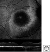
Retinitis pigmentosa GTPase regulator (RPGR)-associated cone-rod dystrophy. Fundus autofluorescence image (top) showing parafoveal hyperautofluorescent ring with corresponding ellipsoid zone disruption on optical coherence tomography (below).
Figure 3.
Figure 3.
Retinal imaging in a female retinitis pigmentosa GTPase regulator (RPGR) retinopathy carrier. Fundus autofluorescence image of the left eye of an asymptomatic 45-yr-old female carrier showing a radial pattern “tapetal” reflex. Optical coherence tomography of the right eye shows preserved retinal lamination (below).

References

    1. Anikina E, Georgiou M, Tee J, Webster AR, Weleber RG, Michaelides M. 2022. Characterization of retinal function using microperimetry-derived metrics in both adults and children with RPGR-associated retinopathy. Am J Ophthalmol 234: 81–90. 10.1016/j.ajo.2021.07.018 - DOI - PMC - PubMed
    1. Beltran WA, Hammond P, Acland GM, Aguirre GD. 2006. A frameshift mutation in RPGR exon ORF15 causes photoreceptor degeneration and inner retina remodeling in a model of X-linked retinitis pigmentosa. Invest Ophthalmol Vis Sci 47: 1669–1681. 10.1167/iovs.05-0845 - DOI - PubMed
    1. Beltran WA, Cideciyan A, Lewin AS, Iwabe S, Khanna H, Sumaroka A, Chiodo VA, Fajardo DS, Román AJ, Deng WT, et al. 2012. Gene therapy rescues photoreceptor blindness in dogs and paves the way for treating human X-linked retinitis pigmentosa. Proc Natl Acad Sci 109: 2132–2137. 10.1073/pnas.1118847109 - DOI - PMC - PubMed
    1. Beltran WA, Cideciyan A, Iwabe S, Swider M, Kosyk MS, McDaid K, Martynyuk I, Ying GS, Shaffer J, Deng WT, et al. 2015. Successful arrest of photoreceptor and vision loss expands the therapeutic window of retinal gene therapy to later stages of disease. Proc Natl Acad Sci 112: E5844–E5853. 10.1073/pnas.1509914112 - DOI - PMC - PubMed
    1. Berson EL, Rosner B, Sandberg MA, Hayes KC, Nicholson BW, Weigel Difranco C, Willett W. 1993. A randomized trial of vitamin A and vitamin E supplementation for retinitis pigmentosa. Arch Opthalmol 111: 761–772. 10.1001/archopht.1993.01090060049022 - DOI - PubMed

LinkOut - more resources