Review
. 2023 Jun;19(6):343-362.
doi: 10.1038/s41584-023-00967-9. Epub 2023 May 15.

Juvenile idiopathic inflammatory myositis: an update on pathophysiology and clinical care

Affiliations
Review

Juvenile idiopathic inflammatory myositis: an update on pathophysiology and clinical care

Charalampia Papadopoulou et al. Nat Rev Rheumatol. 2023 Jun.

Abstract

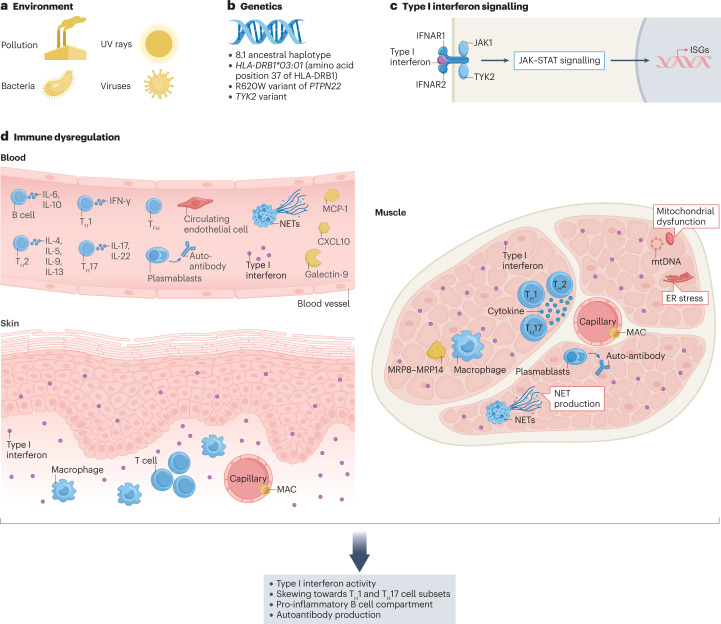
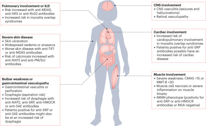
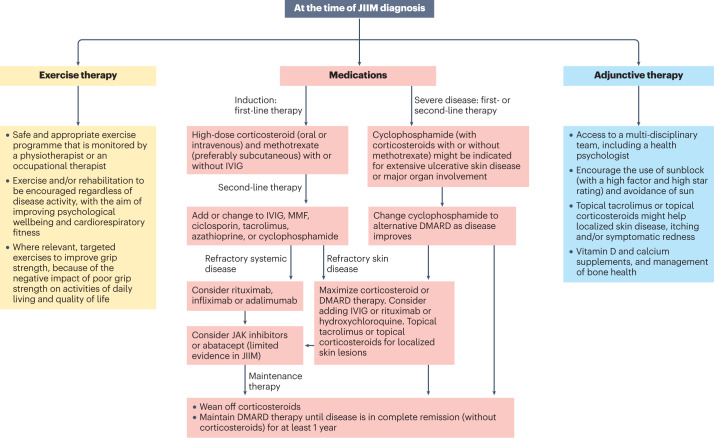
The childhood-onset or juvenile idiopathic inflammatory myopathies (JIIMs) are a heterogenous group of rare and serious autoimmune diseases of children and young people that predominantly affect the muscles and skin but can also involve other organs, including the lungs, gut, joints, heart and central nervous system. Different myositis-specific autoantibodies have been identified that are associated with different muscle biopsy features, as well as with different clinical characteristics, prognoses and treatment responses. Thus, myositis-specific autoantibodies can be used to subset JIIMs into sub-phenotypes; some of these sub-phenotypes parallel disease seen in adults, whereas others are distinct from adult-onset idiopathic inflammatory myopathies. Although treatments and management have much improved over the past decade, evidence is still lacking for many of the current treatments and few validated prognostic biomarkers are available with which to predict response to treatment, comorbidities (such as calcinosis) or outcome. Emerging data on the pathogenesis of the JIIMs are leading to proposals for new trials and tools for monitoring disease.


Conflict of interest statement

All authors declare that the UK JDM Cohort and Biomarker Study (JDCBS) is currently funded by grants from the Great Ormond Street Hospital (GOSH) Children’s Charity, the National Institute for Health Research (NIHR) Biomedical Research Centre at GOSH, Cure JM, Myositis UK, Versus Arthritis and Remission Charity. L.R.W. declares that she is a consultant for Pfizer Inc (with consulting fees going wholly to the University College London). The other authors all declare no competing interests.

Figures

Fig. 1
Fig. 1. Factors implicated in the pathogenesis of juvenile myositis.
The pathogenesis of juvenile idiopathic inflammatory myopathy (JIIM) involves a complex interplay between genetic and environmental factors, leading to immunological, vascular and metabolic dysfunction. a, Environmental triggers of JIIM might include ultraviolet (UV) radiation, pollution and microbial infections. b, Genetic loci in the MHC and non-MHC regions are implicated in disease susceptibility and development. c, Type I interferon signalling is thought to have a central role in the pathological changes seen in various tissues. d, Immune dysregulation within the skin, muscle and blood vessels, as well as in other tissues (not shown), is thought to contribute to disease. Within the muscle, the overexpression of MHC proteins, a hallmark feature thought to be driven by interferons, contributes to endoplasmic reticulum (ER) stress leading to an inflammatory cascade via the nuclear factor kappa B (NF-κB) pathway. Autoreactive B cells are present, as demonstrated by the production of myositis-specific antibodies (MSAs), and regulatory B (Breg) cells have a pro-inflammatory phenotype (including producing elevated levels of IL-6). Circulating inflammatory mediators include Galectin-9 and CXCL10, which correlate with disease activity. Abnormalities in the small blood vessels are reflected by a high number of circulating endothelial cells, which correlates with disease activity; muscle capillary loss and complement deposition on capillaries also frequently occur. T cell dysfunction includes a skewing of the T cell compartment towards a T helper 17 (TH17) cell phenotype, including within the follicular helper T (TFH) cell population. Both neutrophil extracellular trap (NET) formation and mitochondria dysfunction occur in JDM and might be part of a pathological loop that drives interferon production. Overall, the pathogenesis of JIIM involves a complex interplay between innate and adaptive immunity that affects muscle, skin and vascular tissues to drive ongoing inflammation and tissue damage. ISGs, interferon-stimulated genes.
Fig. 2
Fig. 2. Clinical features and autoantibody profile in JIIM indicative of severe disease and/or need for treatment escalation.
Owing to the rarity and heterogeneity of juvenile idiopathy inflammatory myopathy (JIIM), children and young people should be managed by a multidisciplinary team in a specialist centre. To predict the severity of the disease and the potential need for treatment escalation, many factors are considered, as illustrated, including the presence or absence of severe muscle weakness, dysphagia, ulcerative skin disease or major organ involvement. The myositis-specific autoantibody (MSA) and/or myositis-associated autoantibody (MAA) profile might predict the risk of JIIM-related complications, including major organ involvement. Some features associated with specific MSAs or MAAs are shown, but specific complications are not exclusive to patients with these MSA–MAA profiles and not all patients with a particular MSA–MAA profile will demonstrate these complications. CMAS, childhood myositis assessment scale; GI, gastrointestinal; ILD, interstitial lung disease; IMNM, immune-mediated necrotizing myopathy; MMT-8, manual muscle testing in eight muscle groups.
Fig. 3
Fig. 3. Treatment algorithm for JIIM on the basis of current available evidence.
A treatment algorithm for juvenile idiopathic inflammatory myopathy (JIIM) is presented, based on evidence-informed consensus recommendations in UK and Europe,. Treatments need to be individualized and include consideration of the patient age, preferences for oral or parenteral administration of medications, severity of disease and response to treatment. No single approach will be right for every patient and clinicians need to use best judgement on the basis of evidence available. In most cases, with the exception of randomized controlled trials evaluating methotrexate versus ciclosporin, rituximab or exercise in myositis, evidence is limited to case series or cohort studies. More research is needed to compare the efficacy of second-line or third-line treatment options and determine the best treatment approach for myositis-related complications such as interstitial lung disease (ILD) or calcinosis. More evidence is also needed to determine the best treatment for refractory disease, which can be defined as myositis that responds inadequately to at least two immunosuppressant or immunomodulatory drugs given in their full dose for a minimum of 3 months, hindering weaning of corticosteroid. Patients with JIIM should have regular reviews that include measurement of muscle strength, assessment of skin disease and extra-muscular manifestations. Adherence to medication should be checked if patients fail to respond as expected. Treatment should be escalated if patients fail to respond adequately to treatment or are intolerant to the treatment. Exercise therapy and psychological support are important aspects to the management of JIIM in addition to medication. IVIG, intravenous immunoglobulin; JAK, janus kinase; MMF, mycophenolate mofetil.

References

    1. Lundberg IE, et al. European League Against Rheumatism/American College of Rheumatology classification criteria for adult and juvenile idiopathic inflammatory myopathies and their major subgroups. Arthritis Rheumatol. 2017;69:2271–2282. doi: 10.1002/art.40320. - DOI - PMC - PubMed
    1. Patel B, Khan N, Werth VP. Applicability of EULAR/ACR classification criteria for dermatomyositis to amyopathic disease. J. Am. Acad. Dermatol. 2018;79:77–83.e1. doi: 10.1016/j.jaad.2017.12.055. - DOI - PubMed
    1. Bohan A, Peter JB. Polymyositis and dermatomyositis (second of two parts) N. Engl. J. Med. 1975;292:403–407. doi: 10.1056/NEJM197502202920807. - DOI - PubMed
    1. Tansley SL, et al. Autoantibodies in juvenile-onset myositis: their diagnostic value and associated clinical phenotype in a large UK cohort. J. Autoimmun. 2017;84:55–64. doi: 10.1016/j.jaut.2017.06.007. - DOI - PMC - PubMed
    1. Rider LG, et al. The myositis autoantibody phenotypes of the juvenile idiopathic inflammatory myopathies. Medicine. 2013;92:223–243. doi: 10.1097/MD.0b013e31829d08f9. - DOI - PMC - PubMed

Publication types