. 2023 Sep;33(9):712-726.
doi: 10.1038/s41422-023-00818-y. Epub 2023 May 15.

Lin28a maintains a subset of adult muscle stem cells in an embryonic-like state

Affiliations

Lin28a maintains a subset of adult muscle stem cells in an embryonic-like state

Peng Wang et al. Cell Res. 2023 Sep.

Abstract

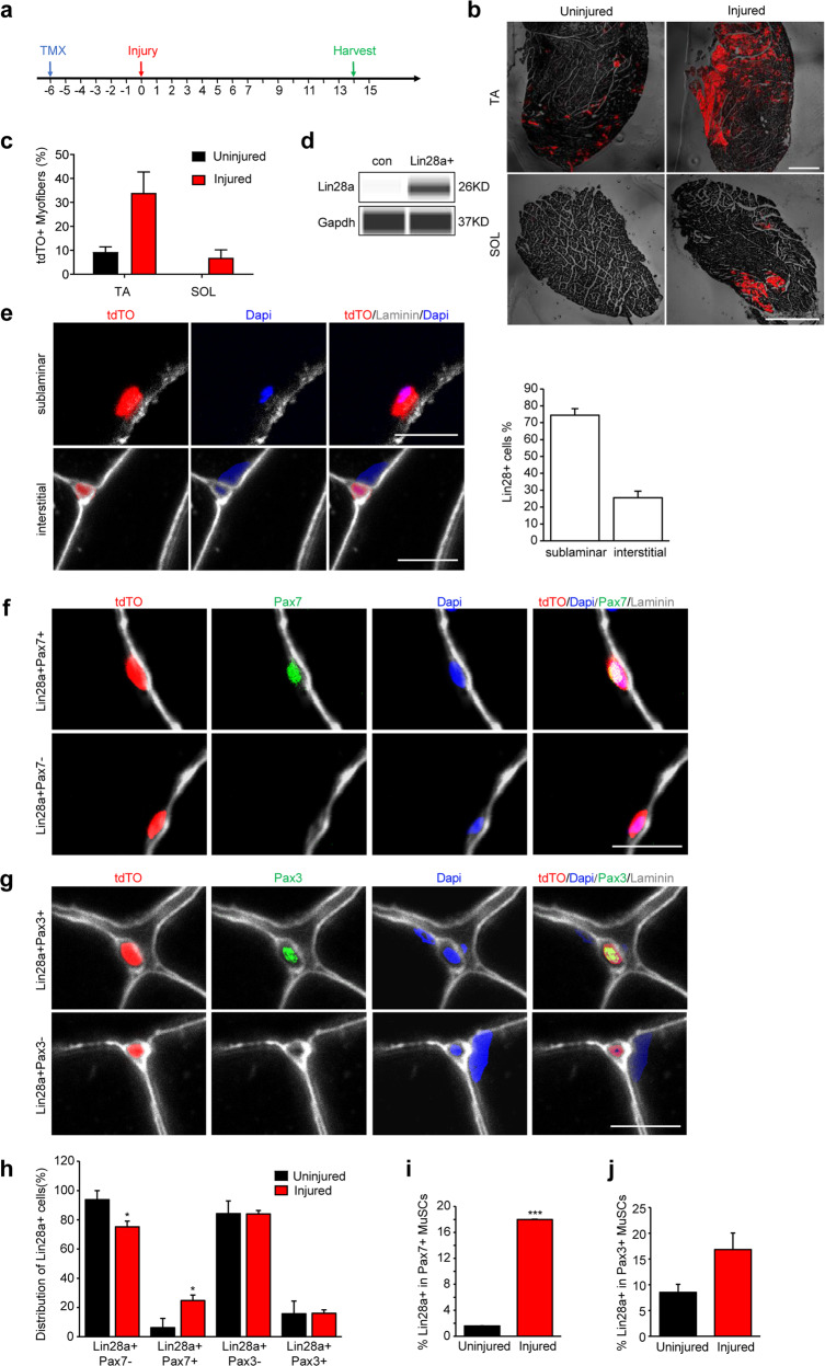
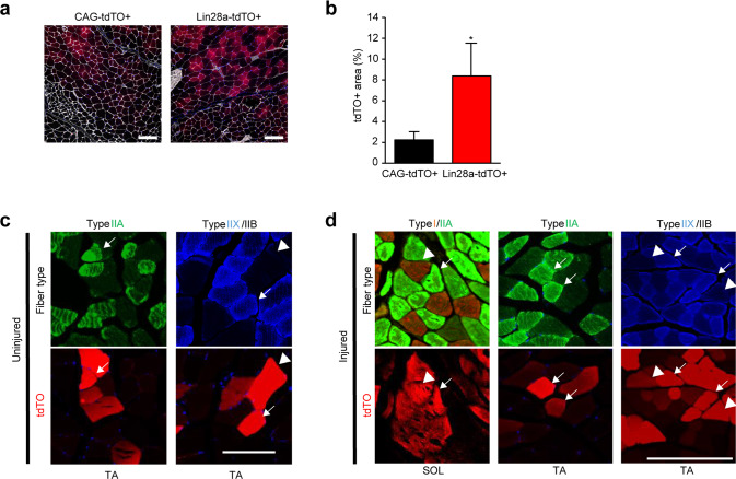
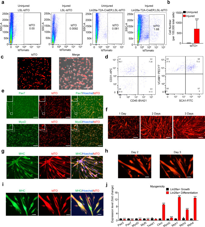
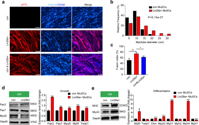
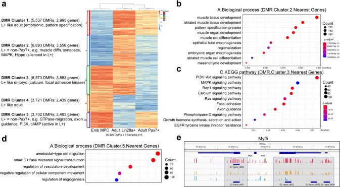
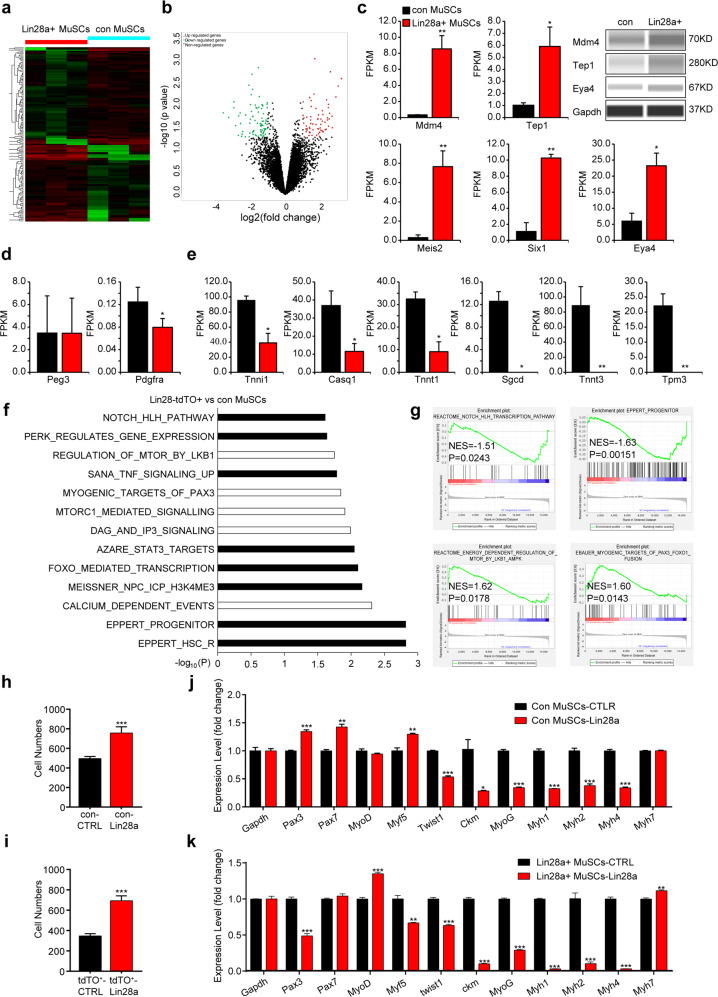
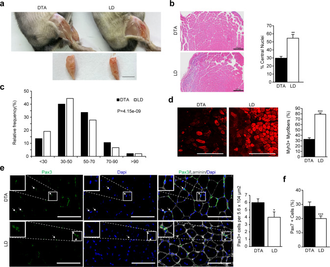
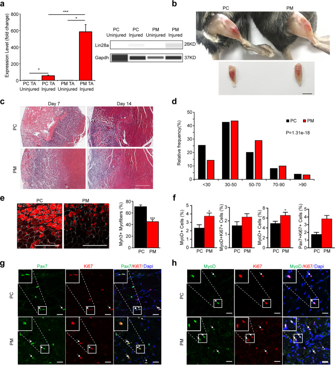
During homeostasis and after injury, adult muscle stem cells (MuSCs) activate to mediate muscle regeneration. However, much remains unclear regarding the heterogeneous capacity of MuSCs for self-renewal and regeneration. Here, we show that Lin28a is expressed in embryonic limb bud muscle progenitors, and that a rare reserve subset of Lin28a+Pax7- skeletal MuSCs can respond to injury at adult stage by replenishing the Pax7+ MuSC pool to drive muscle regeneration. Compared with adult Pax7+ MuSCs, Lin28a+ MuSCs displayed enhanced myogenic potency in vitro and in vivo upon transplantation. The epigenome of adult Lin28a+ MuSCs showed resemblance to embryonic muscle progenitors. In addition, RNA-sequencing revealed that Lin28a+ MuSCs co-expressed higher levels of certain embryonic limb bud transcription factors, telomerase components and the p53 inhibitor Mdm4, and lower levels of myogenic differentiation markers compared to adult Pax7+ MuSCs, resulting in enhanced self-renewal and stress-response signatures. Functionally, conditional ablation and induction of Lin28a+ MuSCs in adult mice revealed that these cells are necessary and sufficient for efficient muscle regeneration. Together, our findings connect the embryonic factor Lin28a to adult stem cell self-renewal and juvenile regeneration.


Conflict of interest statement

The authors declare no competing interests.

Figures

Fig. 1
Fig. 1. Lin28a+ stem cells contribute to satellite cells and myofibers during regeneration.
a Schedule of tamoxifen (TMX) treatment. Lin28a-T2A-CreER;LSL-tdTO mice were injected with TMX, 6 days before cryoinjury, daily for the first 1 week after injury, every other day from the 7th day, and harvested on the 14th day after injury. b TA and SOL muscles of Lin28a-T2A-CreER;LSL-tdTO mice were damaged by cryoinjury and harvested on day 14 post injury. Contralateral TA or SOL muscle was used as the control. Scale bars, 600 μm. c Quantification of the number of tdTO+ muscle fibers per 100 muscle fibers in the TA and SOL muscles of Lin28a-T2A-CreER;LSL-tdTO mice on day 14 post injury and without injury. Quantification was performed on the images in b. For each muscle section, at least 3 whole sections were quantified and averaged. d Low-protein-input capillary-based immunoassays for Lin28a protein expression in Lin28a-tdTO+ and conventional (con) Pax7+ MuSCs. Gapdh protein was used as the loading control. Unprocessed original capillary-based immunoassay results are shown in Supplementary information, Fig. S7. e Cryosections of the TA muscles of Lin28a-T2A-CreER;LSL-tdTO mice that were cryoinjured and harvested on day 14 post injury. Muscle sections were co-stained for laminin (white) and DAPI (blue). Scale bars, 10 μm. Right: quantification of the percentage of sublaminar and interstitial Lin28a+ cells. For each muscle section, at least five different fields were quantified and averaged. f Cryosections of the TA muscle of Lin28a-T2A-CreER;LSL-tdTO mice at 14 days after cryoinjury. Muscle sections were co-stained for laminin (white), DAPI (blue) and Pax7 (green). Scale bar, 10 μm. g Cryosections of the TA muscle of Lin28a-T2A-CreER;LSL-tdTO mice at 14 days after cryoinjury. Muscle sections were co-stained for laminin (white), DAPI (blue) and Pax3 (green). Scale bar, 10 μm. h Quantification of the distribution of Pax3, Pax3+, Pax7 and Pax7+ cells in the Lin28a-tdTO+ pool per field in the TA muscle of Lin28a-T2A-CreER;LSL-tdTO mice on day 14 post injury (red), relative to uninjured muscles (black). For each muscle section, at least five different fields were quantified and averaged. i Quantification of the percentage of Lin28a-tdTO+ cells in Pax7+ satellite cells per field in the TA muscle of Lin28a-T2A-CreER;LSL-tdTO mice on day 14 post injury (red), relative to uninjured muscles (black). For each muscle section, at least five different fields were quantified and averaged. j Quantification of the percentage of Lin28a-tdTO+ cells in Pax3+ MuSCs per field in the TA muscle of Lin28a-T2A-CreER;LSL-tdTO mice on day 14 post injury (red), relative to uninjured muscles (black). For each muscle section, at least five different fields were quantified and averaged. *P < 0.05, ***P < 0.001.
Fig. 2
Fig. 2. Lin28a+ stem cells contribute to myofibers during regeneration.
a Cryosection and laminin immunofluorescence staining of TA muscles of NSG mice at 21 days after 1000 Lin28a-tdTO+ MuSCs or Pax7+ MuSCs (CAG-tdTO+) were freshly sorted by FACS and transplanted orthotopically into cryoinjured TA muscles. Scale bars, 100 μm. b Proportion of tdTO+ myofiber area in the cryoinjured TA region. Data are means ± SEM. n = 3 independent experiments. For each experiment, a total of 20 serial cryosections were counted and averaged. c Immunostaining of transverse sections of uninjured TA muscles obtained from Lin28a-T2A-CreER;LSL-tdTO mice at day 14 of lineage tracing, for type IIA (green), type IIX (blue) and type IIB (black) myofibers, compared to tdTO+ fluorescence (arrowheads). Scale bar, 100 μm. d Immunostaining of transverse sections of injured SOL and TA muscles obtained from Lin28a-T2A-CreER;LSL-tdTO mice at day 14 of lineage tracing, for type I (red), type IIA (green), type IIX (blue) and type IIB (black) myofibers, compared to tdTO+ fluorescence (arrowheads). Scale bar, 200 μm. *P < 0.05.
Fig. 3
Fig. 3. Lin28a+ cells are MuSCs with robust myogenic capacity.
a Flow cytometry analysis for Lin28a-tdTO+ cells in the uninjured and injured muscles of Lin28a-T2A-CreER;LSL-tdTO mice. The control groups were uninjured and injured LSL-tdTO mice. All the mice were injected with TMX, and harvested 14 days after injury. b Quantification of the number of tdTO+ cells in injured or uninjured Lin28a-T2A-CreER;LSL-tdTO mice. n = 6 mice for each group. c Confocal microscopy on freshly FACS-isolated Lin28a-tdTO+ cells. Scale bar, 200 μm. d Flow cytometry analysis for tdTO+ cells. Cells were first labeled with antibodies conjugated with fluorescent dyes for CD31 (APC), CD45 (BV421), VCAM1 (PE), Sca1 (FITC). The Lin28a-tdTO+ cells are mainly VCAM1+CD31+Sca1+CD45 cells. e Immunofluorescence staining (green) revealed that most of the Lin28a-tdTO+ cells cultured in GM become Pax7+MyoD+ progenitors within 24 h of culture in vitro. Scale bars, 250 μm. f Lin28a-tdTO+ cells fused, differentiated and formed multinucleated myotubes starting at 1 and 2 days in myogenic DM. Scale bars, 500 μm. g MHC (green) and Hoechst (blue) staining revealed that the majority of Lin28a-tdTO+ cells expressed MHC after differentiation into myotubes. Scale bars, 80 μm. h Single VCAM1+CD31+Sca1+Lin28a-tdTO+ cells all survived, proliferated, fused, differentiated and formed multinucleated myotubes at 2 and 3 days in DM. Scale bars, 40 μm. i MHC (green) and Hoechst (blue) staining revealed that all the VCAM1+CD31+Sca1+Lin28a-tdTO+ single cell-derived colonies expressed MHC after differentiation into myotubes. Scale bars, 40 μm. j qRT-PCR analysis revealed that myogenic differentiation-related genes, such as MyoG, Ckm, Myh1, Myh2, Myh4, were strongly activated, whereas myogenic progenitor-related genes, such as Pax3, Pax7, MyoD, Myf5, were significantly reduced, when Lin28a-tdTO+ cells were cultured in myogenic DM, relative to undifferentiated Lin28a-tdTO+ cells cultured in GM. ***P < 0.001.
Fig. 4
Fig. 4. Lin28a+ MuSCs show enhanced myogenic potency in vitro.
a Immunofluorescence images of conventional (con) MuSCs (CellTrace Violet-labeled), Lin28a-tdTO+ cells, and a 1:1 mixture of con MuSCs with Lin28a-tdTO+ cells, after FACS-isolation from the TA muscles of Lin28a-T2A-CreER;LSL-tdTO mice 14 days post injury followed by 36-h differentiation into myotubes. Scale bars, 100 μm. b Relative frequency distribution of the diameters of myotubes formed by the fusion of Lin28a-tdTO+ cells and/or con MuSCs. For each bin, 200 myotubes were quantified. P = 6.15 × 10–7. c Quantification of the fusion index of these myotubes. For each group, at least three different fields were quantified. d Western blot analysis (left) for Pax3, Pax7 and MyoD protein expression in Lin28a+ cells. qRT-PCR analysis (right) revealed expression levels of Pax3, Pax7, MyoD, Myf5 and Twist1 in Lin28a+ cells relative to con MuSCs, when the cells were proliferated in GM. Gapdh served as the loading control. Unprocessed original scans of blots are shown in Supplementary information, Fig. S7. e Western blot analysis (left) for MHC and MyoG protein expression in Lin28a+ cell-derived myotubes. qRT-PCR analysis (right) revealed expression levels of Myf5, Twist1, Ckm, MyoG, Myh1/2/4/7 in Lin28a+ cell-derived myotubes, relative to con MuSC-derived myotubes, when they were differentiated in DM. Gapdh served as the loading control. Unprocessed original scans of blots are shown in Supplementary information, Fig. S7. *P < 0.05, **P < 0.01, ***P < 0.001.
Fig. 5
Fig. 5. Epigenomic profiles show that Lin28a+ MuSCs resemble embryonic muscle progenitors.
a Hierarchical clustering analysis (heatmap and dendrogram) for the DNA methylomes of freshly isolated embryonic muscle progenitors, adult Lin28a+ MuSCs, and adult Pax7+ MuSCs. n = 3 mice for each group. DMR differentially methylated region, Emb MPC embryonic muscle progenitors. b GO analysis of Cluster 2 in a. c KEGG analysis of Cluster 3 in a. d GO analysis of Cluster 5 in a. e CpG site methylation level in the region near the Myf5 locus of adult Lin28a+ MuSCs (red), adult Pax7+ MuSCs (yellow), and embryonic muscle progenitors (blue). Myf5 was heavily methylated in adult Lin28a+ MuSCs, partially methylated in adult Pax7+ MuSCs, and demethylated in embryonic muscle progenitors.
Fig. 6
Fig. 6. Transcriptomic profiles show that Lin28a promotes MuSC dedifferentiation.
a Hierarchical clustering analysis for the transcriptomes of freshly isolated Lin28a+ MuSCs and conventional (con) Pax7+ MuSCs. n = 3 mice for each group. b Volcano plot analysis for differentially expressed genes in Lin28a+ MuSCs, relative to con Pax7+ MuSCs. Red, upregulated > 2-fold and P < 0.05. Green, downregulated > 2-fold and P < 0.05. c Expression levels of Mdm4, Tep1 and three primitive limb mesodermal progenitor transcription factors, Meis2, Six1, Eya4 in Lin28a+ MuSCs relative to con Pax7+ MuSCs. FPKM fragments per kilobase of transcript per million mapped reads. Right: low-protein-input capillary-based immunoassays for Eya4, Tep1 and Mdm4 protein expression. Gapdh served as the loading control. d Expression levels of markers expressed in other muscle-resident Pax7-independent progenitors, Peg3 and Pdgfra, in Lin28a+ MuSCs relative to con Pax7+ MuSCs. e Expression levels of myogenic terminal differentiation markers, including many troponins (Tnni1, Tnnt1, Tnnt3), calsequestrin (Casq1), sarcoglycan (Sgcd), and tropomyosin (Tpm3), in Lin28a+ MuSCs relative to con Pax7+ MuSCs. f Signatures enriched in Lin28a+ MuSCs, compared with con Pax7+ MuSCs, as identified by GSEA. Upregulated pathways are shown with black columns, and downregulated pathways are shown with white columns. g Representative GSEA profiles with the normalized enrichment scores (NESs) and nominal P values shown. h, i Cell proliferation rate of con MuSCs (h) and Lin28a-tdTO+ MuSCs (i), after being infected with retroviruses expressing either empty vector (CTRL) or Lin28a. j, k qRT-PCR for myogenic differentiation markers in con MuSCs (j) or Lin28a-tdTO+ MuSCs (k) which overexpressed Lin28a, relative to con MuSCs or Lin28a-tdTO+ MuSCs with the empty vector (CTRL). Data are means ± SEM. n = 3 independent experiments. *P < 0.05, **P < 0.01, ***P < 0.001.
Fig. 7
Fig. 7. Lin28a+ MuSCs are necessary for efficient muscle regeneration.
a Photographs show the resolving of muscle inflammation and muscle regeneration in the TA muscles of adult Rosa26-DTA (DTA) and adult Lin28-CreER;Rosa26-DTA (LD) mice on day 11 post injury. Scale bar, 5 mm. b Hematoxylin and eosin (H&E) staining of TA muscle sections of DTA and LD mice, on day 11 post injury. Scale bars, 200 μm. Right: quantification of the percentage of myofibers that were regenerating with central nuclei. For each muscle section, at least seven different fields were quantified. c Relative frequency distribution of the Feret diameters of myofibers in the TA muscles of DTA vs LD mice. d Immunofluorescence staining for embryonic MHC (Myh3) expression in the injured TA muscles from LD mice relative to DTA mice on day 11 post injury. Right: quantification of the percentage of Myh3+ myofibers. For each muscle section, at least seven different fields were quantified. Scale bars, 200 μm. e Cryosections of the TA muscles of DTA and LD mice that were cryoinjured and harvested on day 11 post injury. Muscle sections were co-stained for Pax3 (green), laminin (white) and DAPI (blue). White arrows indicate Pax3+ cells. Right: quantification of the Pax3+ cell numbers in DTA and LD mice. For each muscle section, at least five different fields were quantified and averaged. Scale bars, 100 μm. f Quantification of the percentage of Pax7+ MuSCs in the TA muscles of DTA and LD mice after cryoinjury. For each muscle section, at least seven different fields were quantified. *P < 0.05, **P < 0.01, ***P < 0.001.
Fig. 8
Fig. 8. Injury-inducible Lin28a is sufficient to enhance adult muscle regeneration.
a qRT-PCR analysis of Lin28a in TA muscles of injured Pax7-CreER (PC) and Pax7-CreER;NF-κB-LSL-Lin28a (PM) mice, relative to uninjured PC mice. Right: low-protein-input capillary-based immunoassays for Lin28a protein expression in Pax7+ MuSCs of TA muscles of the above mice. Gapdh protein was used as the loading control. b Photographs showing the resolving of muscle inflammation and muscle regeneration in the TA muscles of 8-month-old PC and PM mice on day 7 post injury. Scale bar, 5 mm. c H&E staining of TA muscle sections of PM mice, relative to PC mice, on days 7 and 14 post injury. Scale bar, 300 μm. d Relative frequency distribution of the Feret diameters of myofibers in the TA muscles of PC vs PM mice. e Immunofluorescence staining for embryonic MHC (Myh3) expression in the injured TA muscles from PM mice relative to PC mice, on day 11 post injury. Right: quantification of the percentage of Myh3+ myofibers. For each muscle section, at least seven different fields were quantified. Scale bars, 200 μm. f Quantification of the percentage of each type of MuSC and myoblast, after injured TA muscle sections were immunostained for Ki67, MyoD and Pax7. For each muscle section, at least seven different fields were quantified. g Immunofluorescence staining for Pax7 and Ki67 expression in the damaged TA muscles from PM mice, relative to PC mice. White arrows indicate Pax7+Ki67+ cells. Scale bars, 20 μm. h Immunofluorescence staining for MyoD and Ki67 expression in the injured TA muscles from PM mice, relative to PC mice. White arrows indicate MyoD+Ki67+ cells. Scale bars, 20 μm. *P < 0.05, ***P < 0.001.

References

    1. Mauro A. Satellite cell of skeletal muscle fibers. J. Biophys. Biochem. Cytol. 1961;9:493–495. doi: 10.1083/jcb.9.2.493. - DOI - PMC - PubMed
    1. Yin H, Price F, Rudnicki MA. Satellite cells and the muscle stem cell niche. Physiol. Rev. 2013;93:23–67. doi: 10.1152/physrev.00043.2011. - DOI - PMC - PubMed
    1. von Maltzahn J, Chang NC, Bentzinger CF, Rudnicki MA. Wnt signaling in myogenesis. Trends Cell Biol. 2012;22:602–609. doi: 10.1016/j.tcb.2012.07.008. - DOI - PMC - PubMed
    1. Rossi G, Messina G. Comparative myogenesis in teleosts and mammals. Cell Mol. Life Sci. 2014;71:3081–3099. doi: 10.1007/s00018-014-1604-5. - DOI - PMC - PubMed
    1. Kuang S, Chargé SB, Seale P, Huh M, Rudnicki MA. Distinct roles for Pax7 and Pax3 in adult regenerative myogenesis. J. Cell Biol. 2006;172:103–113. doi: 10.1083/jcb.200508001. - DOI - PMC - PubMed

Publication types

LinkOut - more resources