. 2023 Jun;20(6):908-917.
doi: 10.1038/s41592-023-01880-5. Epub 2023 May 15.

Engineered allostery in light-regulated LOV-Turbo enables precise spatiotemporal control of proximity labeling in living cells

Affiliations

Engineered allostery in light-regulated LOV-Turbo enables precise spatiotemporal control of proximity labeling in living cells

Song-Yi Lee et al. Nat Methods. 2023 Jun.

Abstract

The incorporation of light-responsive domains into engineered proteins has enabled control of protein localization, interactions and function with light. We integrated optogenetic control into proximity labeling, a cornerstone technique for high-resolution proteomic mapping of organelles and interactomes in living cells. Through structure-guided screening and directed evolution, we installed the light-sensitive LOV domain into the proximity labeling enzyme TurboID to rapidly and reversibly control its labeling activity with low-power blue light. 'LOV-Turbo' works in multiple contexts and dramatically reduces background in biotin-rich environments such as neurons. We used LOV-Turbo for pulse-chase labeling to discover proteins that traffic between endoplasmic reticulum, nuclear and mitochondrial compartments under cellular stress. We also showed that instead of external light, LOV-Turbo can be activated by bioluminescence resonance energy transfer from luciferase, enabling interaction-dependent proximity labeling. Overall, LOV-Turbo increases the spatial and temporal precision of proximity labeling, expanding the scope of experimental questions that can be addressed with proximity labeling.


Conflict of interest statement

Competing interests

S.-Y.L., J.S.C. and A.Y.T. have filed a patent application covering some aspects of this work (US Provisional Patent Application No. 63/488,940; CZ SF ref. CZB-273S-P1; Stanford ref. S22-487; KT ref. 110221-1361830-009500PR). The remaining authors declare no competing interests.

Figures

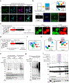
Fig. 1 |
Fig. 1 |. Design and directed evolution of LOV-Turbo.
a, LOV-Turbo has a bluelight-sensitive hLOV1 (ref. 9) domain inserted into its sixth surface-exposed loop (domain structure shown in Supplementary Fig. 1a). In the dark state, clamping by hLOV1 keeps TurboID inactive, while in the light state, clamp release restores TurboID activity. Active LOV-Turbo promiscuously biotinylates nearby proteins, enabling their identification by mass spectrometry. b, Screening of 31 LOV insertion sites in TurboID. Constructs were expressed in the HEK cytosol and labeling was performed for 30 min in light or dark. Relative biotinylation activity was quantified by streptavidin blotting of whole-cell lysates. Multiple blots were quantified parallel by normalizing with TurboID biotinylation intensity. Inset shows sample data. Ribbon structure of BirA (PDB ID 2EWN) shows LOV insertion sites and the result obtained for each. Nonhydrolyzable biotinol-5′-AMP in pink. This experiment was performed once, except the 80/81 and 141/142 constructs, which were performed twice with similar results. c, Truncation of LOV-TurboID at the 80/81 insertion site improves light gating. Amino acids (aa) 75–80 were progressively truncated as shown. The Δ4 construct showed the highest ± light signal ratio and lowest-light leak. This experiment was performed twice with similar results. d, Scheme for directed evolution on the yeast cell surface. PE, phycoerythrin. Ab, antibody. e, Selection conditions used over seven rounds. Positive selection (P) and negative selection (N) gates are shown in d. f, FACS plots comparing LOV-Turbo1 template to postround seven yeast population. Percentages give fractions of active cells (in quadrants I + III) and fractions of expressing cells (in quadrants III + IV). g, Screening of eight evolved mutants in the HEK cytosol. Biotinylation intensity quantified by streptavidin blot of whole-cell lysates (shown in Supplementary Fig. 1f), after labeling for 30 min in light or 5 h in dark. LOV-Tb1, LOV-Turbo1. h, Same as g but with combination mutants C10-C13 as well, after labeling for 30 min in light or 4 h in dark. Blots are shown in Supplementary Fig. 1h. C10 was selected over C11 as our final LOV-Turbo due to its higher expression level in HEK 293T cells.
Fig. 2 |
Fig. 2 |. Characterization of LOV-Turbo.
a, AlphaFold-predicted structure of evolved LOV-Turbo (clone C10). LOV domain in cyan and TurboID in green. L76 and V81 flanking the LOV insertion site are colored orange. The six mutations enriched by directed evolution are colored red. Nonhydrolyzable biotinol-5′-AMP in pink. Close-up views show TurboID’s active site in relation to residues that may be allosterically coupled to the LOV domain. Double-headed arrow shows the distance between indicated backbone loops. b, Light intensity-dependence of LOV-Turbo. HEK 293T cells expressing LOV-Turbo in the cytosol were stimulated with blue light of varying intensities (0.1–1 mW cm−2) for 15 min in the presence of biotin. Lysates were analyzed by streptavidin and anti-V5 blotting. This experiment was performed once. c, Confocal imaging of LOV-Turbo-catalyzed biotinylation in the mitochondrial matrix, outer mitochondrial membrane (OMM), ERM facing the cytosol, nucleus and cytosol. Anti-V5 detects LOV-Turbo expression. Neutravidin-AF647 detects biotinylated proteins, after 30 min of labeling under blue light illumination. TOM20 and calnexin are endogenous mitochondrial and ER markers, respectively. DAPI stains the nucleus. Scale bars, 5 μm. This experiment was performed twice with similar results. d, Streptavidin blotting of HEK 293T lysates expressing LOV-Turbo targeted to various compartments and labeled with biotin and blue light for 30 min. Three different mitochondrial matrix targeting sequences were tested (Methods). This experiment was performed once. e, Comparison of LOV-Turbo (LOV-Tb), TurboID and miniTurbo (miniTb) in HEK mitochondrial matrix, nucleus and cytosol. Labeling time was 30 min. The * indicates auto-labeled LOV-Turbo, TurboID or miniTurbo. This experiment was performed once.
Fig. 3 |
Fig. 3 |. LOV-Turbo is reversible and works in multiple cell types and in the rodent brain.
a, LOV-Turbo labeling is terminated by the removal of light. HEK 293T cells expressing LOV-Turbo in the cytosol were labeled as shown in conditions A–F. Whole-cell lysates were then analyzed by streptavidin blotting. This experiment was performed twice with similar results. b, LOV-Turbo labeling in E. coli. Constructs were expressed in the cytosol of BL21 E. coli and labeling was performed for 4 h. Anti-His6 antibody detects ligase expression. Tb, TurboID. miniT, miniTurbo. This experiment was performed once. c, LOV-Turbo labeling in yeast (BY4741 strain). Labeling was performed for 6 h. This experiment was performed once. d, LOV-Turbo labeling in cultured rat cortical neurons. LOV-Turbo or TurboID was expressed in the cytosol via adeno-associated virus 1/2 (AAV) infection for 5 days before labeling. TurboID shows high background even without exogenous biotin addition. This experiment was performed once. e, LOV-Turbo labeling in the mouse brain. Cytosolic LOV-Turbo was expressed in the mouse cortex via AAV1/2 injection. 2 weeks later, 0.5 μl of 10 mM biotin was injected into the brain while 470 nm light (5 mW cm−2, 10 ms pulses at 10 Hz) was delivered for 1.5 h. Whole-tissue lysate analyzed by streptavidin and anti-V5 blotting. The * indicates endogenously biotinylated proteins. This experiment was performed twice with similar results.
Fig. 4 |
Fig. 4 |. Applications of LOV-Turbo.
a, Reduced background and improved spatial precision with mito-LOV-Turbo in cultured rat cortical neurons. LOV-Turbo or TurboID targeted to the mitochondrial matrix were expressed in cultured rat cortical neurons via AAV1/2 transduction at DIV 6. At DIV 12, neurons were treated with biotin and light for 2 h, then fixed and stained with anti-V5 antibody to detect enzyme expression, and neutravidin-AF647 to detect biotinylated proteins. Scale bar, 10 μm. This experiment was performed once. b, Spatial control of proximity labeling with LOV-Turbo. HEK 293T cells expressing cytosolic LOV-Turbo were labeled with biotin and light for 5 min, while half of the sample was covered with black tape. Images show neutravidin staining in lit area only. DIC, differential interference contrast. Scale bar, 500 μm. This experiment was performed once. c, Pulse-chase labeling with LOV-Turbo. HEK 293T cells expressing nuclear LOV-Turbo were labeled with biotin and light for 10 min, then chased for 8 h in the dark (other chase times shown in Supplementary Fig. 3g). Images show neutravidin staining before and after the chase period. Arrows point to biotinylated proteins in the cytosol. Scale bar, 10 μm. This experiment was performed once. d, Pulse-chase labeling with cytosolic LOV-Turbo following heat shock. After a 2-hour chase at 37 or 42 °C, biotinylated proteins were enriched from purified nuclei. e, Blots showing importin-α and HSP70 from the cytosol detected in the nucleus following heat shock. WCL, whole-cell lysate. Nuc, nuclear fraction. SA, streptavidin. This experiment was performed once. f, Schematic of BRET-based activation of LOV-Turbo with the luciferase NanoLuc. g, Testing a direct fusion of NanoLuc to LOV-Turbo. Addition of NanoLuc’s substrate, furimazine, in the dark is sufficient to activate LOV-Turbo and produce promiscuous biotinylation in the cytosol of HEK 293T cells. This experiment was performed twice with similar results. h, Schematic of LOV-Turbo activation via arrestin recruitment to activated GPCR-NanoLuc fusion. i, Specific proximity labeling by a GPCR-arrestin complex, via BRET-induced LOV-Turbo activation. Arrestin-LOV-Turbo and CCR6-NanoLuc were expressed in HEK 293T cells, and the GPCR (CCR6) was stimulated by its peptide agonist CCL20. Labeling was performed for 1 h in the presence of biotin and furimazine. Anti-Myc detects arrestin-LOV-Turbo expression, and anti-V5 detects CCR6-NanoLuc expression. This experiment was performed three times with similar results.
Fig. 5 |
Fig. 5 |. Mapping proteome dynamics with LOV-Turbo by mass spectrometry.
a, Scheme for pulse-chase labeling with LOV-Turbo localized to the ERM facing the cytosol. After a 2-hour chase, biotinylated proteins were enriched from purified nuclei. See Supplementary Fig. 4c for design of 18-plex proteomic experiment. b, 29 proteins that exhibit increased trafficking from ERM to nucleus following stress induction by tunicamycin. Proteins are ranked by fold-change compared to basal (no stress) condition. Colorings based on GOCC annotation (details in Supplementary Table 2). c, Scheme for pulse-chase labeling with LOV-Turbo at the ERM facing the cytosol. After an 8-h chase, biotinylated proteins are enriched from purified mitochondria. See Supplementary Fig. 5b for design of 17-plex proteomic experiment. d, 16 proteins detected in ERM to mitochondria dataset from c under basal conditions. Proteins ranked by fold-change compared to FCCP control, which inhibits mitochondrial protein import. Coloring reflects GOCC annotations (details in Supplementary Table 3). e, 63 proteins that exhibit increased trafficking from ERM to mitochondria following thapsigargin treatment. Proteins ranked by fold-change compared to basal condition. Colorings based on GOCC annotation (details in Supplementary Table 3). MAM, mitochondria-associated membranes, which includes mitochondria-ER contact site proteins.

Update of

References

    1. Bruder M, Polo G & Trivella DBB Natural allosteric modulators and their biological targets: molecular signatures and mechanisms. Nat. Prod. Rep 37, 488–514 (2020). - PubMed
    1. Wu YI et al. A genetically encoded photoactivatable Rac controls the motility of living cells. Nature 461, 104–108 (2009). - PMC - PubMed
    1. Sanchez MI, Nguyen Q-A, Wang W, Soltesz I & Ting AY Transcriptional readout of neuronal activity via an engineered Ca2+-activated protease. Proc. Natl Acad. Sci. USA 117, 33186–33196 (2020). - PMC - PubMed
    1. Kimberly R & Ranganathan R Hot spots for allosteric regulation on protein surfaces. Cell 147, 1564–1575 (2011). - PMC - PubMed
    1. Branon TC et al. Efficient proximity labeling in living cells and organisms with TurboID. Nat. Biotechnol 36, 880–887 (2018). - PMC - PubMed