Review
. 2023 May 16;14(1):87.
doi: 10.1186/s13244-023-01440-7.

Magnetic resonance imaging for treatment response evaluation and prognostication of hepatocellular carcinoma after thermal ablation

Affiliations
Review

Magnetic resonance imaging for treatment response evaluation and prognostication of hepatocellular carcinoma after thermal ablation

Yun Zhang et al. Insights Imaging. .

Abstract

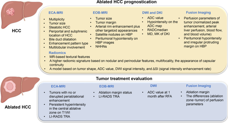
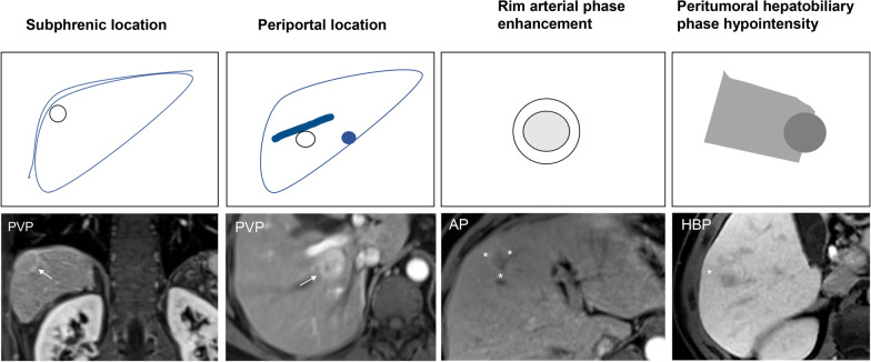
Hepatocellular carcinoma (HCC) accounts for the vast majority of primary liver cancer and constitutes a major global health challenge. Tumor ablation with either radiofrequency ablation (RFA) or microwave ablation (MWA) is recommended as a curative-intent treatment for early-stage HCC. Given the widespread use of thermal ablation in routine clinical practice, accurate evaluation of treatment response and patient outcomes has become crucial in optimizing individualized management strategies. Noninvasive imaging occupies the central role in the routine management of patients with HCC. Magnetic resonance imaging (MRI) could provide full wealth of information with respect to tumor morphology, hemodynamics, function and metabolism. With accumulation of liver MR imaging data, radiomics analysis has been increasingly applied to capture tumor heterogeneity and provide prognostication by extracting high-throughput quantitative imaging features from digital medical images. Emerging evidence suggests the potential role of several qualitative, quantitative and radiomic MRI features in prediction of treatment response and patient prognosis after ablation of HCC. Understanding the advancements of MRI in the evaluation of ablated HCCs may facilitate optimal patient care and improved outcomes. This review provides an overview of the emerging role of MRI in treatment response evaluation and prognostication of HCC patients undergoing ablation. CLINICAL RELEVANCE STATEMENT: MRI-based parameters can help predict treatment response and patient prognosis after HCC ablation and thus guide treatment planning. KEY POINTS: 1. ECA-MRI provides morphological and hemodynamic assessment of ablated HCC. 2. EOB-MRI provides more information for tumor response prediction after ablation. 3. DWI improve the characterization of HCC and optimize treatment decision. 4. Radiomics analysis enables characterization of tumor heterogeneity guidance of clinical decision-making. 5. Further studies with multiple radiologists and sufficient follow-up period are needed.

Keywords: Ablation; Carcinoma; Hepatocellular; Magnetic resonance imaging; Prognosis; Treatment response.


Conflict of interest statement

The authors declare that they have no competing interests.

Figures

Fig. 1
Fig. 1
Published magnetic resonance imaging features for treatment response evaluation and prognostication of hepatocellular carcinoma after thermal ablation. RADCmedian was defined as the ratio of ADCmedian to the mean ADC of the non-lesion area
Fig. 2
Fig. 2
Graphical illustration of some qualitative MR features of HCC associated with poor outcomes after ablation treatment. PVP, portal venous phase. AP, arterial phase. HBP, hepatobiliary phase
Fig. 3
Fig. 3
Gadobenate dimeglumine–enhanced axial MRI scans in a 57-year-old man with hepatitis B virus (A-H). A 1.7-cm steatotic mass is detected in segments VII. The mass shows T1 hypointensity (A), signal intensity reduced on out-of-phase TIWI (arrow in B), moderate T2 hyperintensity (C), marked hyperintensity on DWI (b = 1000 s/mm2) (D), rim enhancement on arterial phase image (E), “wash-out” appearance on portal venous phase (F) and equilibrium phase (G), marked hepatobiliary phase hypointensity (H). The recurrence-free survival for this patient was 150 days
Fig. 4
Fig. 4
Images in a 56-year-old man with chronic hepatitis B virus infection. AD Preoperative gadoxetate disodium–enhanced axial MRI scans show a 2.0-cm mass (NHHN) in segment VII, which shows moderate T2 hyperintensity (A), marked hyperintensity on DWI (b = 800 s/mm2) (B), without obvious enhancement on arterial phase image (C), marked hepatobiliary phase hypointensity (D). EG Enhanced abdominal CT images of the patient 50 days after radiofrequency ablation treatment of tumor. A low-density ablation area was shown (E), and there was not any enhancement tissue in or along the margin of the treated lesion (F, G). Diagnosis was agreed upon by the 2 readers (LR-TR nonviable). HK Gadoxetate disodium–enhanced axial MRI scans 67 days after tumor ablation, which shows T2 hyperintensity (H), marked hyperintensity on DWI (b = 800 s/mm2) (I), irregular thickened enhanced tissue area was found along the margin of the treated lesion (* in J), and marked hepatobiliary phase hypointensity (K). The recurrence-free survival for this patient was 60 days
Fig. 5
Fig. 5
Images in a 62-year-old man with chronic hepatitis B virus infection. AF Gadoxetate disodium–enhanced axial MRI scans show a 2.4-cm mass in segment VII (subphrenic location), which shows T1 hypointensity (A), mild to moderate T2 hyperintensity (B), marked hyperintensity on DWI (b = 800 s/mm2) (C), mild inhomogeneous enhancement on arterial phase image (D), obvious enhancement on portal venous phase (E), and demonstrates marked hepatobiliary phase hypointensity (F). The recurrence-free survival for this patient was 50 days

References

    1. Llovet JM, Kelley RK, Villanueva A, Singal AG, Pikarsky E, Roayaie S, et al. Hepatocellular carcinoma. Nat Rev Dis Prim. 2021;7(1):6. doi: 10.1038/s41572-020-00240-3. - DOI - PubMed
    1. EASL Clinical Practice Guidelines Management of hepatocellular carcinoma. J Hepatol. 2018;69(1):182–236. doi: 10.1016/j.jhep.2018.03.019. - DOI - PubMed
    1. Marrero JA, Kulik LM, Sirlin CB, Zhu AX, Finn RS, Abecassis MM, Roberts LR, Heimbach JK. Diagnosis, staging, and management of hepatocellular carcinoma: 2018 Practice Guidance by the American Association for the Study of Liver Diseases. Hepatology. 2018;68(2):723–750. doi: 10.1002/hep.29913. - DOI - PubMed
    1. Reig M, Forner A, Rimola J, Ferrer-Fàbrega J, Burrel M, Garcia-Criado Á, et al. BCLC strategy for prognosis prediction and treatment recommendation: The 2022 update. J Hepatol. 2022;76(3):681–693. doi: 10.1016/j.jhep.2021.11.018. - DOI - PMC - PubMed
    1. Feng Z, Zhao H, Guan S, Wang W, Rong P. Diagnostic performance of MRI using extracellular contrast agents versus gadoxetic acid for hepatocellular carcinoma: a systematic review and meta-analysis. Liver Int. 2021;41(5):1117–1128. doi: 10.1111/liv.14850. - DOI - PubMed

LinkOut - more resources