. 2023 May 15;21(1):112.
doi: 10.1186/s12964-023-01132-1.

Bacillus cereus extracellular vesicles act as shuttles for biologically active multicomponent enterotoxins

Affiliations

Bacillus cereus extracellular vesicles act as shuttles for biologically active multicomponent enterotoxins

Tanja Buchacher et al. Cell Commun Signal. .

Erratum in

Abstract

Background: Extracellular vesicles (EVs) from Gram-positive bacteria have gained considerable importance as a novel transport system of virulence factors in host-pathogen interactions. Bacillus cereus is a Gram-positive human pathogen, causing gastrointestinal toxemia as well as local and systemic infections. The pathogenicity of enteropathogenic B. cereus has been linked to a collection of virulence factors and exotoxins. Nevertheless, the exact mechanism of virulence factor secretion and delivery to target cells is poorly understood.

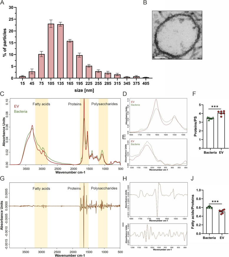
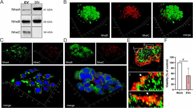
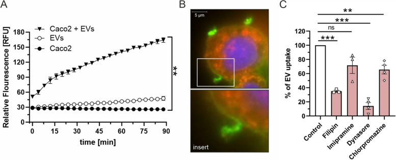
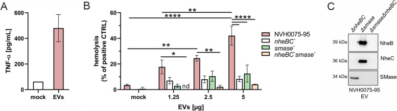
Results: Here, we investigate the production and characterization of enterotoxin-associated EVs from the enteropathogenic B. cereus strain NVH0075-95 by using a proteomics approach and studied their interaction with human host cells in vitro. For the first time, comprehensive analyses of B. cereus EV proteins revealed virulence-associated factors, such as sphingomyelinase, phospholipase C, and the three-component enterotoxin Nhe. The detection of Nhe subunits was confirmed by immunoblotting, showing that the low abundant subunit NheC was exclusively detected in EVs as compared to vesicle-free supernatant. Cholesterol-dependent fusion and predominantly dynamin-mediated endocytosis of B. cereus EVs with the plasma membrane of intestinal epithelial Caco2 cells represent entry routes for delivery of Nhe components to host cells, which was assessed by confocal microscopy and finally led to delayed cytotoxicity. Furthermore, we could show that B. cereus EVs elicit an inflammatory response in human monocytes and contribute to erythrocyte lysis via a cooperative interaction of enterotoxin Nhe and sphingomyelinase.

Conclusion: Our results provide insights into the interaction of EVs from B. cereus with human host cells and add a new layer of complexity to our understanding of multicomponent enterotoxin assembly, offering new opportunities to decipher molecular processes involved in disease development. Video Abstract.

Keywords: 3D-SIM microscopy; Bacillus cereus; Extracellular vesicles; Host–pathogen interaction; Multicomponent toxin; Non-hemolytic enterotoxin; SMase.


Conflict of interest statement

The authors declare no competing interests.

Figures

Fig. 1
Fig. 1
B. cereus releases EVs in vitro. EVs of B. cereus NVH0095-75 were characterized using nanoparticle tracking analysis (NTA), transmission electron microscopy and Fourier transform infrared (FTIR) spectroscopy. A Purified EVs from three biological independent replicates (three technical measurements each) were subjected to NTA for analysis of size distribution. B Resin-embedded TEM, used to determine the shape and confirm the size, revealed round-shaped EVs from B. cereus (scale bar 50 nm). C-I FTIR was used to generate metabolite fingerprints from isolated B. cereus EVs and intact bacterial cells. EVs isolated from six independent bacterial cultures were analyzed by FTIR. Representative full-range normalized spectra are shown (C), highlighting the spectral regions used for subsequent chemometric analysis. D and E provide a zoom-in into the region characteristic for proteins (1720–1500 cm-1) and polysaccharides (1200–900 cm-1), respectively. G As revealed by 2nd derivative subtraction spectral analysis of EVs versus bacterial cells, spectral differences between EVs and bacteria were most pronounced in the spectral regions accounting for proteins (zoom-in see H) and polysaccharides (zoom-in see I). F and H Calculation of spectroscopic ratios of highlighted regions in (C) as described in the method section. Statistical significance is calculated using a two-tailed Student’s t-test (***p < 0.001)
Fig. 2
Fig. 2
Proteomic profiling of B. cereus extracellular vesicles revealed several enterotoxins. The protein cargo of B. cereus secreted extracellular vesicles was determined by LC–MS/MS. Proteins commonly identified in two biologically independent experiments were included in the proteomic analysis. A Subcellular localization of EV proteins predicted with the aid of PsortB and illustrated in proportional numbers. B Prediction of the number of proteins with a secretory signal peptide, using SignalP. C KEGG pathway enrichment analysis of EV proteins. Pathways with a false discovery rate (FDR) < 0.05 were considered as significantly enriched. D Overview of virulence-associated factors identified in B. cereus NVH0095-75 EVs
Fig. 3
Fig. 3
The multicomponent enterotoxin Nhe is packed in B. cereus vesicles and transported through vesicles to human intestinal Caco2 cell. Mouse monoclonal antibodies (anti-NheA IgG1, anti-NheB IgG1κ), anti-NheC IgM) were used for the specific detection of the Nhe components [80]. A As shown by immunoblot analyses, NheA and NheB are detectable in B. cereus-derived extracellular vesicles (EV) and vesicle-free supernatant (SN) while NheC is only detectable in EV. B The presence of NheB (AF488, green) and NheC (AF568, red) in B. cereus vesicle aggregates was confirmed with 3D-SIM microscopy. C-E To study the delivery of Nhe enterotoxin to human intestinal Caco2 cells, cells were treated with 200 µg/mL of B. cereus EVs for 2 h followed by several washing steps. C NheA (AF568; red), NheB (AF488; green) and cell nuclei (DAPI; blue), or (D) NheB (AF488; green), NheC (AF568; red) and cell nuclei (DAPI; blue) were visualized by 3D-SIM microscopy. E 3D-SIM fluorescent images at single cell level showed colocalization of NheB (AF488, green) and NheC (AF568, red) components (I, II) at the edges of Caco2 cells. F Viability of Caco2 cells stimulated with EVs was determined after 24 h using the Vita-Orange Cell Viability Reagent and expressed as a percentage with respect to untreated control cells for three independent biological experiments (mean ± standard error of the mean (SEM). Statistical significance is calculated using two-tailed Student’s t-test (*p < 0.05)
Fig. 4
Fig. 4
Cellular uptake of B. cereus extracellular vesicles is mediated by means of membrane fusion via cholesterol-rich domains and endocytosis. A For B. cereus EV uptake, Rhodamine-R18 labeled B. cereus EVs (5 µg) were applied to Caco2 cells and the fluorescence intensity was measured every two minutes (shown here every six minutes for the sake of clarity) up to 90 min at 37 °C using a microtiter reader. An increase in fluorescence intensity indicates membrane fusion, shown as mean ± standard error of the mean (SEM) for three independent biological experiments. B Vesicles containing NheB were detected with AF488 (green) and found to be aggregated to the edges and surfaces of Caco2 cells indicating fusion (scale bar 5 µm, 3D-SIM images). C Inhibition of vesicle uptake by Caco2 was studied in the presence of either cholesterol-sequestering agents Filipin III (10 µg/ml) and Imipramine (10 mM), or dynamin and clathrin-mediated endocytosis inhibitors Dynasore (80 μM), and chlorpromazine (15 ug/ml; all from Sigma Aldrich, USA), respectively. After 1 h of cell treatment with the inhibitors, EVs were added and cultured for 90 min. The percentage of EV uptake in the presence of inhibitors was normalized to internalization in untreated cells. Mean ± standard error of the mean (SEM) is shown for three or four independent biological experiments. Statistical significance is calculated using two-tailed Student’s t-test (**p < 0.01, ***p < 0.001)
Fig. 5
Fig. 5
Hemolytic and proinflammatory activity of B. cereus extracellular vesicles. A Human monocytes (2.5 × 106 cells/mL) were stimulated with 100 µg/mL of EV for 4 h at 37 °C / 5% CO2 and pro-inflammatory cytokine secretion was assessed using ELISA. Mock stimulation served as control. Concentrations are expressed as mean ± standard error of the mean (SEM) for three independent biological experiments. B Human erythrocytes were stimulated with different concentrations of EVs from wild type strain NVH0075-95 and EVs derived from single ∆nheBC, single smase and double nheBCsmase mutants. After 1 h of stimulation at 37 °C, 5% CO2, erythrocytes were pelleted and the supernatant was transferred into a 96-well microtiter plate. The absorbance was measured at 540 nm using a microplate reader. The hemolytic activity is calculated as percentage of the positive control Triton X-100 and expressed as mean ± standard error of the mean (SEM) for three independent biological experiments. Statistical significance is calculated using the two-way ANOVA with Tukey's multiple comparisons test (*p < 0.05, **p < 0.01, ****p < 0.0001). C Single or double deletion of nheBC and smase in EVs was confirmed by western blotting

References

    1. Ehling-Schulz M, Lereclus D, Koehler TM. The Bacillus cereus Group: Bacillus Species with Pathogenic Potential. Microbiol Spectr. 2019;7(3):1-35. - PMC - PubMed
    1. Dietrich R, Jessberger N, Ehling-Schulz M, Märtlbauer E, Granum PE. The food poisoning toxins of bacillus cereus. Toxins (Basel) 2021;13(2):98. doi: 10.3390/toxins13020098. - DOI - PMC - PubMed
    1. Bottone EJ. Bacillus cereus, a volatile human pathogen. Clin Microbiol Rev. 2010;23(2):382–398. doi: 10.1128/CMR.00073-09. - DOI - PMC - PubMed
    1. Glasset B, Herbin S, Granier SA, Cavalié L, Lafeuille E, Guérin C, et al. Bacillus cereus, a serious cause of nosocomial infections: Epidemiologic and genetic survey. PLoS ONE. 2018;13(5):e0194346. doi: 10.1371/journal.pone.0194346. - DOI - PMC - PubMed
    1. Tschiedel E, Rath PM, Steinmann J, Becker H, Dietrich R, Paul A, et al. Lifesaving liver transplantation for multi-organ failure caused by Bacillus cereus food poisoning. Pediatr Transplant. 2015;19(1):E11–E14. doi: 10.1111/petr.12378. - DOI - PubMed

Publication types

MeSH terms

LinkOut - more resources