. 2023 May 15;21(1):113.
doi: 10.1186/s12964-023-01120-5.

Single-cell RNA sequencing reveals the vascular smooth muscle cell phenotypic landscape in aortic aneurysm

Affiliations

Single-cell RNA sequencing reveals the vascular smooth muscle cell phenotypic landscape in aortic aneurysm

Genmao Cao et al. Cell Commun Signal. .

Abstract

Background and objectives: Phenotypic switching in vascular smooth muscle cells (VSMCs) has been linked to aortic aneurysm, but the phenotypic landscape in aortic aneurysm is poorly understood. The present study aimed to analyse the phenotypic landscape, phenotypic differentiation trajectory, and potential functions of various VSMCs phenotypes in aortic aneurysm.

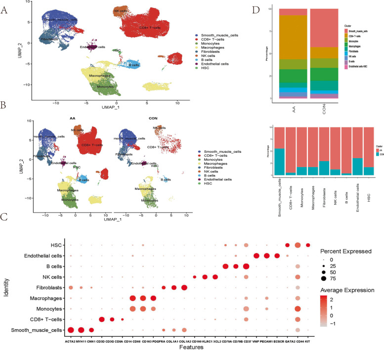
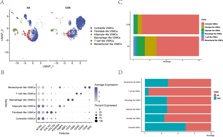
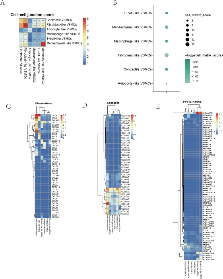
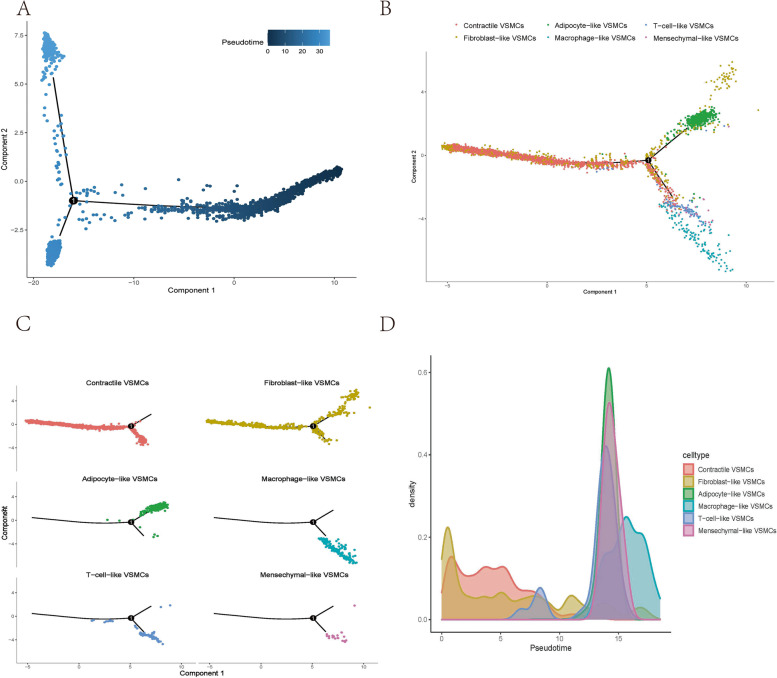
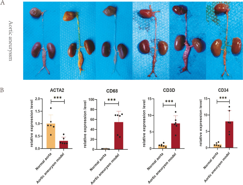
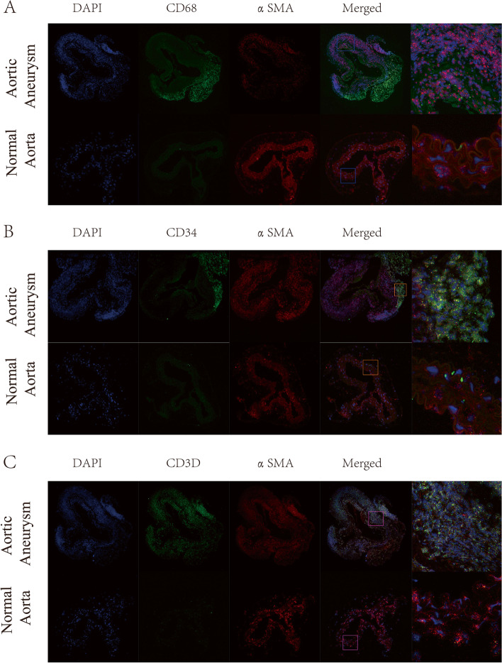
Methods: Single-cell sequencing data of 12 aortic aneurysm samples and 5 normal aorta samples (obtained from GSE166676 and GSE155468) were integrated by the R package Harmony. VSMCs were identified according to the expression levels of ACTA2 and MYH11. VSMCs clustering was determined by the R package 'Seurat'. Cell annotation was determined by the R package 'singleR' and background knowledge of VSMCs phenotypic switching. The secretion of collagen, proteinases, and chemokines by each VSMCs phenotype was assessed. Cell‒cell junctions and cell-matrix junctions were also scored by examining the expression of adhesion genes. Trajectory analysis was performed by the R package 'Monocle2'. qPCR was used to quantify VSMCs markers. RNA fluorescence in situ hybridization (RNA FISH) was performed to determine the spatial localization of vital VSMCs phenotypes in aortic aneurysms.

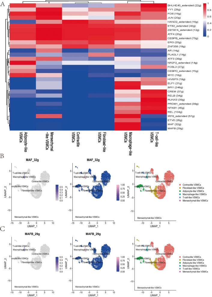
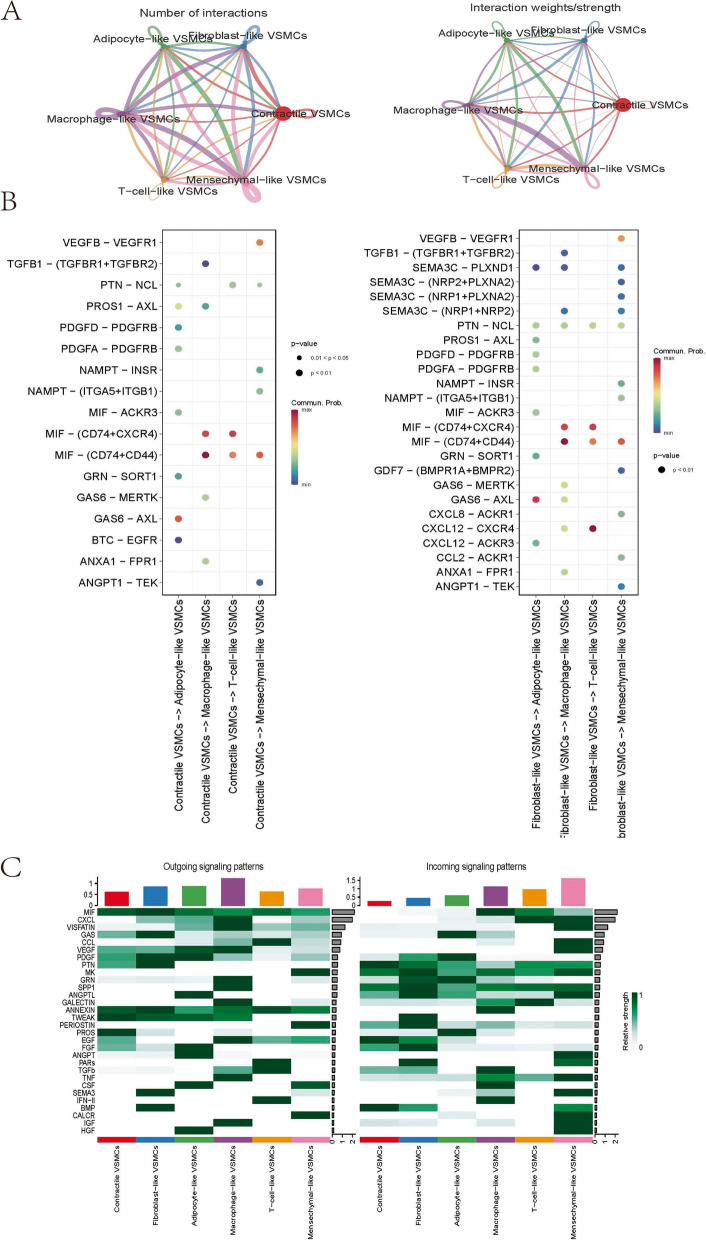
Results: A total of 7150 VSMCs were categorize into 6 phenotypes: contractile VSMCs, fibroblast-like VSMCs, T-cell-like VSMCs, adipocyte-like VSMCs, macrophage-like VSMCs, and mesenchymal-like VSMCs. The proportions of T-cell-like VSMCs, adipocyte-like VSMCs, macrophage-like VSMCs, and mesenchymal-like VSMCs were significantly increased in aortic aneurysm. Fibroblast-like VSMCs secreted abundant amounts of collagens. T-cell-like VSMCs and macrophage-like VSMCs were characterized by high chemokine levels and proinflammatory effects. Adipocyte-like VSMCs and mesenchymal-like VSMCs were associated with high proteinase levels. RNA FISH validated the presence of T-cell-like VSMCs and macrophage-like VSMCs in the tunica media and the presence of mesenchymal-like VSMCs in the tunica media and tunica adventitia.

Conclusion: A variety of VSMCs phenotypes are involved in the formation of aortic aneurysm. T-cell-like VSMCs, macrophage-like VSMCs, and mesenchymal-like VSMCs play pivotal roles in this process. Video Abstract.

Keywords: Aortic aneurysm; Phenotypes; Single-cell transcriptome analysis; Vascular smooth muscle cells.


Conflict of interest statement

The authors declare no conflicts of interest.

Figures

Fig. 1
Fig. 1
General analysis and annotation of all cells from the normal aorta and aortic aneurysm. Uniform manifold approximation and projection (UMAP) representation of the aligned gene expression data from the normal aorta and aortic aneurysm, showing the partitioning of 9 cell types A and groupings by sample source B. The expression of marker genes is shown in the dot plot C. The origin distribution of various cell types and the proportions of various cell types in aortic aneurysm and the normal aorta D
Fig. 2
Fig. 2
Specific analysis and annotation of all VSMCs phenotypes. Uniform manifold approximation and projection (UMAP) representation of the aligned gene expression data of all VSMCs showing the partitioning of 6 annotated VSMCs phenotypes A. The expression of marker genes is shown on the dot plot B (gene expression log-normalized by Seurat). The proportion of VSMCs phenotypes in aortic aneurysm and the normal aorta C and the origin distribution of various VSMCs phenotypes D
Fig. 3
Fig. 3
Functional analysis of various VSMCs phenotypes. The cell‒cell junction scores between any two VSMCs phenotypes A and cell–matrix scores of each VSMCs phenotype B. Heatmap showing chemokine genes C, collagen genes D, and proteinase genes E in various VSMCs phenotypes
Fig. 4
Fig. 4
Cell trajectory analysis of various VSMCs phenotypes. Pseudotime plot showing trajectory analysis of all VSMCs A and various VSMCs phenotypes B-C. Cell density is plotted along the timeline D
Fig. 5
Fig. 5
SCENIC analysis indicated significant regulons for each VSMCs phenotype A. The regulatory activity of MAF (32 g) B and MAFB (29 g) C was projected with a UMAP plot
Fig. 6
Fig. 6
Global cell‒cell communication patterns involve multiple signalling pathways. The line width represents the interaction quantity and interaction strength among various VSMCs phenotypes A. Communication strength of all significant signalling pathways from contractile/fibroblast-like VSMCs to macrophage-like/T-cell-like/adipocyte-like/mesenchymal-like VSMCs B. Significant signalling of each VSMCs phenotype in incoming/outgoing communication patterns C
Fig. 7
Fig. 7
Successful construction of the mouse abdominal aortic aneurysm model A. The relative expression levels of ACTA2. CD68, CD3D, and CD34 were quantified by qPCR B
Fig. 8
Fig. 8
RNA FISH of the normal aorta and aortic aneurysm. Dual hybridization of αSMA and CD68 to identify macrophage-like VSMCs A. Dual hybridization of αSMA and CD34 to identify mesenchymal-like VSMCs B. Dual hybridization of αSMA and CD3D to identify T-cell-like VSMCs C

References

    1. Sonesson B, et al. Infrarenal aortic diameter in the healthy person. Eur J Vasc Surg. 1994;8(1):89–95. doi: 10.1016/S0950-821X(05)80127-6. - DOI - PubMed
    1. Behr Andersen C, et al. Abdominal aortic aneurysms growth is associated with high concentrations of plasma proteins in the intraluminal thrombus and diseased arterial tissue. Arterioscler Thromb Vasc Biol. 2018;38(9):2254–2267. doi: 10.1161/ATVBAHA.117.310126. - DOI - PubMed
    1. Piechota-Polanczyk A, et al. The abdominal aortic aneurysm and intraluminal thrombus: current concepts of development and treatment. Front Cardiovasc Med. 2015;2:19. doi: 10.3389/fcvm.2015.00019. - DOI - PMC - PubMed
    1. Chaikof EL, et al. The society for vascular surgery practice guidelines on the care of patients with an abdominal aortic aneurysm. J Vasc Surg. 2018;67(1):2–77 e2. doi: 10.1016/j.jvs.2017.10.044. - DOI - PubMed
    1. Meijer CA, et al. Doxycycline for stabilization of abdominal aortic aneurysms: a randomized trial. Ann Intern Med. 2013;159(12):815–823. doi: 10.7326/0003-4819-159-12-201312170-00007. - DOI - PubMed

Publication types