. 2023 Mar 29;13(4):615.
doi: 10.3390/biom13040615.

Heterogeneity in Lowe Syndrome: Mutations Affecting the Phosphatase Domain of OCRL1 Differ in Impact on Enzymatic Activity and Severity of Cellular Phenotypes

Affiliations

Heterogeneity in Lowe Syndrome: Mutations Affecting the Phosphatase Domain of OCRL1 Differ in Impact on Enzymatic Activity and Severity of Cellular Phenotypes

Jennifer J Lee et al. Biomolecules. .

Abstract

Lowe Syndrome (LS) is a condition due to mutations in the OCRL1 gene, characterized by congenital cataracts, intellectual disability, and kidney malfunction. Unfortunately, patients succumb to renal failure after adolescence. This study is centered in investigating the biochemical and phenotypic impact of patient's OCRL1 variants (OCRL1VAR). Specifically, we tested the hypothesis that some OCRL1VAR are stabilized in a non-functional conformation by focusing on missense mutations affecting the phosphatase domain, but not changing residues involved in binding/catalysis. The pathogenic and conformational characteristics of the selected variants were evaluated in silico and our results revealed some OCRL1VAR to be benign, while others are pathogenic. Then we proceeded to monitor the enzymatic activity and function in kidney cells of the different OCRL1VAR. Based on their enzymatic activity and presence/absence of phenotypes, the variants segregated into two categories that also correlated with the severity of the condition they induce. Overall, these two groups mapped to opposite sides of the phosphatase domain. In summary, our findings highlight that not every mutation affecting the catalytic domain impairs OCRL1's enzymatic activity. Importantly, data support the inactive-conformation hypothesis. Finally, our results contribute to establishing the molecular and structural basis for the observed heterogeneity in severity/symptomatology displayed by patients.

Keywords: Lowe syndrome; OCRL1; cellular phenotypes; phosphatase activity; rare genetic disease.


Conflict of interest statement

The authors declare no financial, personal or professional competing interests.

Figures

Figure 1
Figure 1
Missense changes affecting OCRL1′s phosphatase domain used in this study. The amino acid sequence of the OCRL1 phosphatase domain is shown with underlined blue residues to indicate those affected by missense mutations; amino acids resulting in such mutations are shown in red. Yellow rectangles enclose regions involved in processing/recognition of the substrate. Substitutions exhibited by variants used in this study are shown; those causing LS, D2, and LS/D2 are shown within red ovals, blue and red vertical rectangles, respectively; mutations with uncertain disease significance are shown within green ovals.
Figure 2
Figure 2
Molecular Dynamics Analysis of OCRL1 variants. (A) Box plots represent the distribution of individual residue fluctuations measured as RMSF throughout the phosphatase domain of OCRL1WT and the indicated OCRL1VAR. All distributions were statistically compared to that corresponding to OCRL1WT, using the Kolmogorov–Smirnov test with p < 0.05; applying Bonferroni’s correction for nine comparisons; αB ≤ 0.005 (*), αB ≤ 0.001 (**) and αB ≤ 0.0001 (***) for individual comparison. NS: Not significant. (B,C) Panels show RMSF (B) and ∆RMSF (C) values for the indicated OCRL1VAR at individual residue positions. For clarity reasons, values for OCRL1VAR were plotted in two graphs shown (left and right) with ≤5 variants per graph bearing substitutions as indicated by the symbol legend. WT distribution (B) is indicated by a dark blue continuous line. Grey shaded vertical areas highlight regions within the phosphatase domain involved in substrate binding or catalysis. See text for more details.
Figure 3
Figure 3
Phosphatase activity displayed by OCRL1WT/VAR. Phosphatase activity of the selected variants was measured utilizing the malachite green assay, as detailed in Materials and Methods, and using Full length OCRL1WT/VAR immunoprecipitated from human cells (A) or the variant’s recombinant, isolated phosphatase domain produced in and purified from bacteria (B). (A) The biologically relevant, full length indicated OCRL1 variants were tested for their ability to catalyze the release of inorganic phosphate from PI(4,5)P2 as a function of time. The phosphatase dead variant pH524R was used as negative control. (B) Experiments using an isolated domain were performed to confirm the detrimental direct effect of the missense mutations on the enzymatic activity of the variant’s catalytic domain. Statistical significance of the differences between normalized catalytic activity of OCRL1VAR and WT was assessed using the t-test with p < 0.05, applying Bonferroni’s correction for six comparisons: αB ≤ 0.008 (*) and αB ≤ 0.001 (**) for individual comparison.
Figure 4
Figure 4
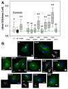
Golgi apparatus fragmentation phenotype induced by OCRL1VAR. (A) Presence and magnitude of this phenotype was assessed by computing the ratio between the area occupied by the Golgi apparatus (GA) and that corresponding to the whole cell (see text for details). Plot depicts combined data from 3 experiments (see Figure 5 for a representative experiment showing individual cell variation). Green dashed line highlights median value obtained for cells expressing OCRL1WT from a distribution with Q1 = 0.034, Q3 = 0.06; IQR = 0.026 (see Materials and Methods). EV: cells transfected with pEGFP empty vector. Statistical significance of the differences between the distributions of WT and variants was assessed using the Wilcoxon test with p < 0.05 applying Bonferroni’s correction for multiple comparisons to αB ≤ 0.005 (*) or αB ≤ 0.001 (**) per comparison NS: Not significant. (B) Representative images showing HK2 OCRL1 KO cells expressing GFP-OCRL1WT or the indicated GFP-OCRL1VAR (green signal) and immunostained for the GA marker TGN46 (red signal). The latter is also reproduced in black and white for clearer visualization. Scale bars: 15 µm. When applicable, additional magnification is indicated (i.e., 1.2×).
Figure 5
Figure 5
Golgi apparatus fragmentation phenotype induced by OCRL1VAR as function of each variant intracellular content. Results in a representative experiment for the evaluation of GA fragmentation induced by the indicated OCRL1VAR. The ratio (Area GA/Area cell) was estimated as described in Material and Methods and Figure 4. 150–200 cells HK2 OCRL1 KO cells expressing only GFP (i.e., transfected with pEGFP empty vector, EV) or GFP-OCRL1WT/VAR were analyzed per sample while also measuring their total fluorescence intensity in arbitrary units reporting total amount of the indicated protein present in the corresponding cells). Green horizontal dashed line depicts the median value obtained for cells only expressing OCRL1WT, while the red reference line corresponds with the median value measured for each specific OCRL1VAR.
Figure 6
Figure 6
Residues affected by missense mutation spatially segregate according to biochemical impairment and phenotype severity. Residues affected by mutations from group 2 (A) and some of those in group 1 (B) (see text for details), were mapped on the 3D structure of the phosphatase domain of OCRL1 (PDB file 4CMN). Panels A and B correspond to two views of the OCRL1 phosphatase domain crystal structure after a rotation of ≈180 degrees.

References

    1. Mehta Z.B., Pietka G., Lowe M. The cellular and physiological functions of the Lowe syndrome protein OCRL1. Traffic. 2014;15:471–487. doi: 10.1111/tra.12160. - DOI - PMC - PubMed
    1. Bökenkamp A., Ludwig M. The oculocerebrorenal syndrome of Lowe: An update. Pediatr. Nephrol. 2016;31:2201–2212. doi: 10.1007/s00467-016-3343-3. - DOI - PMC - PubMed
    1. De Matteis M.A., Staiano L., Emma F., Devuyst O. The 5-phosphatase OCRL in Lowe syndrome and Dent disease 2. Nat. Rev. Nephrol. 2017;13:455–470. doi: 10.1038/nrneph.2017.83. - DOI - PubMed
    1. Recker F., Reutter H., Ludwig M. Lowe syndrome/Dent-2 disease: A comprehensive review of known and novel aspects. J. Pediatr. Genet. 2013;2:53–68. doi: 10.3233/PGE-13049. - DOI - PMC - PubMed
    1. Böckenhauer D., Bökenkamp A., Nuutinen M., Unwin R., Van’t Hoff W., Sirimanna T., Vrljicak K., Ludwig M. Novel OCRL mutations in patients with Dent-2 disease. J. Pediatr. Genet. 2012;1:15–23. doi: 10.3233/PGE-2012-005. - DOI - PMC - PubMed

Publication types

Substances

LinkOut - more resources