

A Multi-Feature Fusion Framework for Automatic Skin Cancer Diagnostics
- PMID: 37189574
- PMCID: PMC10137533
- DOI: 10.3390/diagnostics13081474
A Multi-Feature Fusion Framework for Automatic Skin Cancer Diagnostics
Abstract
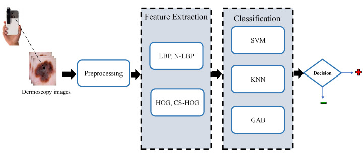
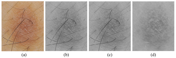
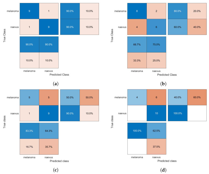
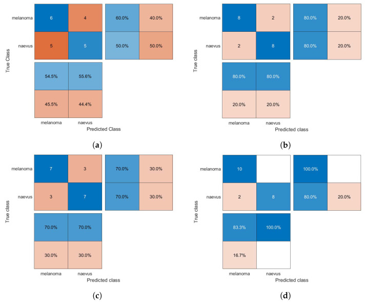
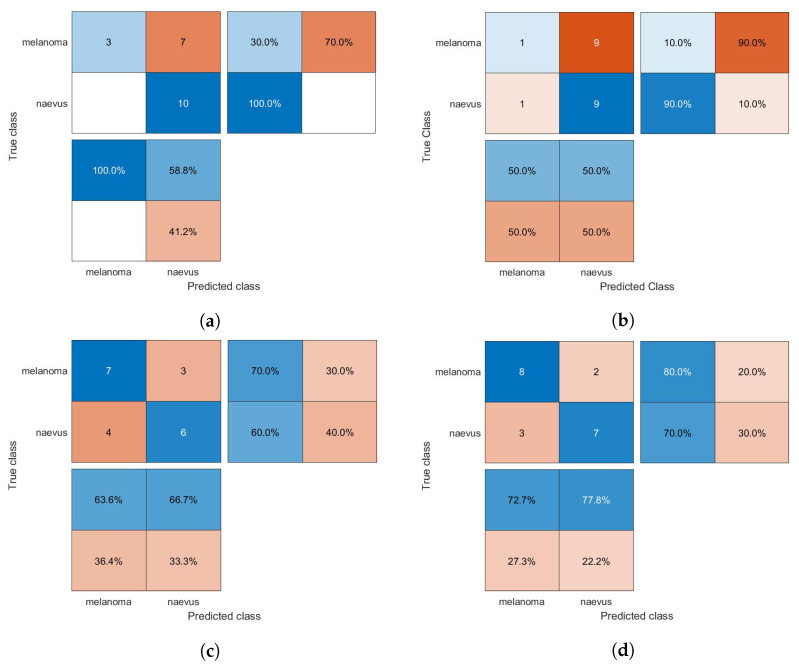
Malignant melanoma is the most invasive skin cancer and is currently regarded as one of the deadliest disorders; however, it can be cured more successfully if detected and treated early. Recently, CAD (computer-aided diagnosis) systems have emerged as a powerful alternative tool for the automatic detection and categorization of skin lesions, such as malignant melanoma or benign nevus, in given dermoscopy images. In this paper, we propose an integrated CAD framework for rapid and accurate melanoma detection in dermoscopy images. Initially, an input dermoscopy image is pre-processed by using a median filter and bottom-hat filtering for noise reduction, artifact removal, and, thus, enhancing the image quality. After this, each skin lesion is described by an effective skin lesion descriptor with high discrimination and descriptiveness capabilities, which is constructed by calculating the HOG (Histogram of Oriented Gradient) and LBP (Local Binary Patterns) and their extensions. After feature selection, the lesion descriptors are fed into three supervised machine learning classification models, namely SVM (Support Vector Machine), kNN (k-Nearest Neighbors), and GAB (Gentle AdaBoost), to diagnostically classify melanocytic skin lesions into one of two diagnostic categories, melanoma or nevus. Experimental results achieved using 10-fold cross-validation on the publicly available MED-NODEE dermoscopy image dataset demonstrate that the proposed CAD framework performs either competitively or superiorly to several state-of-the-art methods with stronger training settings in relation to various diagnostic metrics, such as accuracy (94%), specificity (92%), and sensitivity (100%).
Keywords: computer-aided diagnosis; cross-validation; gentle adaboost; histogram of oriented gradient; k-nearest neighbors; local binary patterns; malignant melanoma; skin cancer; support vector machine.
Conflict of interest statement
The authors declare no conflict of interest.
Figures
References
-
- Singh L., Janghel R.R., Sahu S.P. SLICACO: An automated novel hybrid approach for dermatoscopic melanocytic skin lesion segmentation. Int. J. Imaging Syst. Technol. 2021;31:1817–1833. doi: 10.1002/ima.22591. - DOI
-
- Bakheet S., El-Nagar A. A Deep Neural Approach for Real-Time Malignant Melanoma Detection. Appl. Math. Inf. Sci. 2021;15:89–96.
Grants and funding
LinkOut - more resources
Full Text Sources
Miscellaneous
