. 2023 Mar 29;11(4):1054.
doi: 10.3390/biomedicines11041054.

Streptomyces Bioactive Metabolites Prevent Liver Cancer through Apoptosis, Inhibiting Oxidative Stress and Inflammatory Markers in Diethylnitrosamine-Induced Hepatocellular Carcinoma

Affiliations

Streptomyces Bioactive Metabolites Prevent Liver Cancer through Apoptosis, Inhibiting Oxidative Stress and Inflammatory Markers in Diethylnitrosamine-Induced Hepatocellular Carcinoma

Sana M Alhawsawi et al. Biomedicines. .

Abstract

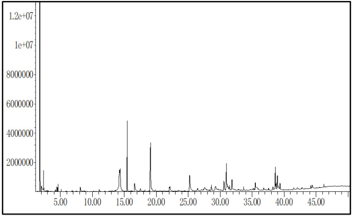
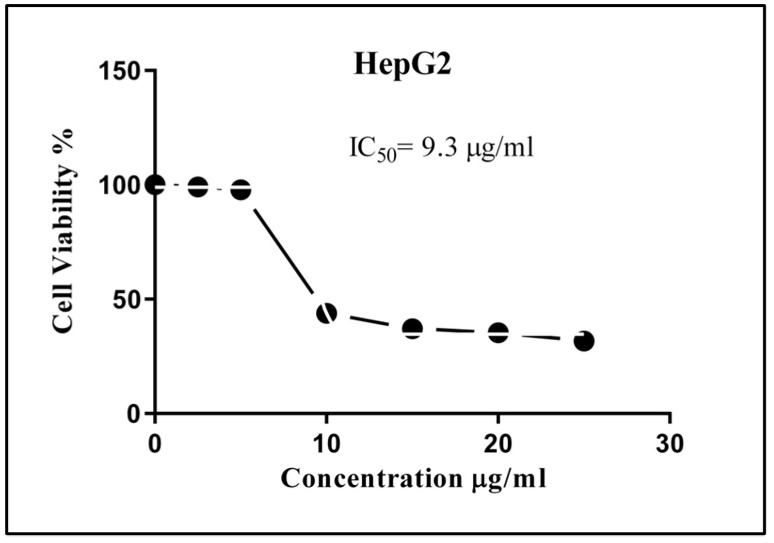
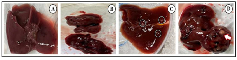
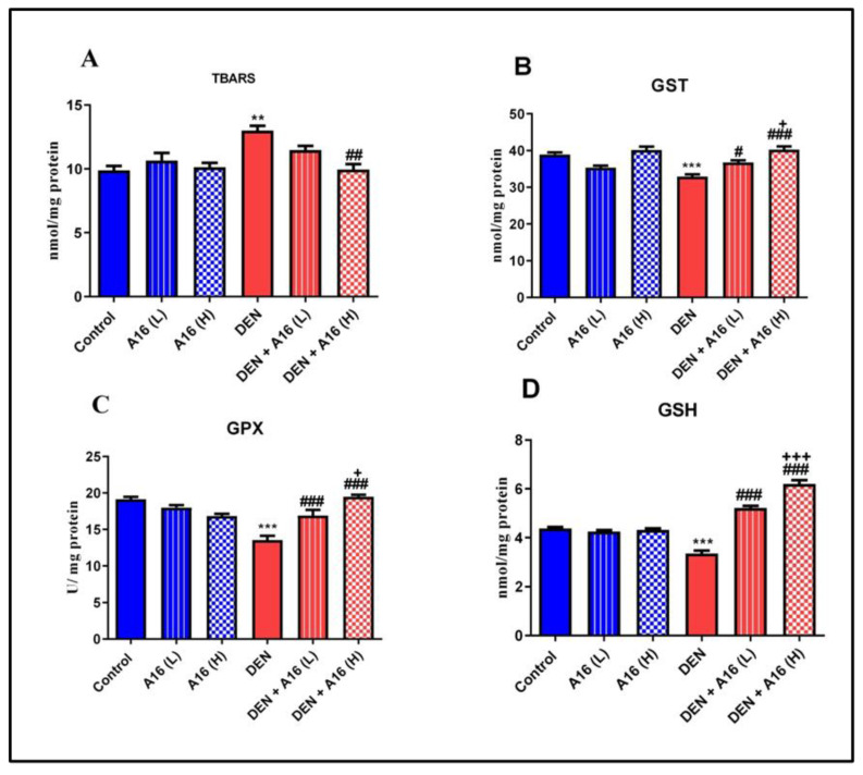
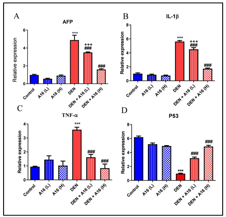
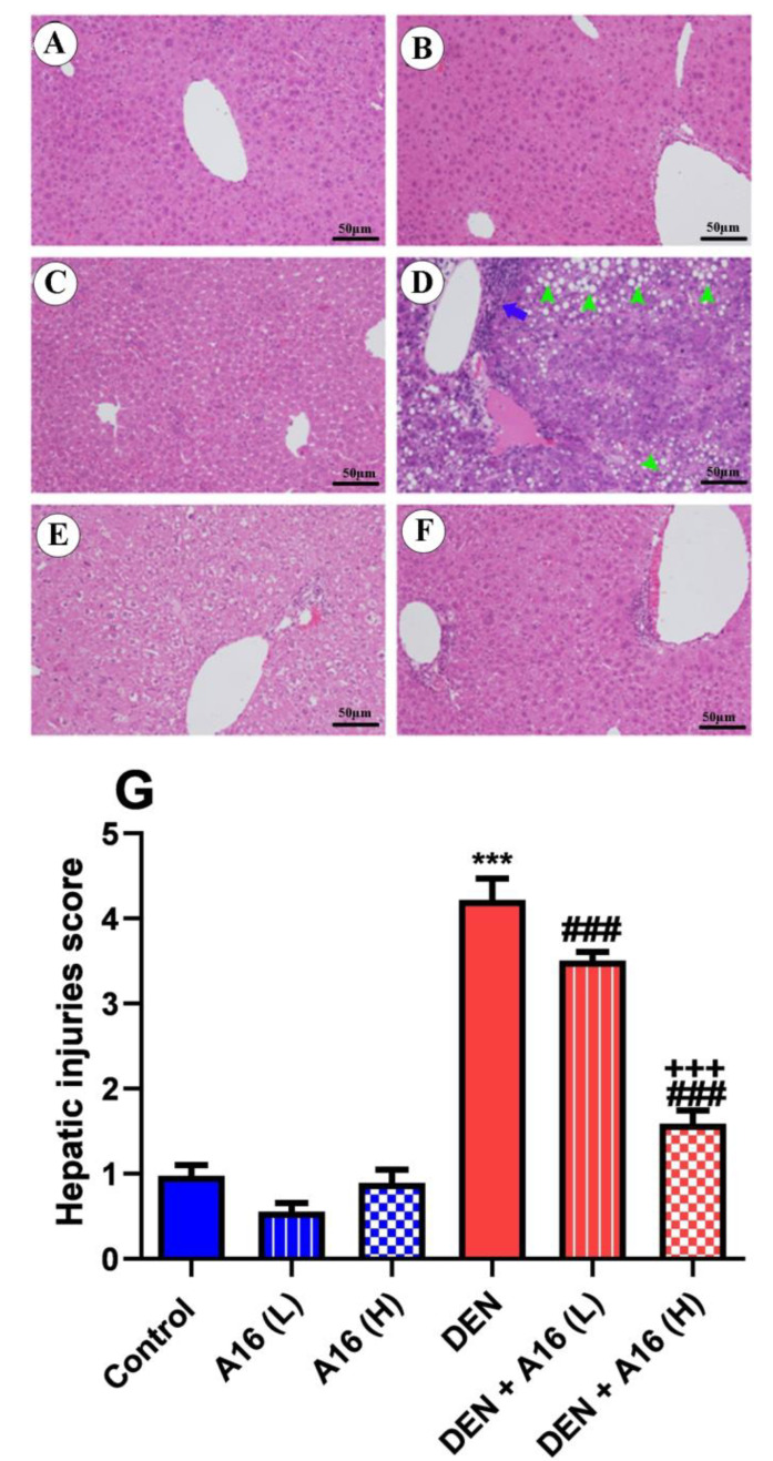
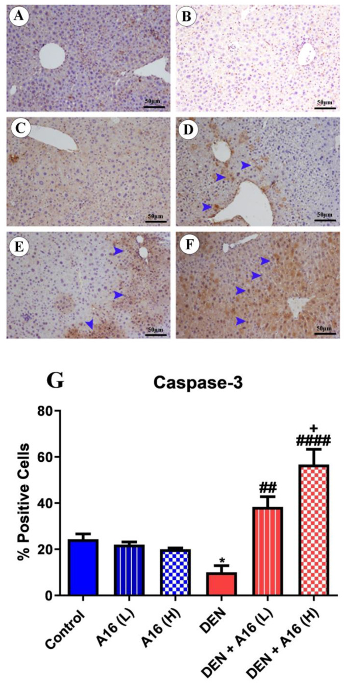
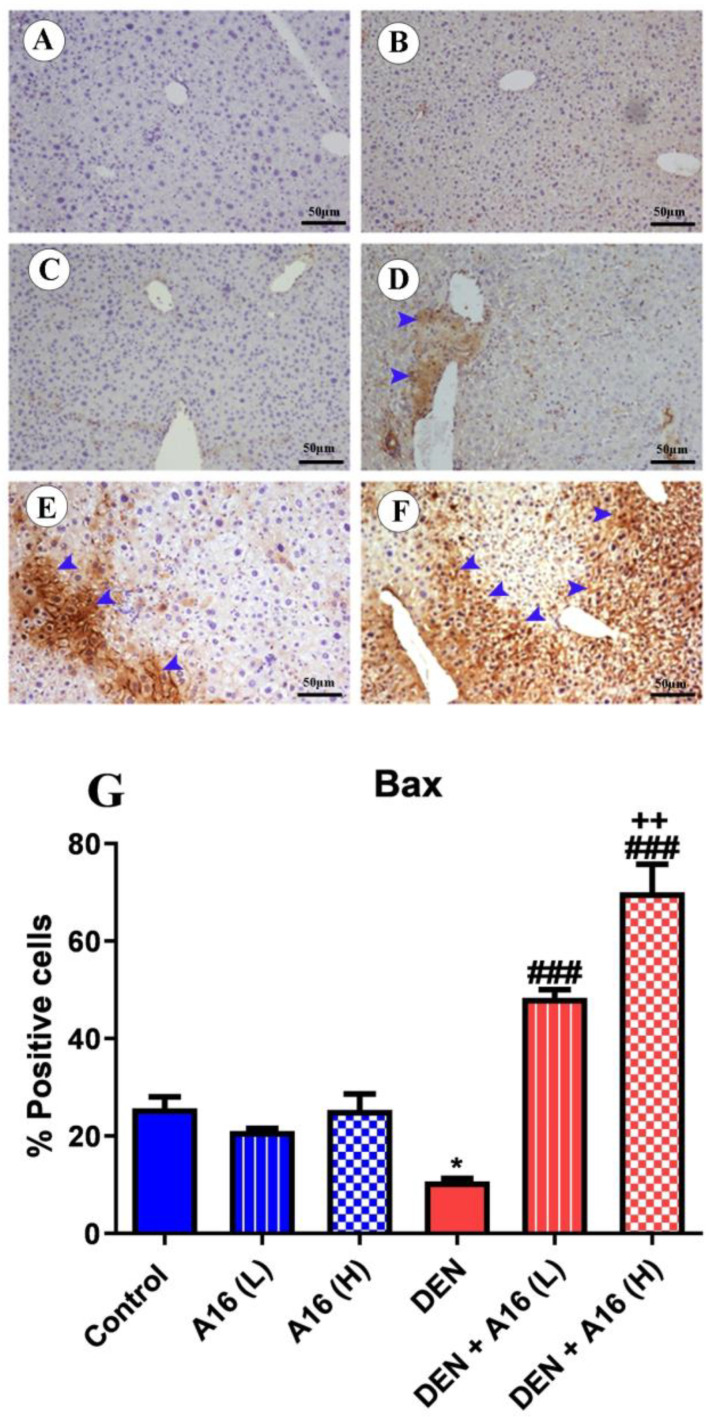
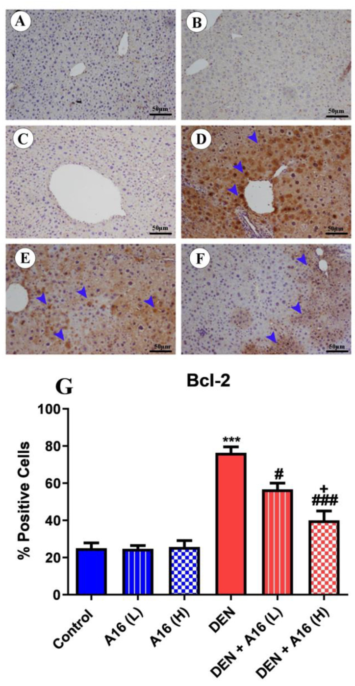
A safe and effective treatment for liver cancer is still elusive despite all attempts. Biomolecules produced from natural products and their derivatives are potential sources of new anticancer medications. This study aimed to investigate the anticancer potential of a Streptomyces sp. bacterial extract against diethylnitrosamine (DEN)-induced liver cancer in Swiss albino mice and explore the underlying cellular and molecular mechanisms. The ethyl acetate extract of a Streptomyces sp. was screened for its potential anticancer activities against HepG-2 using the MTT assay, and the IC50 was also determined. Gas chromatography-mass spectrometric analysis was used to identify the chemical constituents of the Streptomyces extract. Mice were administered DEN at the age of 2 weeks, and from week 32 until week 36 (4 weeks), they received two doses of Streptomyces extract (25 and 50 mg/kg body weight) orally daily. The Streptomyces extract contains 29 different compounds, according to the GC-MS analysis. The rate of HepG-2 growth was dramatically reduced by the Streptomyces extract. In the mice model. Streptomyces extract considerably lessened the negative effects of DEN on liver functions at both doses. Alpha-fetoprotein (AFP) levels were significantly (p < 0.001) decreased, and P53 mRNA expression was increased, both of which were signs that Streptomyces extract was suppressing carcinogenesis. This anticancer effect was also supported by histological analysis. Streptomyces extract therapy additionally stopped DEN-induced alterations in hepatic oxidative stress and enhanced antioxidant activity. Additionally, Streptomyces extract reduced DEN-induced inflammation, as shown by the decline in interleukin-1 beta (IL-1β) and tumor necrosis factor-alpha (TNF-α) levels. Additionally, the Streptomyces extract administration dramatically boosted Bax and caspase-3 levels while decreasing Bcl-2 expressions in the liver according to the Immunohistochemistry examination. In summary, Streptomyces extract is reported here as a potent chemopreventive agent against hepatocellular carcinoma through multiple mechanisms, including inhibiting oxidative stress, cell apoptosis, and inflammation.

Keywords: Streptomyces; anticancer activity; apoptosis; hepatocellular carcinoma; inflammation; oxidative stress.


Conflict of interest statement

The authors declare no conflict of interest.

Figures

Figure 1
Figure 1
Chromatogram of Streptomyces sp. (A16) compounds in extract Ethyl acetate extract. The major compounds were Benzeneacetamide, trans-Cinnamaldehyde, Pyrrole-2-carboxamide and Cinnamamide.
Figure 2
Figure 2
The percentage cytotoxicity on the HepG2 cell line was determined by MTT assay. Cells were cultured in 96-well plates and then treated with different concentration doses of Streptomyces sp. (A16) extract. The data are presented as mean  ±  SEM of three independent experiments and statistically analyzed by the unpaired t-test. The IC50 value was determined.
Figure 3
Figure 3
Representative images of mice livers showing tumor progression over time, control group (A), DEN-treated group after 3 months (B), DEN-treated group after 6 months showing unequal sized gray nodules, which was a signal for HCC formation (C), DEN-treated group after 9 months showing showed liver swelling, cirrhosis, unequal size, white tumor nodules (arrow) (D).
Figure 4
Figure 4
Effects of Streptomyces sp. (A16) extract on antioxidant enzyme activities and lipid peroxidation in mice liver samples following DEN injection, including TBARS (A), GST (B), GPx (C), and GSH (D). Data are presented as mean ± SEM (n = 6) and statistically analyzed by one-way ANOVA followed by the Tukey–Kramer post hoc test. ** p < 0.01, *** p < 0.001 were significant compared with the control group, # p < 0.05, ## p < 0.01, ### p < 0.001 were significant compared with DEN-treated mice and + p < 0.05, +++ p < 0.001 were significant compared with DEN+ A16 (L)-treated mice. Abbreviations: TBARS, thiobarbituric acid reaction substances; GST, glutathione-S-transferase; GPx, glutathione peroxidase; GSH, glutathione.
Figure 5
Figure 5
Assessment of mRNA expressions of AFP (A), IL-1β (B), TNF-α (C) and P53 (D) genes in hepatic tissues following treatment with Streptomyces sp. (A16) extract in DEN-administered mice. Data are presented as mean ± SEM (n = 6) and statistically analyzed by one-way ANOVA followed by the Tukey–Kramer post hoc test. *** p < 0.001 were significant compared with control group, ### p < 0.001 were significant compared with DEN-treated mice and +++ p < 0.001 was significant compared with DEN + A16 (L)-treated mice. Abbreviations: AFP, alpha-fetoprotein; IL-1β, interleukin-1β; TNF-α, tumor necrosis factor-α.
Figure 6
Figure 6
Histological examination of hepatic tissues following treatment with Streptomyces sp. (A16) extract in DEN-administered mice. (H&E, scale bar = 50 µm). (A) Liver tissue from the control group displaying typical hepatic cells. (B,C) Liver sections taken from mice that received a low dose (25 mg/kg body weight) or a high dose (50 mg/kg body weight) of Streptomyces sp. (A16) extract showing normal hepatic cells. (D) Liver tissue from the DEN-treated group exhibited abnormal liver tissue characteristics, such as the destruction of the hepatic lobules’ structural integrity, necrotic hepatic cells with multiple inflammatory infiltrations (blue arrow), fatty change (steatosis) (green head arrows), variable nuclei size, loss of cell membrane, and absence of the hepatic sinusoids’ capillaries. Along with the Kupffer cell hyperplasia, other changes included hyaline changes, bile duct dilatation, and hepatic artery and portal vein enlargement. (E,F) Liver tissue from the DEN+ A16 (L) and DEN + A16 (H) groups showed a significant improvement in liver architecture in the form of decreased hepatic necrosis, fewer inflammatory cells, reduced steatosis, and modest cytoplasmic vacuolization of hepatocytes. (G) Semiquantitative histological scoring of the liver injuries. Data are presented as mean ± SEM (n = 5) and statistically analyzed by the Kruskal-Wallis test and Dunn’s multiple comparison post hoc test. *** p < 0.001 were significant compared with the control group, ### p < 0.001 were significant compared with DEN-treated mice, and +++ p < 0.001 were significant compared with DEN+ A16 (L)-treated mice.
Figure 7
Figure 7
Liver caspase-3 immunohistochemistry. Photomicrographs of sections of liver samples taken from (A) the control. (B,C) Liver sections taken from mice that received a low dose (25 mg/kg body weight) and a high dose (50 mg/kg body weight) of Streptomyces sp. (A16) extract, respectively. (D) DEN-treated group. (E,F) Liver tissue from the DEN+ A16 (L) and DEN + A16 (H) Streptomyces sp. extract. The area percent of positive caspase-3 immunoreactivity (head arrows) was quantified (G). Values are presented as mean ± SEM (n = 3) and statistically analyzed by one-way ANOVA followed by the Tukey–Kramer post hoc test. * p < 0.05 versus control group, ## p < 0.01, #### p < 0.0001 versus DEN-treated mice and + p < 0.05 versus DEN+ A16 (L)-treated mice.
Figure 8
Figure 8
Liver Bax immunohistochemistry. Photomicrographs of sections of liver samples from the following groups: (A) Control group, (B,C) liver sections taken from mice that were given a low dose (25 mg/kg body weight) and a high dose (50 mg/kg body weight) of Streptomyces sp. (A16) extract respectively, (D) DEN-treated group, (E,F) liver tissue from the DEN+ A16 (L) and DEN + A16 (H) Streptomyces sp. extract. The area percent of positive Bax immunoreactivity (head arrows) was quantified (G). Data are expressed as mean ± SEM (n = 3) and statistically analyzed by one-way ANOVA followed by the Tukey–Kramer post hoc test. * p < 0.05 versus control group, ### p < 0.001 versus DEN-treated mice and ++ p < 0.01 versus DEN+ A16 (L)-treated mice.
Figure 9
Figure 9
Liver Bcl-2 immunohistochemistry. Photomicrographs of sections of liver samples from the following groups: (A) Control group, (B,C) liver sections taken from mice that were given a low dose (25 mg/kg body weight) and a high dose (50 mg/kg body weight) of Streptomyces sp. (A16) extract, respectively, (D) DEN-treated group, (E,F) liver tissue from the DEN+ A16 (L) and DEN + A16 (H) Streptomyces sp. extract. The area percent of positive Bcl-2 immunoreactivity (head arrows) was quantified (G). Data are expressed as mean ± SEM (n = 3) and statistically analyzed by one-way ANOVA followed by the Tukey–Kramer post hoc test. *** p < 0.001 versus control group, # p < 0.05, ### p < 0.001 versus DEN-treated mice and + p < 0.05 versus DEN+ A16 (L)-treated mice.

Similar articles

Cited by

References

    1. EASL EASL Clinical Practice Guidelines: Management of hepatocellular carcinoma. J. Hepatol. 2018;69:182–236. doi: 10.1016/j.jhep.2018.03.019. - DOI - PubMed
    1. El-Nakeep S. Molecular and genetic markers in hepatocellular carcinoma: In silico analysis to clinical validation (current limitations and future promises) World J. Gastrointest. Pathophysiol. 2022;13:1. doi: 10.4291/wjgp.v13.i1.1. - DOI - PMC - PubMed
    1. Zajkowska M., Mroczko B. Chemokines in Primary Liver Cancer. Int. J. Mol. Sci. 2022;23:8846. doi: 10.3390/ijms23168846. - DOI - PMC - PubMed
    1. Fujiwara N., Friedman S.L., Goossens N., Hoshida Y. Risk factors and prevention of hepatocellular carcinoma in the era of precision medicine. J. Hepatol. 2018;68:526–549. doi: 10.1016/j.jhep.2017.09.016. - DOI - PMC - PubMed
    1. Poustchi H., Sepanlou S., Esmaili S., Mehrabi N., Ansarymoghadam A. Hepatocellular carcinoma in the world and the middle East. Middle East J. Dig. Dis. 2010;2:31–41. - PMC - PubMed