. 2023 Apr 9;11(4):1133.
doi: 10.3390/biomedicines11041133.

Targeting RNA Polymerase I Transcription Activity in Osteosarcoma: Pre-Clinical Molecular and Animal Treatment Studies

Affiliations

Targeting RNA Polymerase I Transcription Activity in Osteosarcoma: Pre-Clinical Molecular and Animal Treatment Studies

Chang-Won Kang et al. Biomedicines. .

Abstract

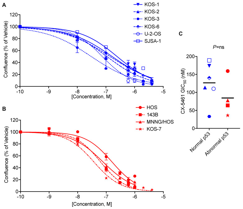
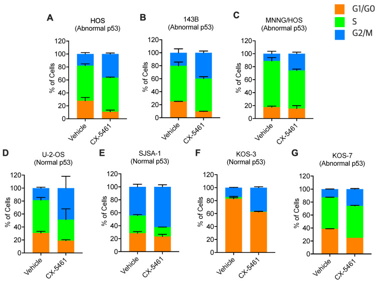
The survival rate of patients with osteosarcoma (OS) has not improved over the last 30 years. Mutations in the genes TP53, RB1 and c-Myc frequently occur in OS and enhance RNA Polymerase I (Pol I) activity, thus supporting uncontrolled cancer cell proliferation. We therefore hypothesised that Pol I inhibition may be an effective therapeutic strategy for this aggressive cancer. The Pol I inhibitor CX-5461 has demonstrated therapeutic efficacy in different cancers in pre-clinical and phase I clinical trials; thus, the effects were determined on ten human OS cell lines. Following characterisation using genome profiling and Western blotting, RNA Pol I activity, cell proliferation and cell cycle progression were evaluated in vitro, and the growth of TP53 wild-type and mutant tumours was measured in a murine allograft model and in two human xenograft OS models. CX-5461 treatment resulted in reduced ribosomal DNA (rDNA) transcription and Growth 2 (G2)-phase cell cycle arrest in all OS cell lines. Additionally, tumour growth in all allograft and xenograft OS models was effectively suppressed without apparent toxicity. Our study demonstrates the efficacy of Pol I inhibition against OS with varying genetic alterations. This study provides pre-clinical evidence to support this novel therapeutic approach in OS.

Keywords: CX-5461; RNA polymerase I transcription; a small molecule inhibitor; osteosarcoma.


Conflict of interest statement

The authors declare no competing interest.

Figures

Figure 1
Figure 1
CX-5461 inhibits human OS cell proliferation irrespective of p53 status. (A) Normal p53 and (B) abnormal p53 osteosarcoma cell lines were treated with the indicated concentrations of CX-5461 or vehicle (50 mM NaH2PO4 (pH 4.5)) for 72 h and assessed for proliferation using the IncuCyte® ZOOM Live Cell Imaging System. Dose–response curves and 50% growth inhibitory concentrations (GIC50) were calculated using non-linear regression analysis, mean +/− SEM of n = 3 biological replicates. (C) Effect of p53 status on GIC50 of CX-5461 with p53 status determined from genomic profiling data (Supplementary Table S1) and p53 protein expression levels in Western blots (Supplementary Figure S1). Normal p53: TP53 wild type with the normal level of p53 protein expression; abnormal p53: TP53 mutant or a dysregulated level of p53 protein expression. Each symbol represents the mean value of n = 3. Statistical analysis was conducted using a Mann–Whitney U test.
Figure 2
Figure 2
CX-5461 suppresses rDNA transcription in p53 normal and abnormal human OS cells. (A) ATCC and KOS (KOS-3 and KOS-7) osteosarcoma cell lines were treated with CX-5461 or vehicle (50 mM NaH2PO4 (pH4.5)) for 1 h, and the rDNA transcription rate was determined by qRT-PCR using the results of the external transcribed spacer (ETS) normalised to β2 microglobulin (β2M). The dose–response curve and tIC50 were estimated using non-linear regression analysis, mean +/− SEM of n = 3 biological replicates. (B) Effect of p53 status on tIC50 of CX-5461. Each symbol represents the mean value of n = 3. Statistical analysis was conducted using a Mann–Whitney U test.
Figure 3
Figure 3
CX-5461 induces G2-phase cell cycle arrest in human OS cells. (AG) ATCC and KOS osteosarcoma cell lines were treated with the respective GIC50 CX-5461 dose (determined from Figure 2) or vehicle (50 mM NaH2PO4 (pH4.5)) for 72 h, and the proportion of cells in each cell cycle phase was determined using BrdU/PI staining and flow cytometric analysis. Mean +/− SD of n = 2 biological replicates. G1/G0 = quiescent and growth phases not involving proliferation; S = DNA synthesis phase; and G2/M = growth and preparation for mitosis and mitosis phases.
Figure 4
Figure 4
CX-5461 reduces tumour growth in human OS xenograft mouse models. (A) Schematic of human OS xenograft mouse model: OS cells were injected subcutaneously into the right flanks of male and female Rag2 KO mice; then, when tumours could be measured using digital callipers, mice were randomised (ensuring equal distribution of sex and tumour size) to vehicle (50 mM NaH2PO4 (pH4.5)) or CX-5461 (30 mg/kg) group thrice weekly for two weeks. (B) 143B-Luc OS cells (abnormal p53; 1 × 10⁶ cells in 50 µL of HBSS) or (C) SJSA-1-Luc OS cells (normal p53; 3 × 10⁶ in 50 µL of HBSS containing 50% Matrigel) were injected. Tumour volume was monitored and expressed in each mouse as the fold change in the tumour size at the start of treatment, with the average of each treatment group depicted. (D,E) The weight of each mouse was monitored during the treatment period, with a 20% reduction in body weight being the ethical end point (dotted line), which was not reached. Mean +/− SEM of biological replicates as indicated. Statistical analysis was performed using two-way ANOVA, * p < 0.05, **** p < 0.0001.
Figure 5
Figure 5
CX-5461 inhibits tumour cell growth in an immune-competent mouse model of osteosarcoma. (A) Schematic of immune-competent murine model of osteosarcoma: K7M2 murine OS cells (1 × 10⁶ in 50 µL of HBSS) were injected subcutaneously into the right flanks of male and female BALB/c mice; then, when tumours could be measured using digital callipers, mice were randomised (ensuring equal distribution of sex and tumour size) to vehicle (50 mM NaH2PO4 (pH4.5)) or CX-5461 (30 mg/kg) group thrice weekly for two weeks. (B) Tumour volume was monitored and expressed in each mouse as the fold-change in the tumour size at the start of treatment, with the average of each treatment group depicted. (C) The weight of each mouse was monitored during the treatment period, with a 20% reduction in body weight being the ethical end point (dotted line), which was not reached. Mean +/− SEM of biological replicates as indicated. Statistical analysis was performed using two-way ANOVA; * p < 0.05, ** p < 0.005, **** p < 0.0001.

References

    1. Gianferante D.M., Mirabello L., Savage S.A. Germline and somatic genetics of osteosarcoma-connecting aetiology, biology and therapy. Nat. Rev. Endocrinol. 2017;13:480–491. doi: 10.1038/nrendo.2017.16. - DOI - PubMed
    1. Meyers P.A., Schwartz C.L., Krailo M., Kleinerman E.S., Betcher D., Bernstein M.L., Conrad E., Ferguson W., Gebhardt M., Goorin A.M., et al. Osteosarcoma: A randomized, prospective trial of the addition of ifosfamide and/or muramyl tripeptide to cisplatin, doxorubicin, and high-dose methotrexate. J. Clin. Oncol. 2005;23:2004–2011. doi: 10.1200/JCO.2005.06.031. - DOI - PubMed
    1. Allison D.C., Carney S.C., Ahlmann E.R., Hendifar A., Chawla S., Fedenko A., Angeles C., Menendez L.R. A meta-analysis of osteosarcoma outcomes in the modern medical era. Sarcoma. 2012;2012:704872. doi: 10.1155/2012/704872. - DOI - PMC - PubMed
    1. Geller D.S., Gorlick R. Osteosarcoma: A review of diagnosis, management, and treatment strategies. Clin. Adv. Hematol. Oncol. 2010;8:705–718. - PubMed
    1. He H., Ni J., Huang J. Molecular mechanisms of chemoresistance in osteosarcoma (Review) Oncol. Lett. 2014;7:1352–1362. doi: 10.3892/ol.2014.1935. - DOI - PMC - PubMed