. 2023 Apr 7;12(8):1101.
doi: 10.3390/cells12081101.

Insights on Host-Parasite Immunomodulation Mediated by Extracellular Vesicles of Cutaneous Leishmania shawi and Leishmania guyanensis

Affiliations

Insights on Host-Parasite Immunomodulation Mediated by Extracellular Vesicles of Cutaneous Leishmania shawi and Leishmania guyanensis

Juliana Inês Weber et al. Cells. .

Abstract

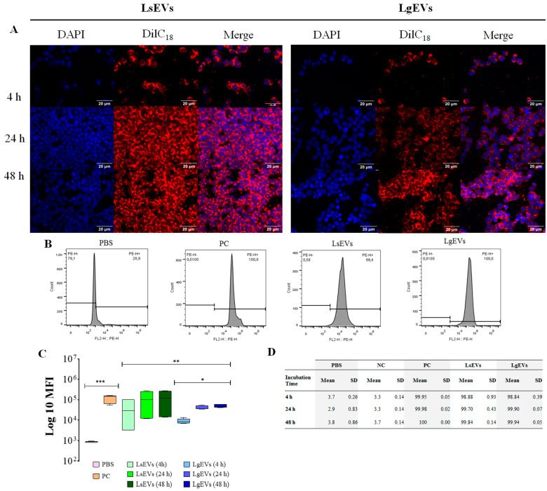
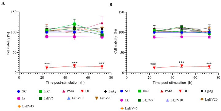
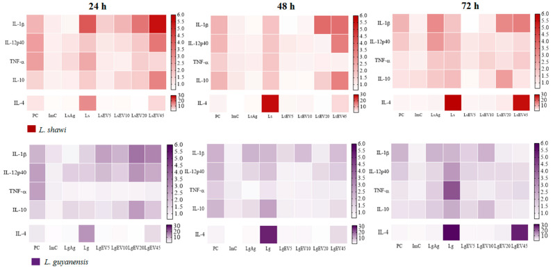
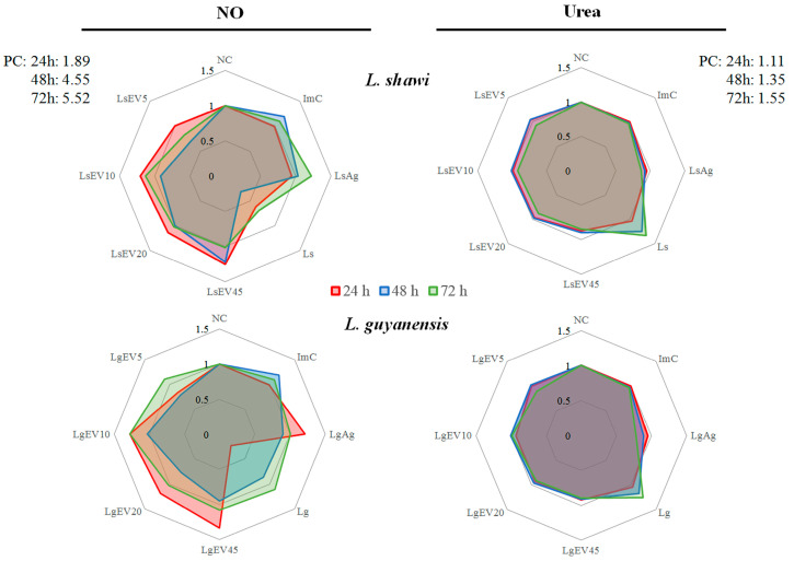
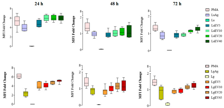
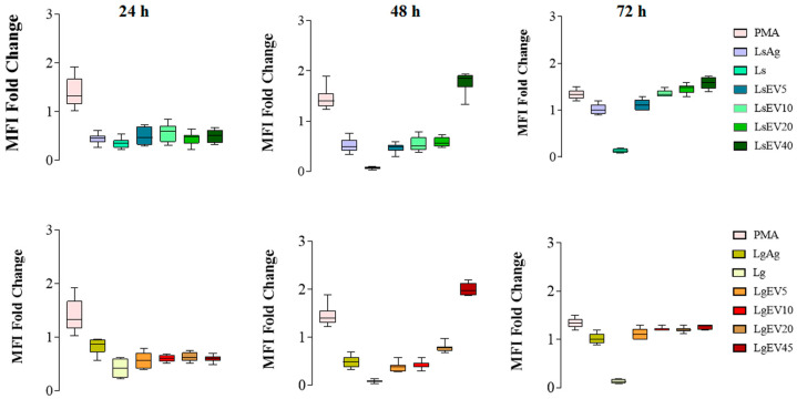
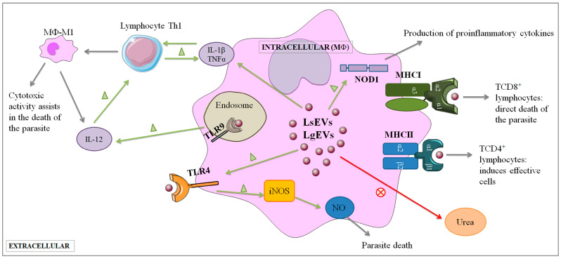
Leishmaniasis is a parasitic disease caused by different species of Leishmania and transmitted through the bite of sand flies vector. Macrophages (MΦ), the target cells of Leishmania parasites, are phagocytes that play a crucial role in the innate immune microbial defense and are antigen-presenting cells driving the activation of the acquired immune response. Exploring parasite-host communication may be key in restraining parasite dissemination in the host. Extracellular vesicles (EVs) constitute a group of heterogenous cell-derived membranous structures, naturally produced by all cells and with immunomodulatory potential over target cells. This study examined the immunogenic potential of EVs shed by L. shawi and L. guyanensis in MΦ activation by analyzing the dynamics of major histocompatibility complex (MHC), innate immune receptors, and cytokine generation. L. shawi and L. guyanensis EVs were incorporated by MΦ and modulated innate immune receptors, indicating that EVs cargo can be recognized by MΦ sensors. Moreover, EVs induced MΦ to generate a mix of pro- and anti-inflammatory cytokines and favored the expression of MHCI molecules, suggesting that EVs antigens can be present to T cells, activating the acquired immune response of the host. Since nano-sized vesicles can be used as vehicles of immune mediators or immunomodulatory drugs, parasitic EVs can be exploited by bioengineering approaches for the development of efficient prophylactic or therapeutic tools for leishmaniasis.

Keywords: Leishmania guyanensis; Leishmania shawi; extracellular vesicles; immunomodulation; leishmaniasis; macrophages.


Conflict of interest statement

The authors declare no conflict of interest.

Figures

Figure 1
Figure 1
Topography of promastigotes and EVs of L. shawi and L. guyanensis. Scanning electron microscopy of cultured L. shawi (A–C) and L. guyanensis (D–F) promastigotes exhibiting protrusions compatible with EVs biogenesis (A,D–F). Free EVs (B,C) with less than 100 nm can also be observed.
Figure 2
Figure 2
Diameter of EVs shed by L. shawi and L. guyanensis promastigotes. The diameter of EVs observed in Figure 1 were analyzed and are shown in table (A). a to g are measurement of EVs from L. shawi and h to n are values obtained for L. guyanensis EVs with Mean and standard error of the mean (SEM). EVs diameter (nm) distribution is shown by the violin plot (B).
Figure 3
Figure 3
Diameter, density, and zeta potential of L. guyanensis and L. shawi EVs. The density and diameter of EVs shed by L. shawi (blue) and L. guyanensis (orange) are represented in a scatter plot (A). The mean and the standard deviation of six independent EV isolations and three readings per sample are indicated in Tables (B,C). Size of L. shawi and L. guyanensis EVs (B) and the zeta potential (C) were analyzed by ZetaSizer. ImC was applied as a control of the EV extraction method. Parametric Student’s t-test (p ≤ 0.05) was used to compare the zeta potential of EVs and ImC.
Figure 4
Figure 4
Protein constitution and proteolytic activity of EVs from L. shawi and L. guyanensis. EVs were evaluated by SDS-PAGE (P) and by zymography (Zy), and images of the strips were acquired. MS: molecular weight size marker. ImC: isolation method control (Supplemented Schneider’s medium extracted with exosome isolation reagent); LsEVs—EVs from L. shawi promastigotes; LgEVs: EVs from L. guyanensis promastigotes; PC: positive control of proteolytic activity (1× trypsin).
Figure 5
Figure 5
Macrophage incorporation of L. shawi and L. guyanensis EVs. EVs of L. shawi (LsEVs) and L. guyanensis (LgEVs) were isolated, stained with DilC18, purified with exosome spin columns (MW3000, Invitrogen, USA), and incubated with MΦ for 4 h, 24 h, and 48 h. Fluorescence microscope images of MΦ stained with DAPI (blue) and incubated with DilC18 (red) labeled EVs were acquired (600× magnification) (A). DilC18 positive cells were analyzed by multiparametric flow cytometry, and median fluorescence intensity (MFI) (B,C) and the frequency of positive stained-MΦ were registered (D). Student’s parametric t-test was used for statistical analysis. * (p < 0.05), ** (p < 0.01) and *** (p < 0.0001) indicate statistical significance. As the negative control (NC) of the assay, 1× PBS was incubated with DilC18, purified through the column, and added to resting (non-stimulated) MΦ. For positive control (PC), DilC18 was directly used to stain the membranes of resting MΦ.
Figure 6
Figure 6
Effect of L. shawi and L. guyanensis EVs on MΦ viability. The viability of MΦ incubated for 24 h, 48 h, and 72 h with 5, 10, 20, and 45 µg·mL−1 of EVs from L. shawi (A) and L. guyanensis (B), parasite antigens, and promastigotes was analyzed by resazurin reduction. NC: negative control (resting MΦ); ImC: control of EV extraction method; PMA: positive control of inflammation (PMA 0.2 µg·mL−1); DC: MΦ death control (2% paraformaldehyde); LsAg: L. shawi antigen; Ls: L. shawi promastigotes; LsEV5: L. shawi EVs at 5 µg·mL−1, LsEV10: L. shawi EVs at 10 µg·mL−1; LsEV20: L. shawi EVs at 20 µg·mL−1; LsEV45: L. shawi EVs at 45 µg·mL−1; LgAg: L. guyanensis antigen; Lg: L. guyanensis promastigotes; LgEV5: L. guyanensis EVs at 5 µg·mL−1; LgEV10: L. guyanensis EVs at 10 µg·mL−1; LgEV20: L. guyanensis EVs at 20 µg·mL−1; LgEV45: L. guyanensis EVs at 45 µg·mL−1. The mean and standard deviation of three independent assays performed in triplicate are represented by dot plots with connecting lines. Student’s parametric t-test was used for statistical analysis. *** indicates significant differences (p ≤ 0.001) when compared to DC.
Figure 7
Figure 7
Effect of L. shawi and L. guyanensis EVs on TLR2, TLR4, TLR9, NOD1, and NOD2 gene expression. MΦ incubated for 24 h, 48 h, and 72 h with 5 (LsEV5; LgEV5), 10 (LsEV10; LgEV10), 20 (LsEV20; LgEV20), and 45 µg·mL−1 (LsEV45; LgEV45) of EVs from L. shawi (red) and L. guyanensis (purple), parasite antigens (LsAg and LgAg), and L. shawi (Ls) and L. guyanensis (Lg) promastigotes were analyzed by RT-qPCR. Results normalized to resting MΦ of three independent assays performed in triplicate are represented by heat maps. PC—positive control (MΦ incubated with PMA); ImC—MΦ incubated with negative control of EV isolation method.
Figure 8
Figure 8
Effect of L. shawi and L. guyanensis EVs on cytokines gene expression. MΦ incubated for 24 h, 48 h, and 72 h with 5 (LsEV5; LgEV5), 10 (LsEV10; LgEV10), 20 (LsEV20; LgEV20), and 45 µg·mL−1 (LsEV45; LgEV45) of EVs from L. shawi (red) and L. guyanensis (purple), parasite antigens (LsAg and LgAg), and L. shawi (Ls) and L. guyanensis (Lg) promastigotes were analyzed by RT-qPCR. Results normalized to resting MΦ are represented by heat maps of three independent assays performed in triplicate. PC—positive control (MΦ incubated with PMA); ImC—MΦ incubated with negative control of EV isolation method.
Figure 9
Figure 9
Effect of L. shawi and L. guyanensis EVs on NO and de novo urea production. MΦ incubated for 24 h, 48 h, and 72 h with 5 (LsEV5; LgEV5), 10 (LsEV10; LgEV10), 20 (LsEV20; LgEV20), and 45 µg·mL−1 (LsEV45; LgEV45) of EVs from L. shawi and L. guyanensis, parasite antigens (LsAg and LgAg), and L. shawi (Ls) and L. guyanensis (Lg) promastigotes were analyzed by colorimetric assays. Fold change (to resting MΦ) results of three independent assays performed in triplicate are represented by radar graph. NC—negative control (resting MΦ); Positive control (PC -; MΦ incubated with PMA).; ImC—MΦ incubated with negative control of EV isolation method.
Figure 10
Figure 10
Effect of L. shawi and L. guyanensis EVs on density of MHCI molecules. MΦ incubated for 24 h, 48 h, and 72 h with 5 (LsEV5; LgEV5), 10 (LsEV10; LgEV10), 20 (LsEV20; LgEV20), and 45 µg·mL−1 (LsEV45; LgEV45) of EVs shed by L. shawi and L. guyanensis, parasite antigens (Ag), and L. shawi (Ls) and L. guyanensis (Lg) promastigotes were analyzed by multiparametric flow cytometry. Fold changes in MHCI molecule density on MΦ are represented by the MFI (median fluorescence intensity) PMA—MΦ incubated with PMA as a positive control.
Figure 11
Figure 11
Effect of L. shawi and L. guyanensis EVs on density of MHCI and MHCII molecules. MΦ incubated for 24 h, 48 h, and 72 h with 5 (LsEV5; LgEV5), 10 (LsEV10; LgEV10), 20 (LsEV20; LgEV20), and 45 µg·mL−1 (LsEV45; LgEV45) of EVs shed by L. shawi and L. guyanensis, parasite antigens (Ag), and L. shawi (Ls) and L. guyanensis (Lg) promastigotes were analyzed by multiparametric flow cytometry. Fold changes in MHC molecules are represented by the MFI (median fluorescence intensity). PMA—MΦ incubated with PMA as a positive control.
Figure 12
Figure 12
Proposed model for the interplay of Leishmania EVs with macrophages. EVs shed by L. shawi and L. guyanensis can interact with MΦ cellular membrane. EVs appear to interact with innate immune receptors, such as TLR4, NOD1, and TLR9, and can be immunogenic and direct MΦ activity. This includes the generation of pro-inflammatory cytokines (IL-12, IL-1β, and TNF-α) and the expansion of MHCI molecules, inducing the activation of cytotoxic immune response in addition to the production of antimicrobial NO, which can promote parasite inactivation. The Figure was partly designed using Servier Medical Art, provided by Servier, licensed under a Creative Commons Attribution 3.0 unported license (https://creativecommons.org/licenses/by/3.0/).

Similar articles

Cited by

References

    1. Pace D. Leishmaniasis. J. Infect. 2014;69:S10–S18. doi: 10.1016/j.jinf.2014.07.016. - DOI - PubMed
    1. Akuffo H., Costa C., van Griensven J., Burza S., Moreno J., Herrero M. New insights into leishmaniasis in the immunosuppressed. PLoS Negl. Trop. Dis. 2018;12:e0006375. doi: 10.1371/journal.pntd.0006375. - DOI - PMC - PubMed
    1. Scorza B.M., Carvalho E.M., Wilson M.E. Cutaneous manifestations of human and murine leishmaniasis. Int. J. Mol. Sci. 2017;18:1296. doi: 10.3390/ijms18061296. - DOI - PMC - PubMed
    1. Ameen M. Cutaneous leishmaniasis: Advances in disease pathogenesis, diagnostics and therapeutics. Clin. Exp. Dermatol. 2010;35:699–705. doi: 10.1111/j.1365-2230.2010.03851.x. - DOI - PubMed
    1. Calvopina M., Aguirre C., Cevallos W., Castillo A., Abbasi I., Warburg A. Case report: Coinfection of Leishmania guyanensis and human immunodeficiency virus-acquired immune deficiency syndrome: Report of a case of disseminated cutaneous leishmaniasis in Ecuador. Am. J. Trop. Med. Hyg. 2017;96:1151–1154. doi: 10.4269/ajtmh.16-0431. - DOI - PMC - PubMed

Publication types

MeSH terms

Substances