. 2023 Apr 8;12(8):1114.
doi: 10.3390/cells12081114.

Induction of miR-665-3p Impairs the Differentiation of Myogenic Progenitor Cells by Regulating the TWF1-YAP1 Axis

Affiliations

Induction of miR-665-3p Impairs the Differentiation of Myogenic Progenitor Cells by Regulating the TWF1-YAP1 Axis

Mai Thi Nguyen et al. Cells. .

Abstract

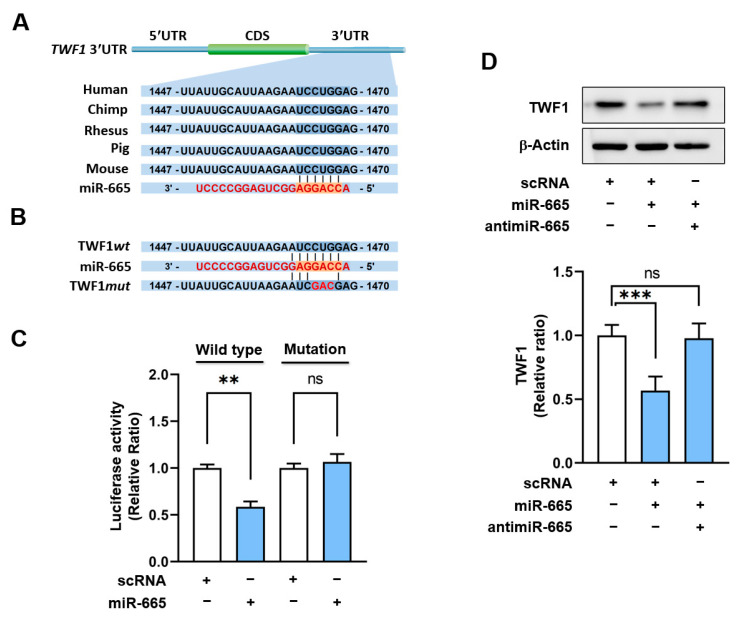
Actin dynamics are known to orchestrate various myogenic processes in progenitor cells. Twinfilin-1 (TWF1) is an actin-depolymerizing factor that plays a crucial role in the differentiation of myogenic progenitor cells. However, little is known about the mechanisms underlying the epigenetic regulation of TWF1 expression and impaired myogenic differentiation in the background of muscle wasting. This study investigated how miR-665-3p affects TWF1 expression, actin filaments' modulation, proliferation, and myogenic differentiation in progenitor cells. Palmitic acid, the most prevalent saturated fatty acid (SFA) in food, suppressed TWF1 expression and inhibited the myogenic differentiation of C2C12 cells while increasing the level of miR-665-3p expression. Interestingly, miR-665-3p inhibited TWF1 expression by targeting TWF1 3'UTR directly. In addition, miR-665-3p accumulated filamentous actin (F-actin) and enhanced the nuclear translocation of Yes-associated protein 1 (YAP1), consequently promoting cell cycle progression and proliferation. Furthermore, miR-665-3p suppressed the expressions of myogenic factors, i.e., MyoD, MyoG, and MyHC, and consequently impaired myoblast differentiation. In conclusion, this study suggests that SFA-inducible miR-665-3p suppresses TWF1 expression epigenetically and inhibits myogenic differentiation by facilitating myoblast proliferation via the F-actin/YAP1 axis.

Keywords: F-actin; YAP1; actin dynamics; differentiation; miR-665-3p; myogenesis; obesity; proliferation; twinfilin-1.


Conflict of interest statement

The authors declare no conflict of interest.

Figures

Figure 1
Figure 1
PA inhibited C2C12 differentiation and upregulated miR-665-3p expression. C2C12 cells were treated with PA (100 μM) or vehicle control for 24 h before inducing differentiation. (A) After differentiation for five days, immunofluorescence staining with MyHC (green) was used to analyze myogenic differentiation. Nuclei were counterstained with Hoechst 33,342 (blue). Scale bar: 50 μm. (B) Myotube formation was quantified based on the results of (A) using MyHC-positive area, differentiation indices, and fusion indices. (C,D) The expressions of myogenic factors and TWF1 were analyzed on day three of differentiation. Expression levels were normalized versus β-actin. (E) The expression of miR-665-3p was analyzed 24 h after PA treatment by qRT-PCR and normalized to U6. (F) The representative immunoblots (6 mice/group) of TWF1 in the gastrocnemius muscle of NFD-fed mice and HFD-fed mice are shown. (G) The expression of miR-665-3p in the gastrocnemius muscle of NFD-fed mice (open column) and HFD-fed mice (closed column) was analyzed by qRT-PCR and normalized to U6. The values are expressed as the relative ratio, where the intensity of the vehicle control was set to one. Data are presented as means ± SEMs (n = 3 except mice experiment (n = 6); * p < 0.05; ** p < 0.01; *** p < 0.001 vs. control or NFD).
Figure 2
Figure 2
MiR-665-3p suppressed TWF1 expression. (A) Sequence conservation analysis of miR-665-3p among different species was conducted using TargetScan and Pictar. (B) Schematic diagram of dual-luciferase reporter plasmids pmirGLO-TWF1 3′UTR-wild type (TWF1wt) with binding sites for the seed sequence of miR-665-3p and its mutant (TWF1mut). (C) Dual luciferase activity was determined 24 h after transfection. (D) Representative immunoblots of TWF1 expression in 200 nM of miR-665-3p, antimir-665, or scRNA-transfected cells after transfection for 24 h. Results are expressed as the means ± SEMs of intensity ratios versus scRNA controls (n = 3; ** p < 0.01; *** p < 0.001 vs. scRNA). ns: no significance.
Figure 3
Figure 3
MiR-665-3p promoted F-actin accumulation and the nuclear localization of YAP1. C2C12 cells were transfected with 200 nM scRNA, siTWF1, or miR-665-3p mimic for 24 h. (A,B) FITC-Phalloidin staining was used to analyze F-actin formation (green). Nuclei were counterstained with Hoechst 33,342 (blue). Scale bar: 25 μm. (C) Representative immunoblots of YAP1 and phosphor-YAP1 (pYAP1) in the nuclear and cytoplasmic fractions. The quality of subcellular fractionation was confirmed by Lamin B and α-Tubulin, which were used as nuclear and cytoplasmic markers, respectively. (D) Densitometry results of YAP1 and pYAP1 expression were normalized versus designated molecules, such as β-Actin, α-Tubulin and YAP1, respectively. Results are expressed as the means ± SEMs of intensity ratios versus scRNA controls (n = 3; ***, p < 0.001 vs. scRNA). ns: no significance.
Figure 4
Figure 4
MiR-665-3p facilitated myoblast proliferation. C2C12 cells were transfected with miR-665 mimic, antimiR-665, siTWF1, or scRNA (200 nM) for 24 h. (A) Cell proliferation was analyzed using an EdU assay. Cells exhibiting DNA replication were labeled with EdU (green) and nuclei with Hoechst 33,342 (blue). Scale bar: 50 μm. (B) Proportions of EdU-positive cells were determined using ImageJ software. (C,D) Cell cycle analysis and percentages of myoblast cells in each cell cycle phase were determined 24 after transfection. (E) mRNA levels of CCNB1, PCNA, and CCND1 were determined using RT-qPCR after normalization versus U6 levels. Results are expressed as the means ± SEMs of intensity ratios versus scRNA controls (n = 3; * p < 0.05; ** p < 0.01; *** p < 0.001 vs. scRNA). ns: no significance.
Figure 5
Figure 5
MiR-665-3p suppressed the expressions of myogenic regulator factors and TWF1. C2C12 cells were transfected with miR-665 mimic, antimiR-665, siTWF1, or scRNA (200 nM) and then differentiated for three days. (A) The expressions of TWF1 and myogenic factors were detected by immunoblotting. (B) Representative immunoblots of TWF1 and myogenic factors’ (MyHC, MyoD, and MyoG) expression as determined by densitometry and normalized versus β-actin. Results are expressed as the means ± SEMs of intensity ratios versus scRNA controls (n = 3; ** p < 0.01; *** p < 0.001 vs. scRNA). ns: no significance.
Figure 6
Figure 6
MiR-665-3p reduced myotube formation. C2C12 myoblasts were transfected with miR-665 mimic, antimiR-665, siTWF1, or scRNA control (200 μM) and then differentiated for five days. (A) Immunofluorescence staining (MyHC, green) was used to assess levels of differentiation of myoblasts. Nuclei were counterstained with Hoechst 33,342 (blue). Scale bar: 50 μm. (B) Myotube formation was quantified based on the results of (A) for MyHC-positivity, differentiation indices, fusion indices, and myotube widths. Results are expressed as the means ± SEMs (n = 3; *** p < 0.001). ns: no significance.

Similar articles

References

    1. Mukund K., Subramaniam S. Skeletal muscle: A review of molecular structure and function, in health and disease. Wiley Interdiscip. Rev. Syst. Biol. Med. 2020;12:e1462. doi: 10.1002/wsbm.1462. - DOI - PMC - PubMed
    1. Chal J., Pourquie O. Making muscle: Skeletal myogenesis in vivo and in vitro. Development. 2017;144:2104–2122. doi: 10.1242/dev.151035. - DOI - PubMed
    1. Sartori R., Romanello V., Sandri M. Mechanisms of muscle atrophy and hypertrophy: Implications in health and disease. Nat. Commun. 2021;12:330. doi: 10.1038/s41467-020-20123-1. - DOI - PMC - PubMed
    1. Akhmedov D., Berdeaux R. The effects of obesity on skeletal muscle regeneration. Front. Physiol. 2013;4:371. doi: 10.3389/fphys.2013.00371. - DOI - PMC - PubMed
    1. Teng S., Huang P. The effect of type 2 diabetes mellitus and obesity on muscle progenitor cell function. Stem Cell Res. Ther. 2019;10:103. doi: 10.1186/s13287-019-1186-0. - DOI - PMC - PubMed

Publication types