. 2023 Apr 11;12(8):1127.
doi: 10.3390/cells12081127.

Distinct Responses to IL4 in Macrophages Mediated by JNK

Affiliations

Distinct Responses to IL4 in Macrophages Mediated by JNK

Luís Arpa et al. Cells. .

Abstract

IL(Interleukin)-4 is the main macrophage M2-type activator and induces an anti-inflammatory phenotype called alternative activation. The IL-4 signaling pathway involves the activation of STAT (Signal Transducer and Activator of Transcription)-6 and members of the MAPK (Mitogen-activated protein kinase) family. In primary-bone-marrow-derived macrophages, we observed a strong activation of JNK (Jun N-terminal kinase)-1 at early time points of IL-4 stimulation. Using selective inhibitors and a knockout model, we explored the contribution of JNK-1 activation to macrophages' response to IL-4. Our findings indicate that JNK-1 regulates the IL-4-mediated expression of genes typically involved in alternative activation, such as Arginase 1 or Mannose receptor, but not others, such as SOCS (suppressor of cytokine signaling) 1 or p21Waf-1 (cyclin dependent kinase inhibitor 1A). Interestingly, we have observed that after macrophages are stimulated with IL-4, JNK-1 has the capacity to phosphorylate STAT-6 on serine but not on tyrosine. Chromatin immunoprecipitation assays revealed that functional JNK-1 is required for the recruitment of co-activators such as CBP (CREB-binding protein)/p300 on the promoter of Arginase 1 but not on p21Waf-1. Taken together, these data demonstrate the critical role of STAT-6 serine phosphorylation by JNK-1 in distinct macrophage responses to IL-4.

Keywords: chemokines; cytokines; inflammation; kinases/phosphatases; monocytes/macrophages.


Conflict of interest statement

The authors declare no conflict of interest.

Figures
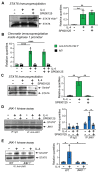
Figure 6
Figure 6
JNK-1 phosphorylates STAT-6 on serine without affecting its capacity to bind DNA. For A to C, macrophages were pretreated with the JNK inhibitor SP600125 (SP) for 1 h and then stimulated with IL-4 for 15 min. (A) Phosphorylation of STAT-6 (Y641) was analyzed by immunoprecipitation of STAT-6 and then via immunoblotting with an antibody, namely, either anti-phospho-Stat6 (Y641) or anti-STAT-6. (B) Chromatin immunoprecipitation assay (CHIP) was performed using the antibodies indicated. The presence of STAT6 Y641P in the Arginase 1 promoter was evaluated using qPCR and normalized with the level of expression of a 36B4 exon and the inputs of each sample as a loading control. (C) Phosphorylation of STAT-6 on serine was analyzed by immunoprecipitating STAT-6 and then via immunoblotting with an antibody against phospho-serine or anti-STAT6. (D) Quiescent macrophages were stimulated with IL-4 for 15 min to reach maximum JNK-1 activity and then total protein extraction was performed. STAT-6 from quiescent macrophages (to avoid any basal kinase activity on STAT) was immunoprecipitated (150 μg of total protein extracts) and used as substrate in an in vitro kinase assay for JNK-1. As control for immunoprecipitation, IgG was used. An immunoblot for JNK1 was performed in parallel as a load control for the kinase assay. (E) An experiment similar to (D) but in which macrophages derived from WT or JNK-1 deficient mice (JNK-1−/−) were used. As a control for charge, a sample of total protein extracts was used for immunoblotting with an antibody (anti-STAT6). The results are shown as the mean ± SD of 3 independent experiments. * p < 0.05, ** p < 0.01, and **** p < 0.0001 in relation to the corresponding treatments after all the independent experiments had been compared. Data were analyzed using Student’s t-test.
Figure 1
Figure 1
Effects of IL-4 on MAPK activation. Bone-marrow-derived macrophages were cultured for 6 days in the presence of M-CSF. Then, to render the cells quiescent, they were deprived of M-CSF for 18 h. At this point, IL-4 (10 ng/mL) or M-CSF (10 ng/mL) was added for the indicated periods of time. (A) JNK-1 activity was studied after immunoprecipitation and then an in vitro kinase assay was performed on recombinant c-Jun. An immunoblot for JNK-1 was performed in parallel as a loading control for the kinase assay. (B,C) Activation of MAPK ERK-1/2 and the phosphorylated form of p38 were analyzed via Western blot using the corresponding antibodies. In parallel, as a loading control, an immunoblot for β-actin was performed. Images on the right depict quantification by densitometry of 3 independent experiments. The results are shown as the mean ± SD. ** p < 0.01 and *** p < 0.001 in relation to the corresponding treatments with IL-4 after all the independent experiments had been compared. Data were analyzed using Student’s t-test.
Figure 2
Figure 2
Effects of IL-4 on MKP expression. Macrophages were treated with M-CSF (control) or IL-4 for the indicated periods of time. MKP expression was analyzed by qPCR. The results are shown as the mean ± SD of 3 independent experiments. * p < 0.05, ** p < 0.01, and *** p < 0.001 in relation to the corresponding treatments after all the independent experiments had been compared. Data were analyzed using Student’s t-test.
Figure 3
Figure 3
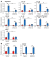
(A,B) Different effects of JNK-1 on IL-4-induced gene expression. Macrophages were pre-incubated for 1 h with the JNK inhibitor SP600125 (5 μM) or vehicle (DMSO) as a control. The cells were then stimulated for 6 h with IL-4 except when gene expression of SOCS1 (3 h) and c-myc and p21Waf1 (1 h) were analyzed by qPCR. The results are shown as the mean ± SD of 3 independent experiments. * p < 0.05, ** p < 0.01, and *** p < 0.001 in relation to the corresponding treatments after all the independent experiments had been compared. Data were analyzed using Student’s t-test.
Figure 4
Figure 4
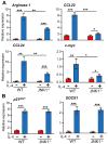
(A,B) Different effects of JNK-1 on IL-4-induced gene expression. Macrophages derived from WT or JNK-1-deficient mice (JNK-1−/−) were stimulated with IL-4 for 6h except when the gene expression of SOCS1 (3 h), p21Waf−1 (1 h), and c-Myc (1 h) was analyzed by qPCR. Control cells from each genotype were left untreated. The results are shown as the mean ± SD of 3 independent experiments. *p < 0.05, ** p < 0.01, and *** p < 0.001 in relation to the corresponding treatments after all the independent experiments had been compared. Data were analyzed using Student’s t-test.
Figure 5
Figure 5
Effects of JNK-1 on the mRNA stability of IL-4-induced genes. Macrophages were pre-incubated with SP600125 for 1 h; then, IL-4 was added, and incubation proceeded for 6 h. At this point, a combination of the RNA synthesis inhibitors 5,6-dichlorobenzimidazole 1-β-D-ribofuranoside (DRB) (20 μg/mL) and actinomycin D (Act D) (5 μg/mL) was added for the indicated periods of time. The levels of gene expression were evaluated using qPCR. To evaluate the rate of mRNA degradation, the mRNA remaining after treatment with inhibitors of RNA synthesis was calculated as a percentage of the expression of the gene in the cells stimulated with IL-4 (+/− SP600125) in the absence of RNA synthesis inhibitors. These experiments were performed three times, and the results from the mean are shown. Data were analyzed using Student’s t-test, and no significant differences were found.
Figure 7
Figure 7
JNK-1 activity is required for the binding of the cofactor CBP/p300 to the Arginase 1 promoter in response to IL-4 but not to the p21Waf−1 promoter. Quiescent macrophages were treated with IL-4 for 15 min. In A, the cells were pretreated for 1 h with the JNK inhibitor SP600125 (SP) or the vehicle (DMSO) before the addition of IL-4. (A,B) Chromatin immunoprecipitation assay was performed with the antibodies indicated. The expression of the promoters was evaluated by quantitative PCR and normalized with the level of expression of a 36B4 exon and the inputs of each sample as a control for loading. The results are shown as the mean ± SD of 3 independent experiments. ** p < 0.01, and *** p < 0.001 in relation to the corresponding treatments after all the independent experiments had been compared. Data were analyzed using Student’s t-test.

References

    1. Nelms K., Keegan A.D., Zamorano J., Ryan J.J., Paul W.E. The IL-4 receptor: Signaling mechanisms and biologic functions. Annu. Rev. Immunol. 1999;17:701–738. doi: 10.1146/annurev.immunol.17.1.701. - DOI - PubMed
    1. Murray P.J. Macrophage Polarization. Annu. Rev. Physiol. 2017;79:541–566. doi: 10.1146/annurev-physiol-022516-034339. - DOI - PubMed
    1. Wynn T.A., Vannella K.M. Macrophages in Tissue Repair, Regeneration, and Fibrosis. Immunity. 2016;44:450–462. doi: 10.1016/j.immuni.2016.02.015. - DOI - PMC - PubMed
    1. Gordon S., Martinez F.O. Alternative Activation of Macrophages: Mechanism and Functions. Immunity. 2010;32:593–604. doi: 10.1016/j.immuni.2010.05.007. - DOI - PubMed
    1. Wick K.R., Berton M.T. IL-4 induces serine phosphorylation of the STAT6 transactivation domain in B lymphocytes. Mol. Immunol. 2000;37:641–652. doi: 10.1016/S0161-5890(00)00088-2. - DOI - PubMed

Publication types