. 2023 Apr 12;12(8):1142.
doi: 10.3390/cells12081142.

Drosophila Models Reveal Properties of Mutant Lamins That Give Rise to Distinct Diseases

Affiliations

Drosophila Models Reveal Properties of Mutant Lamins That Give Rise to Distinct Diseases

Sydney G Walker et al. Cells. .

Abstract

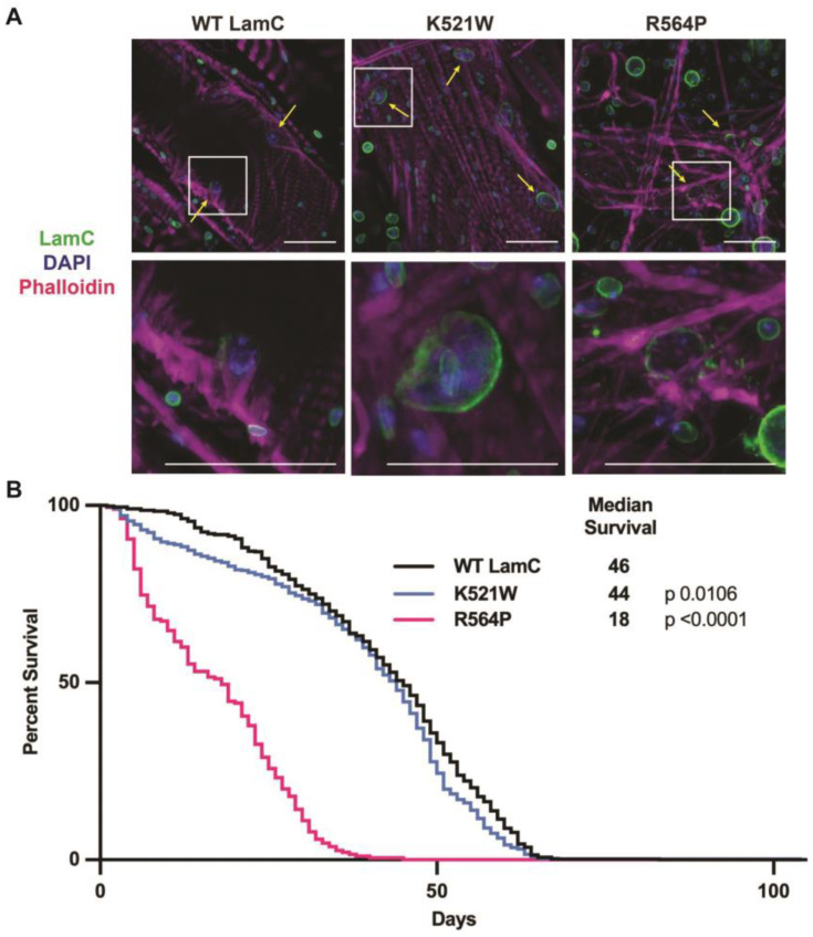
Mutations in the LMNA gene cause a collection of diseases known as laminopathies, including muscular dystrophies, lipodystrophies, and early-onset aging syndromes. The LMNA gene encodes A-type lamins, lamins A/C, intermediate filaments that form a meshwork underlying the inner nuclear membrane. Lamins have a conserved domain structure consisting of a head, coiled-coil rod, and C-terminal tail domain possessing an Ig-like fold. This study identified differences between two mutant lamins that cause distinct clinical diseases. One of the LMNA mutations encodes lamin A/C p.R527P and the other codes lamin A/C p.R482W, which are typically associated with muscular dystrophy and lipodystrophy, respectively. To determine how these mutations differentially affect muscle, we generated the equivalent mutations in the Drosophila Lamin C (LamC) gene, an orthologue of human LMNA. The muscle-specific expression of the R527P equivalent showed cytoplasmic aggregation of LamC, a reduced larval muscle size, decreased larval motility, and cardiac defects resulting in a reduced adult lifespan. By contrast, the muscle-specific expression of the R482W equivalent caused an abnormal nuclear shape without a change in larval muscle size, larval motility, and adult lifespan compared to controls. Collectively, these studies identified fundamental differences in the properties of mutant lamins that cause clinically distinct phenotypes, providing insights into disease mechanisms.

Keywords: Drosophila; Dunnigan type; Emery–Dreifuss muscular dystrophy; cardiomyopathy; familial partial lipodystrophy; intermediate filaments; laminopathy; lamins; nuclear envelope; nuclear pore.


Conflict of interest statement

The authors declare no conflict of interest.

Figures

Figure 1
Figure 1
A-type lamins have three conserved protein domains. (A) Diagram of lamin A/C showing the head, coiled-coil rod, and tail domain that possesses a nuclear localization sequence (NLS) and an Ig-like fold domain. (B) Ribbon diagram of the Ig-like fold domain (PDB: 1IVT) showing the position of the two amino acid substitutions studied here (arrows). (C) Amino acid sequence comparison of human lamin A/C and Drosophila LamC is shown for the sequences surrounding the amino acid substitutions studied here. Amino acid R482 in human lamin A/C is a conserved K521 in Drosophila LamC. Amino acid R527 in human lamin A/C is identical between the two species and corresponds to R564 in Drosophila LamC.
Figure 2
Figure 2
LamC K521W and R564P show different patterns of LamC immunostaining. (A) Larval body wall muscles expressing either wild-type or mutant LamC were stained with phalloidin (magenta), DAPI (blue), and antibodies to LamC (green). Note the abnormal nuclear morphology caused by LamC K521W and the cytoplasmic lamin aggregation caused by LamC R564P. The white box outlines the magnified region shown on the right. The scale bar represents 30 µm. (B) A graph of the average percent of nuclear LamC in each genotype is shown. The subcellular location of LamC was quantified based on the intensity of immunofluorescent staining in the nucleus compared to the total intensity from three-channel microscopy images of larval body wall muscles. (C) A graph of the quantification of the overlap of LamC and DAPI in larval body wall muscles is shown. The Manders’ coefficient was calculated using JACoP for the fraction of staining in the green channel (LamC) that overlaps with staining in the blue channel (DAPI). A total of 25 to 38 nuclei from three individual larvae per genotype were analyzed. Error bars represent the standard deviation of the mean. The values in the graph are expressed as mean ± standard deviation. A total of 15-34 nuclei were analyzed from three larvae per genotype. For panels (B,C), statistical significance was determined using a one-way ANOVA multiple comparisons analysis followed by Dunnett’s correction (GraphPad Prism version 9.5.0, GraphPad Software, San Diego, CA, USA) and is indicated by: not significant (ns), p > 0.05; **, p < 0.01; ****, p < 0.0001.
Figure 3
Figure 3
LamC K521W and R564P alter the localization of nuclear envelope proteins TMEM43 and Otefin. (A) Larval body wall muscles expressing either wild-type or mutant LamC were stained with phalloidin (magenta), DAPI (blue), and antibodies to Otefin (green). Note the cytoplasmic aggregation caused by both mutants. The white box outlines the magnified region shown on the right. (B) Larval body wall muscles expressing either wild-type or mutant LamC were stained with phalloidin (magenta), DAPI (blue), and antibodies to TMEM43 (green). Note the clustering of TMEM43 foci caused by LamC K521W and the cytoplasmic aggregation caused by LamC R564P. The scale bar represents 30 µm.
Figure 4
Figure 4
LamC R564P, but not LamC K521W, reduces larval motility and muscle size. (A) An image of a muscle filet from a hand-dissected third instar larvae stained with phalloidin (magenta) is shown. The diagram on the right shows the numbered muscles used for measurements. (B,C) The width and length of larval body wall muscles were measured for four different muscles diagrammed in A. Values are expressed as mean ± standard deviation with 33–53 muscles from 8–10 larvae analyzed per genotype. Statistical significance was determined using a one-way ANOVA analysis followed by Dunnett’s multiple comparisons test (GraphPad Prism version 9.5.0, GraphPad Software, San Diego, CA, USA) No statistical difference, ns; p > 0.05; * p < 0.05; ** p < 0.01; *** p < 0.001; **** p < 0.0001. (D) A diagram of the assay used to measure larval motility (see Materials and Methods for details) (created using BioRender.com; access date 11/2022). (E,F) Larval velocity and distance per larval contraction were measured by analyzing two-minute videos of larvae crawling on a thin layer of water in a petri dish. Values are expressed as the mean ± standard deviation, and 9–29 individual larvae were analyzed per genotype. Statistical significance was determined using a one-way ANOVA multiple comparisons analysis followed by Dunnett’s correction (GraphPad Prism version 9.5.0, GraphPad Software, San Diego, CA, USA). No statistical difference, ns; p > 0.05; * p < 0.05; *** p < 0.001; **** p < 0.0001.
Figure 5
Figure 5
Larval body wall muscle-specific expression of mutant LamC reduces adult viability. (A) Graphical representation of the Drosophila lifecycle stages is shown at the top (created with BioRender.com, access date 12/2022). Vials containing larvae with larval body wall muscle-specific expression of either wild-type or mutant LamC are shown. Red bars indicate the larval movement up the side of the vial. (B) The percentage adult viability is represented as # living adults# living adults+# dead pupae× 100. The average percentage ± the standard deviation of the mean are shown. Total progeny (255–326) was counted from multiple individual crosses for each genotype. Statistical significance was determined using the Fisher’s exact test (GraphPad Prism version 9.5.0, GraphPad Software, San Diego, CA, USA). No statistical difference, ns; p < 0.001; **** p < 0.0001.
Figure 6
Figure 6
LamC K521W and R564P cause nuclear and cytoplasmic abnormalities in larval fat body tissue. (A) Larval fat body tissue expressing either wild-type or mutant LamC were stained with phalloidin (magenta), DAPI (blue), and antibodies to LamC (green). Note that LamC K521W localizes to the nuclear envelope and causes nuclear lobulations. By contrast, LamC R564P localizes within the cytoplasm and the nucleus remains spherical. (B) Larval fat body tissue expressing either wild-type or mutant LamC were stained with Oil Red O and DAPI. Scale bar represented 30 µm. (C) The average area per adipose cell for each genotype was determined and plotted. Note the reduced size of the fat body cells expressing LamC K521W compared to that of the control. (D) The percent area of lipid droplets was calculated and plotted. Three fat bodies from three larvae of similar age were analyzed per genotype. Error bars represent the standard deviation of the mean. Statistical significance was determined using ANOVA multiple comparisons analysis followed by Dunnett’s correction (GraphPad Prism version 9.5.0, GraphPad Software, San Diego, CA). No statistical difference, ns; p > 0.05; * p < 0.05; ** p < 0.01; *** p < 0.001; **** p < 0.0001.
Figure 7
Figure 7
Cardiac-specific expression of LamC R564P, but not LamC K521W, reduces adult lifespan. (A) Drosophila hearts expressing either wild-type or mutant LamC were stained with phalloidin (magenta), DAPI (blue), and antibodies to LamC (green). The yellow arrows indicate cardiomyocytes. The scale bar represents 30 µm. (B) Adult longevity was determined for flies expressing either wild-type or mutant LamC in the heart. The graph shows the percent of living adults per day, which were maintained in fresh vials. The median survival time and the statistical analysis using the Mantel–Cox test was calculated using GraphPad Prism version 9.5.0, GraphPad Software, San Diego, CA, USA.
Figure 8
Figure 8
A model showing the differential effects of the two mutant lamins on the localization of nuclear proteins. (Left) Muscle expressing wild-type LamC (green) exhibit a lamin meshwork that lines the inner nuclear membrane. Nuclear pores (dark gray), TMEM43 (purple) and Otefin (red) are confined to the nuclear envelope. The DNA is represented in blue and the nucleolus organizer in gray. (Middle) Muscle expressing LamC K521W exhibits an uneven distribution of the lamin meshwork and nuclear lobulations with nuclear pores and TMEM43 confined to the nuclear envelope. By contrast, a portion of Otefin is mislocalized to the cytoplasm. As a result of the nuclear shape change, the genomic DNA (blue) has an atypical distribution. (Right) Muscles expressing LamC R564P exhibit abnormal localization of LamC, FG-repeat nuclear pore proteins, TMEM43, and Otefin throughout the cytoplasm.

Similar articles

Cited by

References

    1. Verbrugge S.A.J., Schonfelder M., Becker L., Yaghoob Nezhad F., Hrabe de Angelis M., Wackerhage H. Genes Whose Gain or Loss-Of-Function Increases Skeletal Muscle Mass in Mice: A Systematic Literature Review. Front. Physiol. 2018;9:553. doi: 10.3389/fphys.2018.00553. - DOI - PMC - PubMed
    1. Salari N., Fatahi B., Valipour E., Kazeminia M., Fatahian R., Kiaei A., Shohaimi S., Mohammadi M. Global prevalence of Duchenne and Becker muscular dystrophy: A systematic review and meta-analysis. J. Orthop. Surg. Res. 2022;17:96. doi: 10.1186/s13018-022-02996-8. - DOI - PMC - PubMed
    1. Moser H. Duchenne muscular dystrophy: Pathogenetic aspects and genetic prevention. Hum. Genet. 1984;66:17–40. doi: 10.1007/BF00275183. - DOI - PubMed
    1. Emery A.E. Emery-Dreifuss syndrome. J. Med. Genet. 1989;26:637–641. doi: 10.1136/jmg.26.10.637. - DOI - PMC - PubMed
    1. Zatz M., Vainzof M., Passos-Bueno M.R. Limb-girdle muscular dystrophy: One gene with different phenotypes, one phenotype with different genes. Curr. Opin. Neurol. 2000;13:511–517. doi: 10.1097/00019052-200010000-00002. - DOI - PubMed

Publication types

LinkOut - more resources