. 2023 Apr 13;12(8):1147.
doi: 10.3390/cells12081147.

Establishment of Skeletal Myogenic Progenitors from Non-Human Primate Induced Pluripotent Stem Cells

Affiliations

Establishment of Skeletal Myogenic Progenitors from Non-Human Primate Induced Pluripotent Stem Cells

June Baik et al. Cells. .

Abstract

Pluripotent stem (PS) cells enable the scalable production of tissue-specific derivatives with therapeutic potential for various clinical applications, including muscular dystrophies. Given the similarity to human counterparts, the non-human primate (NHP) is an ideal preclinical model to evaluate several questions, including delivery, biodistribution, and immune response. While the generation of human-induced PS (iPS)-cell-derived myogenic progenitors is well established, there have been no data for NHP counterparts, probably due to the lack of an efficient system to differentiate NHP iPS cells towards the skeletal muscle lineage. Here, we report the generation of three independent Macaca fascicularis iPS cell lines and their myogenic differentiation using PAX7 conditional expression. The whole-transcriptome analysis confirmed the successful sequential induction of mesoderm, paraxial mesoderm, and myogenic lineages. NHP myogenic progenitors efficiently gave rise to myotubes under appropriate in vitro differentiation conditions and engrafted in vivo into the TA muscles of NSG and FKRP-NSG mice. Lastly, we explored the preclinical potential of these NHP myogenic progenitors in a single wild-type NHP recipient, demonstrating engraftment and characterizing the interaction with the host immune response. These studies establish an NHP model system through which iPS-cell-derived myogenic progenitors can be studied.

Keywords: RNA sequencing; induced pluripotent stem cells; muscle regeneration; muscular dystrophy; myogenesis; non-human primate; stem cell therapy.


Conflict of interest statement

RCRP is cofounder and holds equity in Myogenica. All other authors declare no conflict of interest.

Figures

Figure 1
Figure 1
Generation of NHP iPS cells and their in vitro myogenic differentiation: (A) Outline of experimental design. PAX7 expression was induced on day 5 of differentiation by adding dox to the medium; (B) representative images show the morphology of NHP iPAX7 iPS cell colonies (left), day-4 embryoid bodies (EBs; center), and expanded PAX7+ myogenic progenitors (right); (C) immunostaining for PAX7 expression in GFP+-sorted myogenic progenitors from day-12 cultures. PAX7 is shown in red and DAPI stains nuclei in blue; (D) representative images show terminal differentiation of PAX7 myogenic progenitors into myotubes. Staining for MHC is shown in red and nuclei in blue. Scale bars, 100 μm.
Figure 2
Figure 2
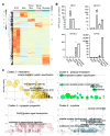
Transcriptional characterization of differentiating NHP iPAX7 iPS cells: (A) Heatmap representing unsupervised clustering of differentially expressed transcripts identified during the skeletal myogenic specification of NHP iPAX7 iPS cell lines. Clusters are enumerated from 1 to 5. Pluripotent stem cells: iPSC; uncommitted mesoderm: Mes; paraxial mesoderm: Parax Mes; myogenic progenitors: Myog Prog; myotubes: Myotube; (B) expression of selected genes characterizing the clusters identified in panel (A). Dots represent replicates; (C) functional classification based on gene ontology (GO) analysis using DAVID. Genes from clusters 2 to 5 were analyzed to identify significantly enriched categories, displayed according to −log(adj p-value) and z-score. The results are shown as a Bubbleplot generated using the R package GOplot. CPM: counts per million reads.
Figure 3
Figure 3
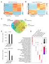
Comparison of NHP and human myogenesis shows high similarity in vitro: (A,B) Heatmap representing differentially expressed transcripts between myogenic progenitors (Myog Prog) and myotubes (Myotube) derived from NHP and human iPAX7 iPS cell cultures, respectively; (C) Venn diagram representing the number of shared genes (blue squares) between human and NHP myogenic cultures; (D) expression levels of MYOG and MYH8 in Myog Prog (MP) and Myotube (MT) from NHP and human datasets; (E) identification and comparison of biological processes enriched in the different list of genes identified in the Venn diagram from panel (C). Gene lists were analyzed using the R package enrichGO. Abbreviation hum-NHP refers to human and NHP. Graph reports the top 5 categories for each cluster. CPM: counts per million reads.
Figure 4
Figure 4
NHP iPAX7 iPS-cell-derived myogenic progenitors contribute to in vivo muscle regeneration in mice: (A,B) In vivo regenerative capacity of CyMN.1 iPAX7 myogenic progenitors in mice: (A) Representative engraftment resulting from intramuscular transplantation of CyMN.1 iPS-cell-derived myogenic progenitors in immunodeficient (NSG) mice. PBS-injected muscle served as a negative control. Injected muscles were analyzed using primate-specific antibodies for DYSTROPHIN (DYS—white) and LAMIN A/C (red). Nuclei (blue). Scale bar is 100 µm; (B) quantification of muscle fiber engraftment. Dots represent average engraftment on each TA and bars represent mean ± standard error. CyMN.1 (n = 5, 10 TAs); (C,D) donor-derived RFP-labeled CyMN.2 PAX7+ myogenic progenitors are identified in the CTX-injured TA muscles of mice. PBS-injected muscles served as a negative control: (C) Transplanted muscles were analyzed using primate-specific antibodies for DYSTROPHIN (DYS—white) and RFP (red). Nuclei (blue). Scale bar is 100 µm; (D) quantification of myofiber engraftment. Dots represent each TA and bars represent mean ± standard error. NSG (n = 7) and FKRP-NSG (n = 7).
Figure 5
Figure 5
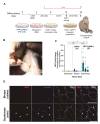
NHP iPAX7 iPS-cell-derived myogenic progenitors show engraftment capacity in NHP: (A) Schematic representation of the derivation of myogenic progenitors and transplantation in an NHP; (B) cells were suspended and injected IM at CTX-pre-injured sites using a grid pattern; (C,D) donor-derived RFP-labeled CyMN.2 iPAX7 myogenic progenitors are identified in the quadriceps of NHP recipient using qPCR and immunofluorescent assays: (C) Dot plot depicting fold change in RFP signal detected in skeletal muscle sites collected upon necropsy, where diaphragm represents an unmanipulated negative control. Biceps received an intramuscular injection of saline (negative control), whereas the quadriceps received an intramuscular injection of 4 × 10⁷ RFP-labeled CyMN.2 myogenic progenitors. Each bar represents a separate muscle biopsy collected at necropsy. Each dot represents one technical replicate. n = 3 independent replicates, with 3–5 technical replicates each. Two-way ANOVA, with Tukey’s multiple-comparison tests, where * p = 0.0307 for diaphragm v. quad, **** p < 0.0001 for biceps v. quad. Diaphragm v. biceps was n.s. (p = 0.43). Error bars depict the mean with standard error of the mean (s.e.m) for all; (D) representative images show muscle section from saline-injected biceps muscle of NHP recipient (upper panel). Donor-derived RFP+ nuclei (RFP; red) counterstained with DAPI (DAPI; blue) and DYSTROPHIN (DYS—white) myofibers are identified as donor-derived RFP-labeled CyMN.2 PAX7+ myogenic progenitors in muscle section from quadriceps muscle of NHP recipient (lower panel). White arrowheads indicate RFP+DAPI+ nuclei. (Scale bar, 100 μm).

Similar articles

Cited by

References

    1. Takahashi K., Yamanaka S. Induction of Pluripotent Stem Cells from Mouse Embryonic and Adult Fibroblast Cultures by Defined Factors. Cell. 2006;126:663–676. doi: 10.1016/j.cell.2006.07.024. - DOI - PubMed
    1. Azzag K., Bosnakovski D., Tungtur S., Salama P., Kyba M., Perlingeiro R.C.R. Transplantation of PSC-derived myogenic progenitors counteracts disease phenotypes in FSHD mice. NPJ Regen. Med. 2022;7:43. doi: 10.1038/s41536-022-00249-0. - DOI - PMC - PubMed
    1. Azzag K., Ortiz-Cordero C., Oliveira N.A.J., Magli A., Selvaraj S., Tungtur S., Upchurch W., Iaizzo P.A., Lu Q.L., Perlingeiro R.C.R. Efficient engraftment of pluripotent stem cell-derived myogenic progenitors in a novel immunodeficient mouse model of limb girdle muscular dystrophy 2I. Skelet. Muscle. 2020;10:10. doi: 10.1186/s13395-020-00228-3. - DOI - PMC - PubMed
    1. Darabi R., Arpke R.W., Irion S., Dimos J.T., Grskovic M., Kyba M., Perlingeiro R.C. Human ES- and iPS-derived myogenic progenitors restore DYSTROPHIN and improve contractility upon transplantation in dystrophic mice. Cell Stem Cell. 2012;10:610–619. doi: 10.1016/j.stem.2012.02.015. - DOI - PMC - PubMed
    1. Dhoke N.R., Kim H., Selvaraj S., Azzag K., Zhou H., Oliveira N.A., Tungtur S., Ortiz-Cordero C., Kiley J., Lu Q.L., et al. A universal gene correction approach for FKRP-associated dystroglycanopathies to enable autologous cell therapy. Cell Rep. 2021;36:109360. doi: 10.1016/j.celrep.2021.109360. - DOI - PMC - PubMed

Publication types

LinkOut - more resources