. 2023 Apr 13;12(8):1155.
doi: 10.3390/cells12081155.

IKKβ Inhibition Attenuates Epithelial Mesenchymal Transition of Human Stem Cell-Derived Retinal Pigment Epithelium

Affiliations

IKKβ Inhibition Attenuates Epithelial Mesenchymal Transition of Human Stem Cell-Derived Retinal Pigment Epithelium

Srinivasa R Sripathi et al. Cells. .

Abstract

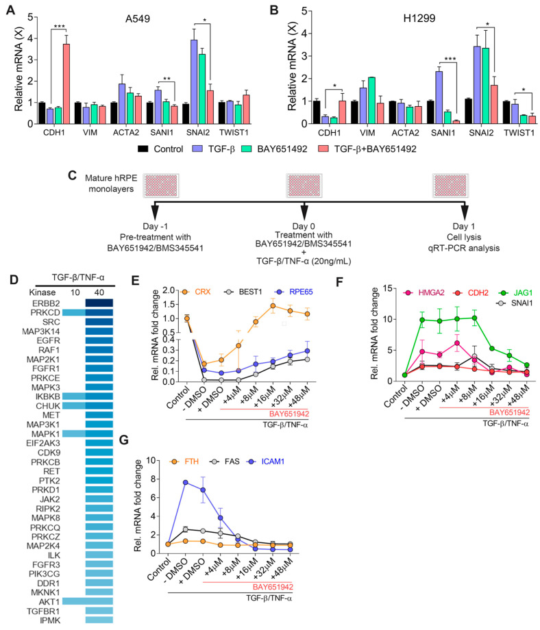
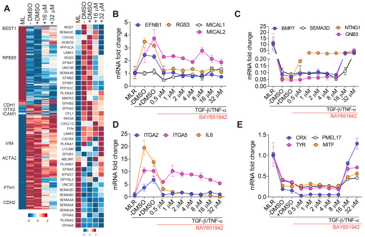
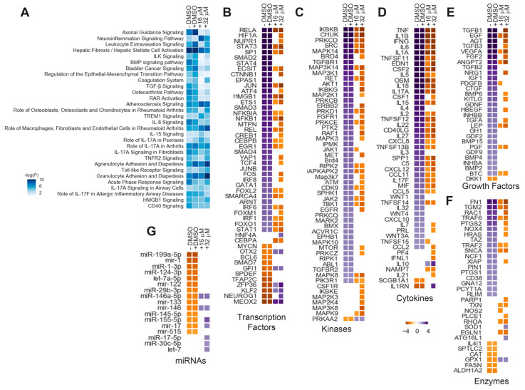
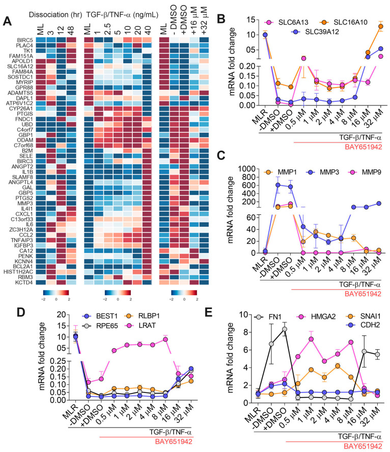
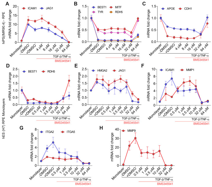
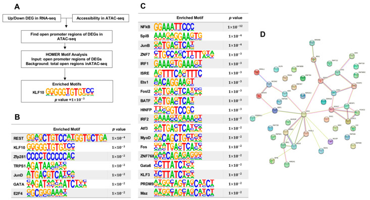
Epithelial-mesenchymal transition (EMT), which is well known for its role in embryonic development, malignant transformation, and tumor progression, has also been implicated in a variety of retinal diseases, including proliferative vitreoretinopathy (PVR), age-related macular degeneration (AMD), and diabetic retinopathy. EMT of the retinal pigment epithelium (RPE), although important in the pathogenesis of these retinal conditions, is not well understood at the molecular level. We and others have shown that a variety of molecules, including the co-treatment of human stem cell-derived RPE monolayer cultures with transforming growth factor beta (TGF-β) and the inflammatory cytokine tumor necrosis factor alpha (TNF-α), can induce RPE-EMT; however, small molecule inhibitors of RPE-EMT have been less well studied. Here, we demonstrate that BAY651942, a small molecule inhibitor of nuclear factor kapa-B kinase subunit beta (IKKβ) that selectively targets NF-κB signaling, can modulate TGF-β/TNF-α-induced RPE-EMT. Next, we performed RNA-seq studies on BAY651942 treated hRPE monolayers to dissect altered biological pathways and signaling events. Further, we validated the effect of IKKβ inhibition on RPE-EMT-associated factors using a second IKKβ inhibitor, BMS345541, with RPE monolayers derived from an independent stem cell line. Our data highlights the fact that pharmacological inhibition of RPE-EMT restores RPE identity and may provide a promising approach for treating retinal diseases that involve RPE dedifferentiation and EMT.

Keywords: PVR and AMD; TGF–β/–α; differentiation; epithelial-mesenchymal transition; kinase inhibitors; retinal pigment epithelium; stem cells; transcriptomics.


Conflict of interest statement

RCT is an employee of Caris Life Sciences.

Figures

Figure 1
Figure 1
Identification of small molecule kinase inhibitors that modulate TGF–β/TNF–α associated RPE–EMT. qPCR validation of altered EMT-associated factors in (A) non-small cell lung cancer A549 and (B) H1299 cells. (C) Schematic representation of kinase inhibitors treatment and RPE–EMT induction. (D) Heatmap of potential small molecule kinase inhibitors identified by IPA-generated upstream regulator analysis. qPCR analysis shows differential expression of (E) RPE genes, (F) EMT and (G) NF-κB factors from TGF–β/TNF–α-induced EMT were altered by BAY651942 treatment. Error bars represent SD of at least three biological replicates and statistically significant mean differences (p ≤ 0.05 by ANOVA). * p ≤ 0.05; ** p ≤ 0.01; *** p ≤ 0.001.
Figure 2
Figure 2
Transcriptomic analysis of BAY651942 modulated RPE–EMT in TGF–β/TNF–α model. (A) Hierarchical clustered heatmaps of log2-transformed ratios. Average abundances of DEG showing significant differences from DMSO and BAY651942 treatment after TGF–β/TNF–α-induced EMT in hiPS–RPE. Heatmap of axon guidance genes that were altered by BAY651942 treatment compared TGF–β/TNF–α plus DMSO treatment. (BE) qPCR validation of altered multiple axon guidance genes (BD), RPE genes (E) by BAY651942 treatment followed by TGF–β/TNF–α-induced EMT. Error bars represent SD of at least three biological replicates and statistically significant mean differences (p < 0.05 by ANOVA).
Figure 3
Figure 3
BAY651942 regulated transcriptome dynamics of TGF–β/TNF–α-induced RPE. (A) Top canonical pathways were predicted based on the alteration of highly-enriched genes that changed in abundance (activated or inhibited) from DMSO and BAY651942 treated hiPS-RPE monolayers prior to TGF–β/TNF–α-induced EMT (BG) heatmaps of IPA-generated upstream regulator analysis from DMSO- and BAY651942-treated hiPS RPE monolayers. IPA uses activation Z-score as a statistical measure of the match between expected relationship direction and observed changes in the gene expression regulator such as different transcription factors; (TFs) (B), kinases (C), cytokines (D), growth factors (E), enzymes (F), and miRNAs (G) were predicted to be activated (violet) or inhibited (yellow) after DMSO and BAY651942. Z-scores of >2 (activated) or <−2 (inhibited) were considered significant. Only genes with statistically significant changes at an FDR of 5% (p < 0.05) were included in the analysis.
Figure 4
Figure 4
Differential expression of AMD-associated risk factor modulated by BAY651942 treatment. (A) Heatmaps of dysregulated AMD-associated risk factors altered due to dissociation, TGF–β/TNF–α-induced EMT and differentially regulated with the treatment for BAY651942 from TGF–β/TNF–α. (BE) qPCR validation of multiple AMD-associated risk factors (B,C), RPE factors and EMT factors (D) by BAY651942 treatment followed by TGF–β/TNF–α-induced EMT. Error bars represent SD of at least three biological replicates and statistically significant mean differences (p < 0.05 by ANOVA).
Figure 5
Figure 5
qPCR validation of TGF–β/TNF–α-induced RPE–EMT by IKKβ inhibitor BMS345541. (AC) Differential expression of RPE-EMT-associated factors were measured after treatment with BMS345541 in TGF–β/TNF–α-induced EMT in hiPS (IMR90.4) RPE. (DH). Differential expression of RPE–EMT-associated factors were measured after treatment with BMS345541 in TGF–β/TNF–α-induced EMT in hES (H7) RPE.
Figure 6
Figure 6
Hierarchical analysis of transcription factors impacted by NFκB pathway inhibition. (A) Flowchart for identifying potential regulators of differentially expressed genes (DEGs). (B) Enriched binding motifs from up-regulated DEGs. (C) Enriched binding motifs from down-regulated DEGs. (D) Connectivity diagram of DEGs.

Similar articles

Cited by

References

    1. Strauss O. The retinal pigment epithelium in visual function. Physiol. Rev. 2005;85:845–881. doi: 10.1152/physrev.00021.2004. - DOI - PubMed
    1. Sparrow J.R., Hicks D., Hamel C.P. The retinal pigment epithelium in health and disease. Curr. Mol. Med. 2010;10:802–823. doi: 10.2174/156652410793937813. - DOI - PMC - PubMed
    1. Yang S., Zhou J., Li D. Functions and Diseases of the Retinal Pigment Epithelium. Front. Pharmacol. 2021;12:727870. doi: 10.3389/fphar.2021.727870. - DOI - PMC - PubMed
    1. Zhou M., Geathers J.S., Grillo S.L., Weber S.R., Wang W., Zhao Y., Sundstrom J.M. Role of Epithelial-Mesenchymal Transition in Retinal Pigment Epithelium Dysfunction. Front. Cell Dev. Biol. 2020;8:501. doi: 10.3389/fcell.2020.00501. - DOI - PMC - PubMed
    1. Shu D.Y., Butcher E., Saint-Geniez M. EMT and EndMT: Emerging Roles in Age-Related Macular Degeneration. Int. J. Mol. Sci. 2020;21:4271. doi: 10.3390/ijms21124271. - DOI - PMC - PubMed

Publication types

MeSH terms

Substances