. 2023 Apr 14;12(8):1164.
doi: 10.3390/cells12081164.

Timing of Interleukin-4 Stimulation of Macrophages Determines Their Anti-Microbial Activity during Infection with Salmonella enterica Serovar Typhimurium

Affiliations

Timing of Interleukin-4 Stimulation of Macrophages Determines Their Anti-Microbial Activity during Infection with Salmonella enterica Serovar Typhimurium

Natascha Brigo et al. Cells. .

Abstract

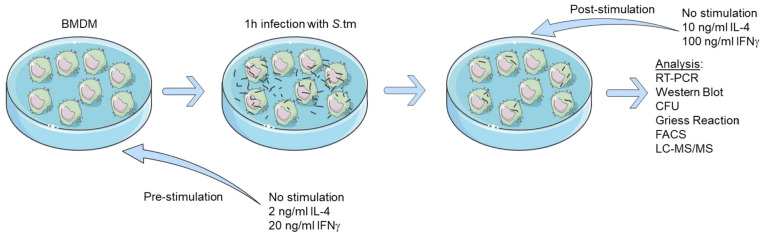
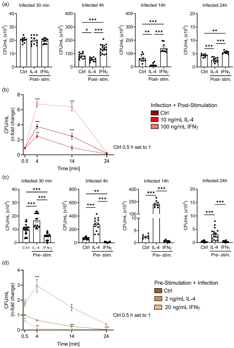
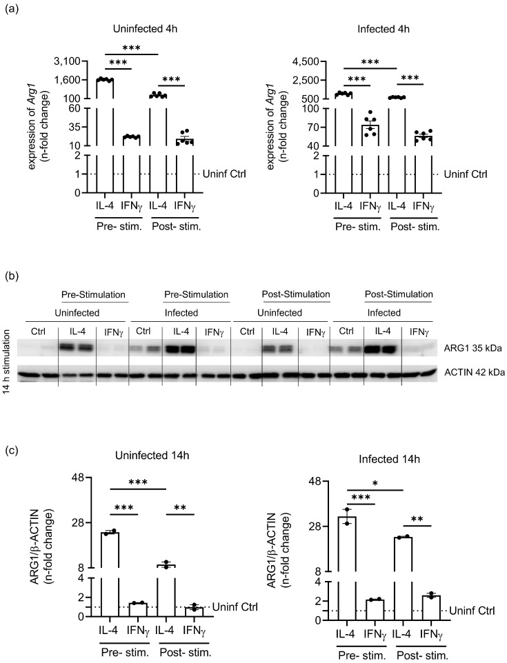
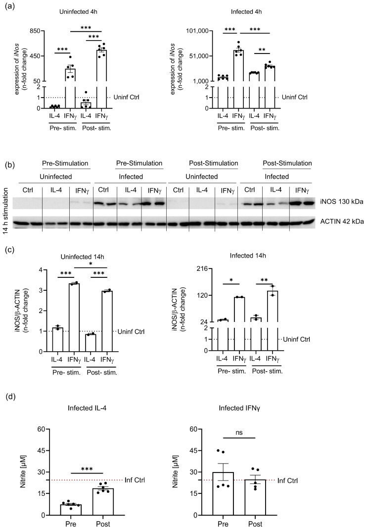
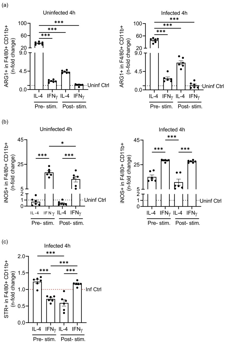
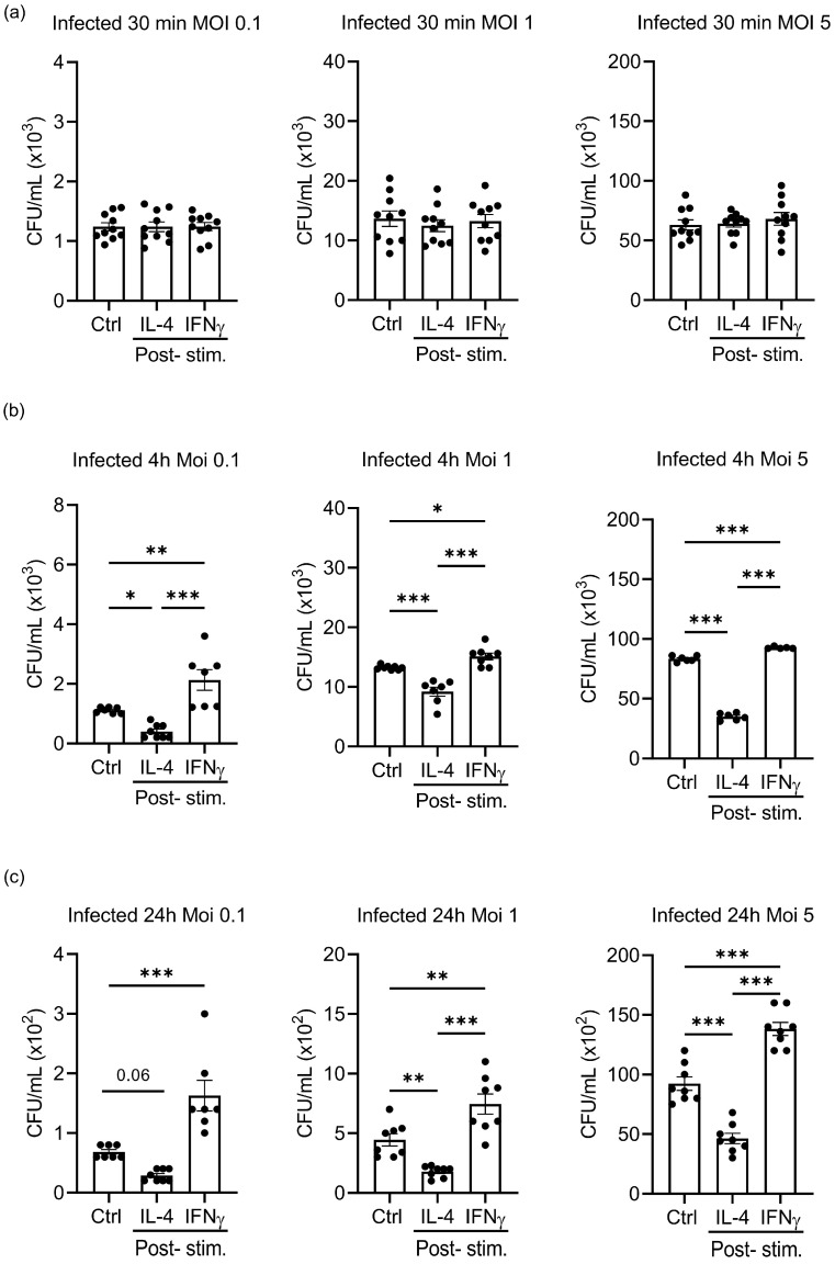
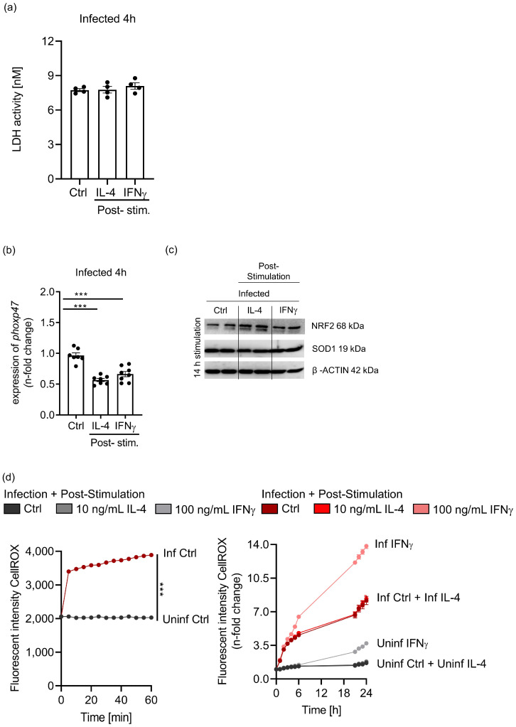
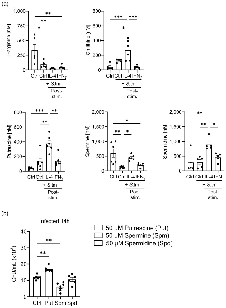
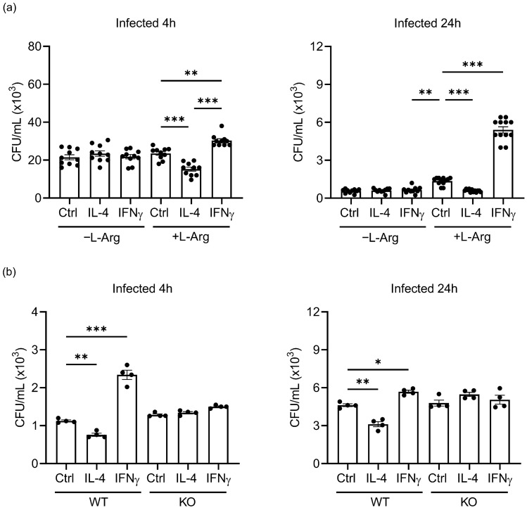
Priming of macrophages with interferon-gamma (IFNγ) or interleukin-4 (IL-4) leads to polarisation into pro-inflammatory or anti-inflammatory subtypes, which produce key enzymes such as inducible nitric oxide synthase (iNOS) and arginase 1 (ARG1), respectively, and in this way determine host responses to infection. Importantly, L-arginine is the substrate for both enzymes. ARG1 upregulation is associated with increased pathogen load in different infection models. However, while differentiation of macrophages with IL-4 impairs host resistance to the intracellular bacterium Salmonella enterica serovar Typhimurium (S.tm), little is known on the effects of IL-4 on unpolarised macrophages during infection. Therefore, bone-marrow-derived macrophages (BMDM) from C57BL/6N, Tie2Cre+/-ARG1fl/fl (KO), Tie2Cre-/-ARG1fl/fl (WT) mice were infected with S.tm in the undifferentiated state and then stimulated with IL-4 or IFNγ. In addition, BMDM of C57BL/6N mice were first polarised upon stimulation with IL-4 or IFNγ and then infected with S.tm. Interestingly, in contrast to polarisation of BMDM with IL-4 prior to infection, treatment of non-polarised S.tm-infected BMDM with IL-4 resulted in improved infection control whereas stimulation with IFNγ led to an increase in intracellular bacterial numbers compared to unstimulated controls. This effect of IL-4 was paralleled by decreased ARG1 levels and increased iNOS expression. Furthermore, the L-arginine pathway metabolites ornithine and polyamines were enriched in unpolarised cells infected with S.tm and stimulated with IL-4. Depletion of L-arginine reversed the protective effect of IL-4 toward infection control. Our data show that stimulation of S.tm-infected macrophages with IL-4 reduced bacterial multiplication via metabolic re-programming of L-arginine-dependent pathways.

Keywords: Salmonella enterica serovar Typhimurium; arginase 1; inducible nitric oxide synthase; interferon-gamma; interleukin-4; intracellular bacteria; macrophages; ornithine; polyamines.


Conflict of interest statement

The authors declare no conflict of interest.

Figures

Figure 1
Figure 1
Schematic representation of in vitro stimulation and infection of BMDM with S.tm.
Figure 2
Figure 2
IL-4 stimulation of macrophages before or after infection determines the control of Salmonella infection. Unpolarised BMDM and pre-stimulated polarised BMDM (2 ng/mL IL-4 or 20 ng/mL IFNγ) were infected with S.tm for 1 h. Unpolarised cells were then stimulated (post-stimulation) with 10 ng/mL IL-4, 100 ng/mL IFNγ as indicated or left unstimulated. Differences between colony-forming units (CFU) were analysed. (a) Uptake of S.tm (Infected 30 min) and quantification of intracellular CFU in post-stimulated macrophages (Infected 4 h, 14 h, and 24 h). (b) Normalized intracellular CFU over time in post-stimulated BMDM. (c) Uptake of S.tm in pre-stimulated BMDM and killing capacity over the course of 24 h. (d) Normalized intracellular CFU over time in pre-stimulated BMDM. Statistical significance was determined by one-way ANOVA with Tukey post hoc test (a + c) and by two-way ANOVA with Tukey post hoc tests (b + d). * p-value < 0.05; ** p-value < 0.01; *** p-value < 0.001. Representative data (mean ± SEM) from three independent experiments performed in technical triplicates or quadruplicates are shown.
Figure 3
Figure 3
Priming of macrophages before S.tm infection shows differences in ARG1 regulation. Unpolarised BMDM and pre-stimulated polarised BMDM (2 ng/mL IL-4 or 20 ng/mL IFNγ) were infected with S.tm for 1 h. Unpolarised cells were then stimulated (post-stimulation) with 10 ng/mL IL-4, 100 ng/mL IFNγ or left unstimulated for 4 h or 14 h. ARG1 mRNA and protein levels in pre- and post-stimulation conditions were analysed in uninfected and infected macrophages. (a) Arg1 gene expression depending on stimulation (IL-4 or IFNγ) and infection. Arg1 transcript levels were determined by quantitative real-time PCR and normalized to Hypoxanthine phosphoribosyltransferase (Hprt) mRNA levels using the ΔΔCT method. (b) Western blot analysis of ARG1 and β-ACTIN. (c) Densidometrical quantification of immunoblotting results relative to β-ACTIN expression. Statistical significance was determined by one-way ANOVA with Tukey post hoc test (a + c). * p-value < 0.05; ** p-value < 0.01; *** p-value < 0.001. Representative data from three independent experiments (mean ± SEM) performed in technical duplicates are shown. Data were normalised to the unstimulated, uninfected Ctrl.
Figure 4
Figure 4
Priming of macrophages before S.tm infection shows differences in iNOS regulation. Unpolarised BMDM and pre-stimulated polarised BMDM (2 ng/mL IL-4 or 20 ng/mL IFNγ) were infected with S.tm for 1 h. Unpolarised cells were then stimulated (post-stimulation) with 10 ng/mL IL-4, 100 ng/mL IFNγ or left unstimulated for 4 h or 14 h. iNOS mRNA and protein levels in pre- and post-stimulation conditions were analysed in uninfected and infected macrophages. (a) iNos gene expression depending on stimulation (IL-4 or IFNγ) and infection. iNos transcript levels were determined by quantitative real-time PCR and normalised to Hypoxanthine phosphoribosyltransferase (Hprt) mRNA levels using the ΔΔCT method. (b) Western blot analysis of iNOS and β-ACTIN. (c) Densidometrical quantification of immunoblotting results relative to β-ACTIN expression. (d) Nitrite levels of pre- and post-stimulated samples after 14 h determined via Griess reaction. Statistical significance was determined by one-way ANOVA with Tukey post hoc test (a + c). * p-value < 0.05; ** p-value < 0.01; *** p-value < 0.001; ns = not statistically significant. Representative data from three independent experiments (mean ± SEM) performed in technical duplicates are shown. Data were normalised to the unstimulated uninfected Ctrl.
Figure 5
Figure 5
Pre- or post-stimulation of macrophages with IL-4 and IFNγ impacts the amount of S.tm phagocytosed by F4/80+CD11b+ macrophages. Unpolarised BMDM and pre-stimulated polarised BMDM (2 ng/mL IL-4 or 20 ng/mL IFNγ) were infected with S.tm expressing RFP (STR) for 1 h. Unpolarised cells were then stimulated (post-stimulation) with 10 ng/mL IL-4, 100 ng/mL IFNγ or left unstimulated for 4 h. Percentages of ARG1, iNOS and STR in macrophages were analysed via flow cytometry. (a) Percentage of ARG1-positive cells in F4/80+CD11b+ cells in pre-stimulated compared to post-stimulated BMDM. Data shown for uninfected (left) and infected (right) cells. (b) Percentage of iNOS positive cells in F4/80+CD11b+ in pre-stimulated compared to post-stimulated BMDM. Data shown for uninfected (left) and infected (right) cells. (c) Percentage of STR positive cells in F4/80+CD11b+ cells in infected pre-stimulated compared to post-stimulated BMDM. Statistical significance was determined by one-way ANOVA with Tukey post hoc test. * p-value < 0.05; *** p-value < 0.001. Representative data (mean ± SEM) from three independent experiments with two technical replications are shown. Data were normalised to the unstimulated uninfected Ctrl or, as indicated, to the unstimulated infected Ctrl.
Figure 6
Figure 6
Different multiplicities of infection do not alter the effects of IL-4 and IFNγ stimulation on the growth of S.tm in macrophages. Unpolarised BMDM were infected with S.tm for 1 h. Afterwards, cells were stimulated (post-stimulation) with 10 ng/mL IL-4, 100 ng/mL IFNγ or left unstimulated as indicated. (a) CFU analysis after 0.5 h (a), 4 h (b) and 24 h (c) of infection with several multiplicities of infection (MOI) of S.tm in post-stimulated BMDM. Statistical significance was determined by one-way ANOVA with Tukey post hoc test: * p-value < 0.05; ** p-value < 0.01; *** p-value < 0.001. Representative data (mean ± SEM) from three independent experiments with 2–3 technical replications are shown.
Figure 7
Figure 7
IL-4 stimulation of unpolarised S.tm-infected BMDM does not affect cellular viability, radical formation and expression of radical detoxifying enzymes. Unpolarised BMDM were infected with S.tm for 1 h. Afterwards, cells were stimulated (post-stimulation) with 10 ng/mL IL-4, 100 ng/mL IFNγ or left unstimulated as indicated. (a) Determination of cell viability by analysing LDH activity after 4 h post-stimulation. (b) Phoxp47 transcript levels due to infection and 4 h post-stimulation were determined by quantitative real-time PCR and normalized to Hypoxanthine phosphoribosyltransferase (Hprt) mRNA levels using the ΔΔCT method. Data were normalised to the infected Ctrl. (c) Western blot analysis of SOD1, NRF2 and β-ACTIN expression after 14 h. (d) Quantification of ROS formation using the CellROXTM reagent, during a 1 h infection period (left) and during a gentamicin neutralisation assay and post-stimulation over the course of 24 h. Time course data were normalised to the levels of the 30 min unstimulated uninfected Ctrl. Statistical significance was determined by one-way ANOVA with Tukey post hoc test for three groups and two-way ANOVA with Tukey post hoc tests for more groups: *** p-value < 0.001. Representative data (mean ± SEM) from two to three independent experiments with two technical replications are shown.
Figure 8
Figure 8
IL-4 stimulation of unpolarised macrophages after infection with S.tm alters L-arginine metabolic pathways with direct impact on the growth of Salmonella. Unpolarised BMDM were infected with S.tm for 1 h. Afterwards, cells were stimulated (post-stimulation) with 10 ng/mL IL-4, 100 ng/mL IFNγ or left unstimulated as indicated. (a) Concentrations of L-arginine and related metabolites determined by LC-MS/MS in 24 h post-stimulated BMDM cell lysates. (b) Growth of S.tm after 14 h in media supplemented with either 50 µM spermine, 50 µM spermidine or 50 µM putrescine. Statistical significance was determined by one-way ANOVA with Tukey post hoc test. * p-value < 0.05; ** p-value < 0.01; *** p-value < 0.001. Representative data (mean ± SEM) of three independent experiments with 2–4 technical replications are shown.
Figure 9
Figure 9
IL-4 exerts its inhibitory effect on S.tm proliferation via L-arginine-mediated metabolic pathways. Effects of L-arginine depletion and deletion of ARG1 on S.tm survival in macrophages was analysed. BMDM generated from C57BL/6N mice were cultured in L-arginine-free medium. Moreover, BMDM of Tie2Cre+/−ARG1fl/fl (KO) and Tie2Cre−/−ARG1fl/fl (WT) were generated. Macrophages were infected with S.tm and stimulated (post-stimulation) with 10 ng/mL IL-4, 100 ng/mL IFNγ or left unstimulated as indicated. (a) Effect of arginine-depleted medium (− L-Arg) on survival of S.tm in BMDM compared to S.tm survival in medium supplemented with L-arginine (+ L-Arg). (b) S.tm CFU in Tie2Cre−/−ARG1fl/fl (WT) BMDM compared to Tie2Cre+/−ARG1fl/fl (KO) BMDM after post-stimulation as indicated. Statistical significance was determined by one-way ANOVA with Tukey post hoc test: * p-value < 0.05; ** p-value < 0.01; *** p-value < 0.0001. Representative data (mean ± SEM) of two to three independent experiments with 2–4 technical replications are shown.

References

    1. Bosurgi L., Cao Y.G., Cabeza-Cabrerizo M., Tucci A., Hughes L.D., Kong Y., Weinstein J.S., Licona-Limon P., Schmid E.T., Pelorosso F., et al. Macrophage function in tissue repair and remodeling requires IL-4 or IL-13 with apoptotic cells. Science. 2017;356:1072–1076. doi: 10.1126/science.aai8132. - DOI - PMC - PubMed
    1. Nairz M., Theurl I., Swirski F.K., Weiss G. “Pumping iron”-how macrophages handle iron at the systemic, microenvironmental, and cellular levels. Pflug. Arch. Eur. J. Physiol. 2017;469:397–418. doi: 10.1007/s00424-017-1944-8. - DOI - PMC - PubMed
    1. Mosser D.M., Edwards J.P. Exploring the full spectrum of macrophage activation. Nat. Rev. Immunol. 2008;8:958–969. doi: 10.1038/nri2448. - DOI - PMC - PubMed
    1. Gordon S., Martinez-Pomares L. Physiological roles of macrophages. Pflug. Arch. Eur. J. Physiol. 2017;469:365–374. doi: 10.1007/s00424-017-1945-7. - DOI - PMC - PubMed
    1. Murray P.J. Macrophage Polarization. Annu. Rev. Physiol. 2017;79:541–566. doi: 10.1146/annurev-physiol-022516-034339. - DOI - PubMed

Publication types