Review
. 2023 Apr 12;15(8):2256.
doi: 10.3390/cancers15082256.

A Recent Review on Cancer Nanomedicine

Affiliations
Review

A Recent Review on Cancer Nanomedicine

Paras Mani Giri et al. Cancers (Basel). .

Abstract

Cancer is one of the most prevalent diseases globally and is the second major cause of death in the United States. Despite the continuous efforts to understand tumor mechanisms and various approaches taken for treatment over decades, no significant improvements have been observed in cancer therapy. Lack of tumor specificity, dose-related toxicity, low bioavailability, and lack of stability of chemotherapeutics are major hindrances to cancer treatment. Nanomedicine has drawn the attention of many researchers due to its potential for tumor-specific delivery while minimizing unwanted side effects. The application of these nanoparticles is not limited to just therapeutic uses; some of them have shown to have extremely promising diagnostic potential. In this review, we describe and compare various types of nanoparticles and their role in advancing cancer treatment. We further highlight various nanoformulations currently approved for cancer therapy as well as under different phases of clinical trials. Finally, we discuss the prospect of nanomedicine in cancer management.

Keywords: cancer; chemotherapy; inorganic nanoparticles; liposomes; nanoparticles; targeted drug delivery; theranostics.


Conflict of interest statement

The authors declare no conflict of interest.

Figures

Figure 1
Figure 1
Nanocarrier-mediated tumor targeting. (A) Passive tumor targeting, (B) ligand-based active tumor targeting, and (C) TME-responsive drug delivery. Created with BioRender.com.
Figure 2
Figure 2
Types of nanocarriers frequently used for cancer therapy. Created with BioRender.com.
Figure 3
Figure 3
The in vitro cell viability of (A) MCF-7 and (B) A549 cells at various drug concentrations of the drug after 72 h treatment. (C) Plasma concentration–time profile of resveratrol (RSV) in rats with intravenously administered free RSV, RSV-NLCs and RSV-FA-NLCs; inset shows representative HPLC chromatogram of RSV in rat plasma. Source: reprinted from Poonia, N.; Kaur Narang, J.; Lather, V.; Beg, S.; Sharma, T.; Singh, B.; Pandita, D., Resveratrol loaded functionalized nanostructured lipid carriers for breast cancer targeting: Systematic development, characterization and pharmacokinetic evaluation. Colloids and Surfaces B: Biointerfaces 2019, 181, 756–766 [53]. With permission from Elsevier.
Figure 4
Figure 4
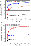
(A) Zeta potential as a function of different pH (pH 5.0, 6.5, and 7.4) for DOX@MMSN−SS−PEI−cit. (B) Cumulative release of DOX from DOX@MMSN−SS−PEI−cit nanoplatform in PBS (pH 7.4) with different GSH concentrations (0, 1, and 10 mM) in shaking table at 37 °C. Data are shown as mean ± SD, n = 3 per treatment. Source: reprinted from Wan, L.; Chen, Z.; Deng, Y.; Liao, T.; Kuang, Y.; Liu, J.; Duan, J.; Xu, Z.; Jiang, B.; Li, C., A novel intratumoral pH/redox-dual-responsive nanoplatform for cancer MR imaging and therapy. Journal of Colloid and Interface Science 2020, 573, 263–277 [87]. With permission from Elsevier.
Figure 5
Figure 5
4T1-Luc tumor-bearing mice treated with different samples (PBS, DOX@MSNs, and DOX@MSNs-CAIX) for 11 days. The tumors’ bioluminescence imaging before (A) and after (B) intervention with the above samples. (C) The tumors’ bioluminescence intensity before (the 0th day) and after (the 11th day) intervention with the above samples. (D) The tumors’ photographs from the scarified mice. (E) The final average tumor weight. (F) The variation curves of average tumor volume. (* p < 0.05 as compared with PBS group.) Source: reprinted from Chen, M.; Hu, J.; Wang, L.; Li, Y.; Zhu, C.; Chen, C.; Shi, M.; Ju, Z.; Cao, X.; Zhang, Z., Targeted and redox-responsive drug delivery systems based on carbonic anhydrase IX-decorated mesoporous silica nanoparticles for cancer therapy. Scientific Reports 2020, 10, (1), 14447 [112] under an open access Creative Commons CC BY 4.0 license.
Figure 6
Figure 6
Distribution of nanoprobes in vivo. (A) In vivo continuous observations (48 h) of mice administered SWNT-CY7-IGF-1Ra via the tail vein. The black dotted circle represents the location of the pancreatic carcinoma in situ. (B) Ex vivo imaging of tumor and major organs. H: heart. Li: liver. P: pancreas. T: tumor. S: spleen. Lu: lung. K: kidney. In: intestine. (C) Comparison of TBR (TBR = average fluorescence intensity of the tumor area/average fluorescence intensity, with the ear as the background area) profiles of the nanoprobes. The peak was at 18 h post-injection, which implied an optimal experimental window. (D) Fluorescence intensity of different tissues. Data represent mean ± SD of triplicate experiments. (E) The accumulation of as-prepared nanotubes along the tumor blood vessels and at the normal–tumor tissue junction at 18 h post-injection. The as-prepared nanotubes appear as green fluorescent dots in 488 nm and the blood vessels are shown as red fluorescent regions in 660 nm. The scale bar is 20 μm. * p < 0.05, ** p < 0.01, *** p < 0.001. Source: Lu, G.-H.; Shang, W.-T.; Deng, H.; Han, Z.-Y.; Hu, M.; Liang, X.-Y.; Fang, C.-H.; Zhu, X.-H.; Fan, Y.-F.; Tian, J., Targeting carbon nanotubes based on IGF-1R for photothermal therapy of orthotopic pancreatic cancer guided by optical imaging. Biomaterials 2019, 195, 13–22 [124]. With permission from Elsevier.
Figure 7
Figure 7
In vitro delivery efficacies of folic acid and 2-(Diisopropylamino) ethyl methacrylate dual-functionalized trimethyl chitosan nanoparticles (FTD NPs). (A) Cellular uptake of doxorubicin (DOX) and pDNA in 4T1 cells after 4 h incubation with NPs. Indicated values are mean ± SD (n = 3). * p < 0.05, ** p < 0.01, *** p < 0.001. (B) CLSM images showing the nuclear transport of FITC-pDNA (green) and DOX (red) loaded into FTD NPs in 4T1 cells after 2, 4, and 8 h incubation. The nuclei were stained with Hoechst 33,258 (blue). Bar represents 10 μm. (C) Intracellular distribution of DOX and pDNA in 4T1 cells following treatment with FTD/DOX/sgSurvivin pDNA NPs for 2, 4, and 8 h. Indicated values are mean ± SD (n = 3). (D) Fluorescence microscope images and relative fluorescence intensity of pEGFP in 4T1 cells transfected for 48 h. Indicated values are mean ± SD (n = 3). Bar represents 50 μm. * p < 0.05, ** p < 0.01, *** p < 0.001. Source: reprinted from Li, Q.; Lv, X.; Tang, C.; Yin, C., Co-delivery of doxorubicin and CRISPR/Cas9 or RNAi-expressing plasmid by chitosan-based nanoparticle for cancer therapy. Carbohydrate Polymers 2022, 287, 119315 [151]. With permission from Elsevier.
Figure 8
Figure 8
The inhibition of metastases growth in mouse lungs upon exosome-loaded paclitaxel (exoPTX) treatment. C57BL/6 mice were i.v. injected with 8FlmC-FLuc-3LL-M27 (red) cells to establish pulmonary metastases. After 48 h, mice were treated with exoPTX, or Taxol, or saline, or empty sonicated exosomes as a control, and the treatment was repeated every other day, totally, seven times. Representative IVIS images were taken at day 21 (A). Statistical significance of metastases levels from IVIS images in lungs of treated animals compared to control mice is shown by asterisk (* p < 0.05; ** p < 0.005) (B). At the endpoint, 21 days later, mice were sacrificed, perfused, and lung slides were examined by confocal microscopy (C). The bar: 10 μm. Source: reprinted from Kim, M. S.; Haney, M. J.; Zhao, Y.; Mahajan, V.; Deygen, I.; Klyachko, N. L.; Inskoe, E.; Piroyan, A.; Sokolsky, M.; Okolie, O.; Hingtgen, S. D.; Kabanov, A. V.; Batrakova, E. V., Development of exosome-encapsulated paclitaxel to overcome MDR in cancer cells. Nanomedicine: nanotechnology, biology, and medicine 2016, 12, (3), 655–664 [168]. With permission from Elsevier.

References

    1. Sung H., Ferlay J., Siegel R.L., Laversanne M., Soerjomataram I., Jemal A., Bray F. Global Cancer Statistics 2020: GLOBOCAN Estimates of Incidence and Mortality Worldwide for 36 Cancers in 185 Countries. CA A Cancer J. Clin. 2021;71:209–249. doi: 10.3322/caac.21660. - DOI - PubMed
    1. Debela D.T., Muzazu S.G., Heraro K.D., Ndalama M.T., Mesele B.W., Haile D.C., Kitui S.K., Manyazewal T. New approaches and procedures for cancer treatment: Current perspectives. SAGE Open Med. 2021;9:20503121211034366. doi: 10.1177/20503121211034366. - DOI - PMC - PubMed
    1. Altun İ., Sonkaya A. The Most Common Side Effects Experienced by Patients Were Receiving First Cycle of Chemotherapy. Iran. J. Public Health. 2018;47:1218–1219. - PMC - PubMed
    1. Danhier F., Feron O., Préat V. To exploit the tumor microenvironment: Passive and active tumor targeting of nanocarriers for anti-cancer drug delivery. J. Control. Release Off. J. Control. Release Soc. 2010;148:135–146. doi: 10.1016/j.jconrel.2010.08.027. - DOI - PubMed
    1. Din F.U., Aman W., Ullah I., Qureshi O.S., Mustapha O., Shafique S., Zeb A. Effective use of nanocarriers as drug delivery systems for the treatment of selected tumors. Int. J. Nanomed. 2017;12:7291–7309. doi: 10.2147/IJN.S146315. - DOI - PMC - PubMed

LinkOut - more resources