. 2023 Apr 17;15(8):2326.
doi: 10.3390/cancers15082326.

VISTA Ligation Reduces Antitumor T-Cell Activity in Pancreatic Cancer

Affiliations

VISTA Ligation Reduces Antitumor T-Cell Activity in Pancreatic Cancer

David Digomann et al. Cancers (Basel). .

Abstract

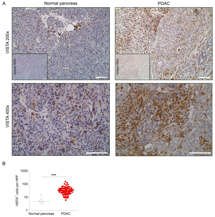
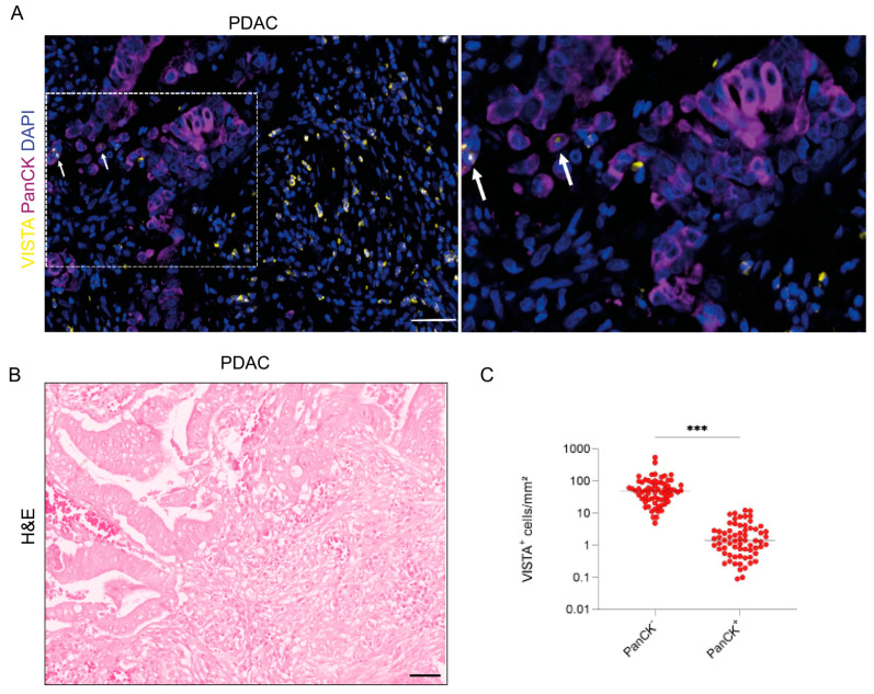
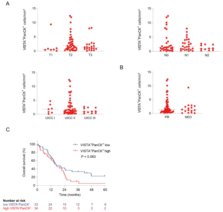
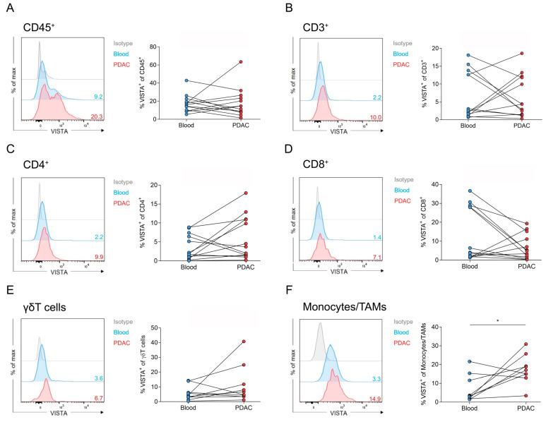
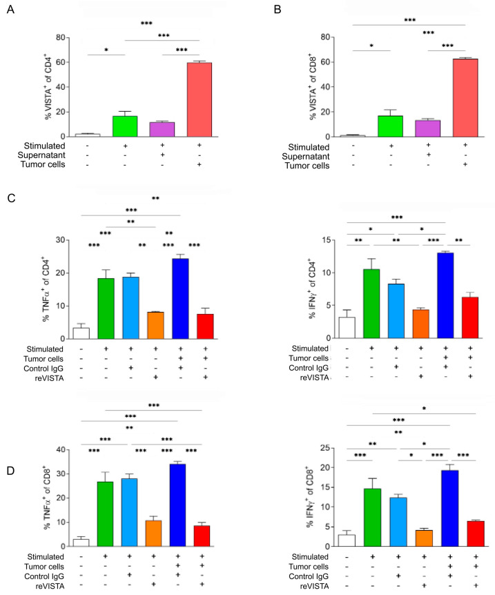
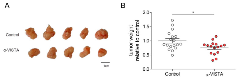
Immunotherapy has shown promising results in multiple solid tumors and hematological malignancies. However, pancreatic ductal adenocarcinoma (PDAC) has been largely refractory to current clinical immunotherapies. The V-domain Ig suppressor of T-cell activation (VISTA) inhibits T-cell effector function and maintains peripheral tolerance. Here, we determine VISTA expression in nontumorous pancreatic (n = 5) and PDAC tissue using immunohistochemistry (n = 76) and multiplex immunofluorescence staining (n = 67). Additionally, VISTA expression on tumor-infiltrating immune cells and matched blood samples (n = 13) was measured with multicolor flow cytometry. Further, the effect of recombinant VISTA on T-cell activation was investigated in vitro, and VISTA blockade was tested in an orthotopic PDAC mouse model in vivo. PDAC showed significantly higher VISTA expression compared to that of a nontumorous pancreas. Patients with a high density of VISTA-expressing tumor cells had reduced overall survival. The VISTA expression of CD4+ and CD8+ T cells was increased after stimulation and particularly after a coculture with tumor cells. We detected a higher level of proinflammatory cytokine (TNFα and IFNγ) expression by CD4+ and CD8+ T cells, which was reversed with the addition of recombinant VISTA. A VISTA blockade reduced tumor weights in vivo. The VISTA expression of tumor cells has clinical relevance, and its blockade may be a promising immunotherapeutic strategy for PDAC.

Keywords: VISTA; cytokines; pancreatic cancer; prognosis.


Conflict of interest statement

The authors declare no conflict of interest.

Figures

Figure 1
Figure 1
VISTA is expressed in pancreatic ductal adenocarcinoma. (A) Representative IHC staining for VISTA in normal pancreatic tissue and pancreatic adenocarcinoma with isotypic control (200× and 400× magnification); scale bar is 100 µm. (B) VISTA+ cells per high-power field (HPF) in pancreatic adenocarcinoma (n = 76) compared to normal pancreatic tissue (n = 5; *** p < 0.001; Mann–Whitney test).
Figure 2
Figure 2
VISTA is expressed by cancer (PanCK+) and noncancer (PanCK) cells located at the tumor site. (A) Representative multiplex immunofluorescence staining (VISTA in yellow, PanCK in purple, and DAPI in blue) of pancreatic adenocarcinoma with a magnified section. Double positive cells (PanCK+/VISTA+) cells are marked with arrows; scale bar is 50 µm. (B) Hematoxylin and eosin (H&E) stain to isualize the tumor area from (A); scale bar is 50 µm. (C) Noncancer cells (PanCK) displayed significantly higher VISTA expression compared to that of cancer cells (PanCK+; *** p < 0.001; Mann–Whitney test).
Figure 3
Figure 3
Impact of VISTA expression on clinicopathologic features. (A) VISTA expression on PanCK+ was not associated with tumor size, lymph-node metastasis, or UICC stage (n = 67; Kruskal–Wallis test with Dunn’s statistics). (B) Neoadjuvant therapy did not influence VISTA expression (n = 67; Mann–Whitney test). (C) Survival analysis of PDAC patients according to their VISTA+ PanCK+ expression (median) shown as Kaplan–Meier curves (n = 67; overall survival: p = 0.063; log-rank test).
Figure 4
Figure 4
Tumor-infiltrating immune cells highly express VISTA in human PDAC. (AF) The expression of VISTA on different tumor-infiltrating immune cells in human PDAC tissue samples was measured and quantified with flow cytometry. Significant differences were found between the blood and tumor in monocytes/TAMs (* p = 0.015; paired t-test; n = 13).
Figure 5
Figure 5
Tumor cells promote VISTA expression on T cells, while VISTA leads to the inhibition of cytokine production. (A,B) VISTA expression of CD4+ and CD8+ T cells increased significantly after stimulation and after coculturing with AsPC-1 cells (* p < 0.05, *** p < 0.001; one-way ANOVA with Tukey statistics). (C,D) The cytokine production (TNFα and IFNγ) of CD4+ and CD8+ T cells significantly increased after stimulation (one-way ANOVA with Tukey statistics). Treatment with recombinant VISTA significantly reversed cytokine production in stimulated and AsPC-1-cocultured conditions (* p < 0.05, ** p < 0.01, *** p < 0.001; one-way ANOVA with Tukey statistics).
Figure 6
Figure 6
VISTA mAb treatment suppressed tumors in an orthotopic PDAC mouse model. (A) Representative tumors of the orthotopic mouse model treated with α-VISTA or the control. (B) Tumor weight was significantly reduced when treated with α-VISTA compared to the control (* p < 0.05; unpaired t-test).

References

    1. Sung H., Ferlay J., Siegel R.L., Laversanne M., Soerjomataram I., Jemal A., Bray F. Global Cancer Statistics 2020: GLOBOCAN Estimates of Incidence and Mortality Worldwide for 36 Cancers in 185 Countries. CA A Cancer J. Clin. 2021;71:209–249. doi: 10.3322/caac.21660. - DOI - PubMed
    1. Siegel R.L., Miller K.D., Fuchs H.E., Jemal A. Cancer Statistics, 2022. CA Cancer J. Clin. 2022;72:7–33. doi: 10.3322/caac.21708. - DOI - PubMed
    1. Ryan D.P., Hong T.S., Bardeesy N. Pancreatic Adenocarcinoma. N. Engl. J. Med. 2014;371:1039–1049. doi: 10.1056/NEJMra1404198. - DOI - PubMed
    1. Seifert A.M., Reiche C., Heiduk M., Tannert A., Meinecke A.-C., Baier S., von Renesse J., Kahlert C., Distler M., Welsch T., et al. Detection of Pancreatic Ductal Adenocarcinoma with Galectin-9 Serum Levels. Oncogene. 2020;39:3102–3113. doi: 10.1038/s41388-020-1186-7. - DOI - PMC - PubMed
    1. Le D.T., Durham J.N., Smith K.N., Wang H., Bartlett B.R., Aulakh L.K., Lu S., Kemberling H., Wilt C., Luber B.S., et al. Mismatch Repair Deficiency Predicts Response of Solid Tumors to PD-1 Blockade. Science. 2017;357:409–413. doi: 10.1126/science.aan6733. - DOI - PMC - PubMed