Review
. 2023 Nov;96(1151):20230028.
doi: 10.1259/bjr.20230028. Epub 2023 Jun 7.

The role of CT in arrhythmia management-treatment planning and post-procedural imaging surveillance

Affiliations
Review

The role of CT in arrhythmia management-treatment planning and post-procedural imaging surveillance

Giulia Cundari et al. Br J Radiol. 2023 Nov.

Abstract

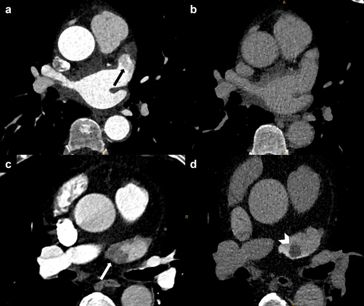
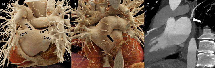
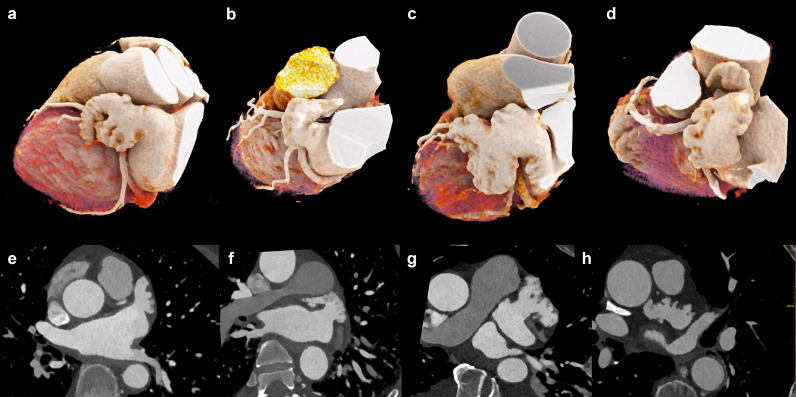
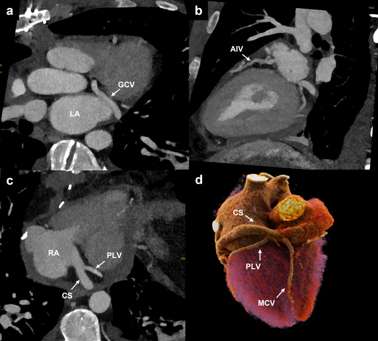
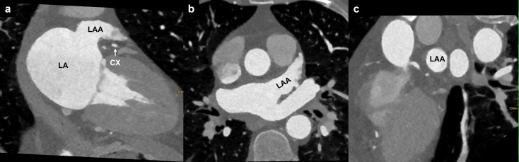
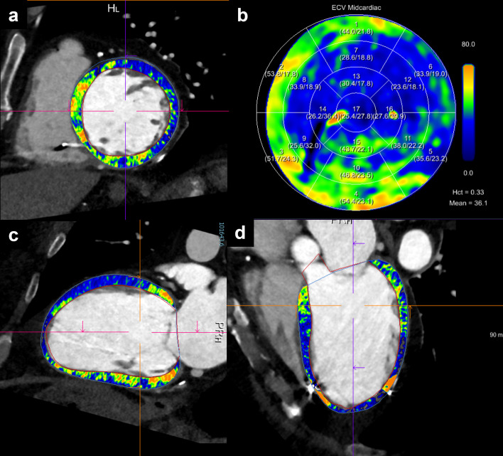
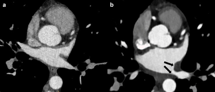
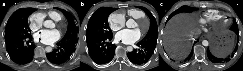
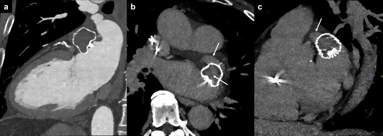
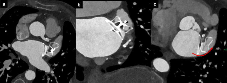
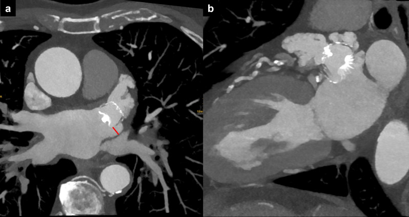
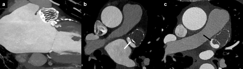
Several interventional treatment options exist in patients with atrial and ventricular arrhythmia. Cardiac CT is routinely performed prior to occlusion of the left atrial appendage, pulmonary vein isolation, and cardiac device implantation. Besides the evaluation of coronary artery disease, cardiac CT provides isotropic, high-resolution CT images of the cardiac anatomy with the possibility of multiplanar reformations and three-dimensional reconstructions which are helpful to guide interventional treatment. In addition, cardiac CT is increasingly used to rapidly evaluate periprocedural complications and for the routine post-procedural imaging surveillance in patients after interventions. This review article will discuss current applications of pre- and post-interventional CT imaging in patients with arrhythmia.


Figures

Figure 1.
Figure 1.
Ruling out LAA thrombus. (a) Filling defect of the LAA in the arterial phase being suspicious of thrombus (black arrow), (b) delayed phase after 60 s showing normal opacification of the LAA. (c) Filling defect of the LAA in the arterial phase (white arrow). (d) Delayed phase after 60 s showing a 1 cm thrombus in the LAA (white arrowhead). LAA, left atrial appendage
Figure 2.
Figure 2.
PV anatomy. (a) Cinematic 3D rendering of the heart showing the posterior aspect of the LA with normal PV anatomy: two veins to the right (RSPV and RIPV) and two veins to the left (LSPV and LIPV) joining the LA. (b) Cinematic 3D rendering showing a single PV ostium for the superior and inferior left pulmonary veins (conjoined vein, black arrow). (c) white arrow highlighting an accessory pulmonary vein (right top vein). LA, left atrium; LIPV, left inferior pulmonary vein; LSPV, left superior pulmonary vein; RIPV, right inferior pulmonary vein; RSPV, right superior pulmonary vein; PV, pulmonary vein.
Figure 3.
Figure 3.
Types of left atrial appendage configuration. Cinematic 3D rendering (a–d) and multiplanar reformations (e–h) of left atrial appendage morphology types. (a, e) Windsock variant. (b, f) Chicken-wing variant. (c, g) Cauliflower variant. (d, h) Cactus variant.
Figure 4.
Figure 4.
Cardiac vein anatomy. (a) Oblique axial reformation showing the GCV running in the atrioventricular sulcus. (b) Oblique sagittal reformation illustrating the anterior interventricular vein, which runs in the anterior interventricular sulcus from the apex towards the base of the heart. (c) Oblique axial reformation demonstrating the CS draining into the RA after receiving the PLV. (d) Cinematic 3D rendering showing the posterior aspect of the heart with the MCV running in the posterior interventricular sulcus and draining directly into the RA. CS and PLV are also represented. CS, coronary sinus; GCV, great cardiac vein; MCV, middle cardiac vein; LA, left atrium; PLV, posterolateral vein; RA, right atrium.
Figure 5.
Figure 5.
Evaluation of LAA anatomy prior to the before occlusion procedure. (a) Oblique sagittal LAA reformation (both long axis LAA and circumflex artery are visualized). (b) Orthogonal plane (axial to the LAA walls). (c) Oblique plane to evaluate the LAA orifice. CX: circumflex artery; LA, left atrium; LAA, left atrial appendage.
Figure 6.
Figure 6.
Scar detection. A 60-year-old female patient with ischemic cardiomyopathy, reduced ejection fraction and ICD implantation. Ablation was planned due to an episode of ventricular tachycardia with cardiogenic shock. Cardiac photon-counting detector CT with acquisition of a dual-energy late enhancement phase (after 5 min.) revealed an extensive post-ischemic scar in anterior and anteroseptal segments of the basal and mid-ventricular left ventricle (a, c, d), with associated increased extracellular volume fraction (b). Successful radiofrequency ablation confirmed left ventricular tachycardia associated with the scar, with mid-diastolic potentials in anteroseptal, anterior and anterolateral segments from basal to midventricular planes. ICD, implantable cardioverter defibrillator
Figure 7.
Figure 7.
PV stenosis after radiofrequency catheter ablation. (a) PV ostium before procedure. (b) Ostium wall thickening with PV stenosis after RFCA (black arrows). PV, pulmonary vein; RFCA, radiofrequency catheter ablation.
Figure 8.
Figure 8.
Atrioesophageal fistula after radiofrequency catheter ablation. Air bubbles and thrombus inside the left atrium (a, b) and the left ventricle (c).
Figure 9.
Figure 9.
Fabric permeability. LAA patency after Watchman device implantation. (a) The arterial phase CT indicates apparent complete occlusion of the LAA. (b, c) The very delayed phase (5 min) reveals increased attenuation in the LAA and within the device itself (white arrows), suggestive for LAA patency due to fabric permeability. LAA, left atrial appendage.
Figure 10.
Figure 10.
Malapposition of device for left atrial appendage occlusion. Malposition of the proximal segment of the device lobe in axial (a), oblique coronal (b), and oblique axial (c) reformations, with persistent opacification of the left atrial appendage via the posterior quadrant (red curved arrow).
Figure 11.
Figure 11.
Peridevice leak. (a) Axial and (b) oblique sagittal reformation of the LAA showing its complete opacification after LAA occlusion with the Watchman device, due to a peridevice leak (>5 mm, red line). LAA, left atrial appendage
Figure 12.
Figure 12.
Device-related thrombus. (a) Normal LAA occluder position; (b) low-grade HAT (<3 mm, white arrow); (c) high grade HAT (>3 mm, black arrow). HAT, hypoattenuated thickening; LAA, left atrial appendage

References

    1. Hindricks G, Potpara T, Dagres N, Arbelo E, Bax JJ, Blomström-Lundqvist C. 2020 ESC guidelines for the diagnosis and management of atrial fibrillation developed in collaboration with the European Association for Cardio-Thoracic surgery (EACTS). Eur Heart J 2021; 42: 373–498. Available from: https://academic.oup.com/eurheartj/article/42/5/373/5899003 - PubMed
    1. Zeppenfeld K, Tfelt-Hansen J, de Riva M, Winkel BG, Behr ER, Blom NA, et al. . 2022 ESC guidelines for the management of patients with ventricular arrhythmias and the prevention of sudden cardiac death. Eur Heart J 2022; 43: 3997–4126. doi: 10.1093/eurheartj/ehac262 - DOI - PubMed
    1. Hell MM, Achenbach S. CT support of cardiac structural interventions. Br J Radiol 2019; 92(): 20180707. doi: 10.1259/bjr.20180707 - DOI - PMC - PubMed
    1. Lewis MA, Pascoal A, Keevil SF, Lewis CA. Selecting a CT scanner for cardiac imaging: The heart of the matter. Br J Radiol 2016; 89(): 20160376. doi: 10.1259/bjr.20160376 - DOI - PMC - PubMed
    1. Clayton B, Roobottom C, Morgan-Hughes G. CT coronary angiography in atrial fibrillation: A comparison of radiation dose and diagnostic confidence with retrospective gating vs prospective gating with systolic acquisition. Br J Radiol 2015; 88(): 20150533. doi: 10.1259/bjr.20150533 - DOI - PMC - PubMed

MeSH terms