. 2023 Sep;25(9):100897.
doi: 10.1016/j.gim.2023.100897. Epub 2023 May 13.

Biallelic variants in ribonuclease inhibitor (RNH1), an inflammasome modulator, are associated with a distinctive subtype of acute, necrotizing encephalopathy

Collaborators, Affiliations

Biallelic variants in ribonuclease inhibitor (RNH1), an inflammasome modulator, are associated with a distinctive subtype of acute, necrotizing encephalopathy

Vandana Shashi et al. Genet Med. 2023 Sep.

Abstract

Purpose: Mendelian etiologies for acute encephalopathies in previously healthy children are poorly understood, with the exception of RAN binding protein 2 (RANBP2)-associated acute necrotizing encephalopathy subtype 1 (ANE1). We provide clinical, genetic, and neuroradiological evidence that biallelic variants in ribonuclease inhibitor (RNH1) confer susceptibility to a distinctive ANE subtype.

Methods: This study aimed to evaluate clinical data, neuroradiological studies, genomic sequencing, and protein immunoblotting results in 8 children from 4 families who experienced acute febrile encephalopathy.

Results: All 8 healthy children became acutely encephalopathic during a viral/febrile illness and received a variety of immune modulation treatments. Long-term outcomes varied from death to severe neurologic deficits to normal outcomes. The neuroradiological findings overlapped with ANE but had distinguishing features. All affected children had biallelic predicted damaging variants in RNH1: a subset that was studied had undetectable RNH1 protein. Incomplete penetrance of the RNH1 variants was evident in 1 family.

Conclusion: Biallelic variants in RNH1 confer susceptibility to a subtype of ANE (ANE2) in previously healthy children. Intensive immunological treatments may alter outcomes. Genomic sequencing in children with unexplained acute febrile encephalopathy can detect underlying genetic etiologies, such as RNH1, and improve outcomes in the probands and at-risk siblings.

Keywords: Acute demyelinating encephalopathy; Acute necrotizing encephalopathy; Inflammasome; RANBP2; RNH1.


Conflict of interest statement

Conflict of Interest Rebecca Ganetzky is a paid consultant for Minovia Therapeutics and Nurture Genomics. Neal Sondheimer is employed by Synlogic, Inc. All other authors declare no conflicts of interest.

Figures

Figure 1
Figure 1. Pedigrees of families with biallelic rare variants in RNH1.
Filled shapes, affected individuals; unfilled shapes, healthy individuals; square, male; circle, female. Segregation of RNH1 variants as confirmed by bidirectional Sanger sequencing are indicated in blue. WT, wild type.
Figure 2
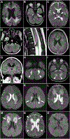
Figure 2. Representative magnetic resonance imaging from affected individuals harboring biallelic RNH1 variants
Images (A-I) illustrate the location of lesions in 5 individuals. Axial DWI of individual 1-II-1 at age 3 years 6 months (A), axial T2-weighted images from individual 1-II-3 at age 6 months (B-D), sagittal T2-weighted image of the spinal cord in individual 2-II-3 at 4 months of age (E), axial postcontrast T1 SPGR of individual 4-II-1 at age 17 years (F), coronal postcontrast T1 SPGR of individual 4-II-2 at age 12 years (G), and axial DWI (H) and T2-weighted (I) images from individual 4-II-2 at 8 months are shown. Involvement of deep and periventricular white matter was common (A, F, and G). Involvement of the dentate nuclei of the cerebellum (B, H, and I) was also frequently observed. Less commonly observed areas included the dorsal pons (B), thalami (C), medulla (D), and spinal cord (E). Images (J-O) illustrate the temporal evolution of lesions in individual 4-II-2. Axial T2 (J) and DWI (K) images at 8 months of age show areas of T2 hyperintensity and swelling in the periventricular white matter associated with restricted diffusion (arrows in K). Repeat T2 (L) and DWI (M) imaging performed 4 years later shows new lesions in the frontal periventricular and deep white matter (arrows in M). Cavitation is seen at the site of the prior lesions. Follow-up T2 (N) and DWI (O) imaging performed 15 years after the initial scan shows no new lesions, and cavitation of all prior lesions (arrowheads in N). DWI, diffusion-weighted imaging; SPGR, spoiled gradient.
Figure 3
Figure 3. Gene and protein schematics showing localization of RNH1 variants
A. Schematic representation of RNH1 on chromosome 11. Blue boxes, coding exons; unfilled boxes, untranslated regions; lines, introns. Exon number is listed below each box. Variants named according to GenBank ID: NM_203387.3 transcript are indicated with black (presumed truncating) and red (missense) lollipops. B. Schematic representation of RNH1 amino acid sequence. Green boxes, leucine-rich repeat domains. Amino acid number is listed below the schematic; variants named according to NP_976321.1 are indicated with black (presumed truncating) and red (missense) lollipops. C. Conservation of human RNH1 missense variants as compared with 8 vertebrate species (blue box). Clustal Omega (v1.2.4) was used to generate a multiple sequence alignment using human (NP_976321.1), chimpanzee (NP_001009060.1), beluga whale (XP_022421109.1), horse (XP_001488525.1), mouse (NP_660117.2), platypus (XP_028917039.1), Chinese softshell turtle (XP_006124919.1), common wall lizard (XP_028591537.1), and chicken (XP_040556805.1) amino acid sequences. D. Three-dimensional structure of human RNH1. AlphaFold (version 2022-11-01) was used to generate a predicted model; 2 different orientations are shown with localization of missense variants indicated. LRR, leucine-rich repeat.
Figure 4
Figure 4. Missense variants in family 1 and family 2 result in RNH1 protein degradation
A. Pedigrees indicating individual of origin for skin punch biopsies that were used to establish primary dermal fibroblasts. See Figure 1 for further details. B. Immunoblotting of RNH1 in 20 μg/lane total protein harvested from primary fibroblast cultures. β-actin was used as a loading control. See Supplemental Figure 1 for full blot images. C. Quantification of signal shown in panel B. RNH1 quantity was normalized to β-actin and shown in terms of arbitrary units. Affected individuals have nearly undetectable RNH1 protein compared with heterozygous parents.

References

    1. Imataka G, Kuwashima S, Yoshihara S. A comprehensive review of pediatric acute encephalopathy. J Clin Med 2022;11(19):5921. 10.3390/jcm11195921 - DOI - PMC - PubMed
    1. Partanen T, Chen J, Lehtonen J, et al. Heterozygous TLR3 mutation in patients with Hantavirus encephalitis. J Clin Immunol 2020;40(8):1156–1162. 10.1007/s10875-020-00834-2 - DOI - PMC - PubMed
    1. Jiang J, Wang YE, Palazzo AF, Shen Q. Roles of nucleoporin RanBP2/Nup358 in acute necrotizing encephalopathy Type 1 (ANE1) and viral infection. Int J Mol Sci 2022;23(7):3548. 10.3390/ijms23073548 - DOI - PMC - PubMed
    1. Hedberg-Oldfors C, Mitra S, Molinaro A, et al. Ribonuclease inhibitor 1 (RNH1) deficiency cause congenital cataracts and global developmental delay with infection-induced psychomotor regression and anemia. Eur J Hum Genet Published online March 20, 2023. 10.1038/s41431-023-01327-7 - DOI - PMC - PubMed
    1. Bashiri FA, Al Johani S, Hamad MH, et al. Acute necrotizing encephalopathy of childhood: a multicenter experience in Saudi Arabia. Front Pediatr 2020;8:526. 10.3389/fped.2020.00526 - DOI - PMC - PubMed

Publication types