. 2023 Jun 15;11(3):e0014823.
doi: 10.1128/spectrum.00148-23. Epub 2023 May 16.

Macrolides Decrease the Proinflammatory Activity of Macrolide-Resistant Streptococcus pneumoniae

Affiliations

Macrolides Decrease the Proinflammatory Activity of Macrolide-Resistant Streptococcus pneumoniae

Hisanori Domon et al. Microbiol Spectr. .

Abstract

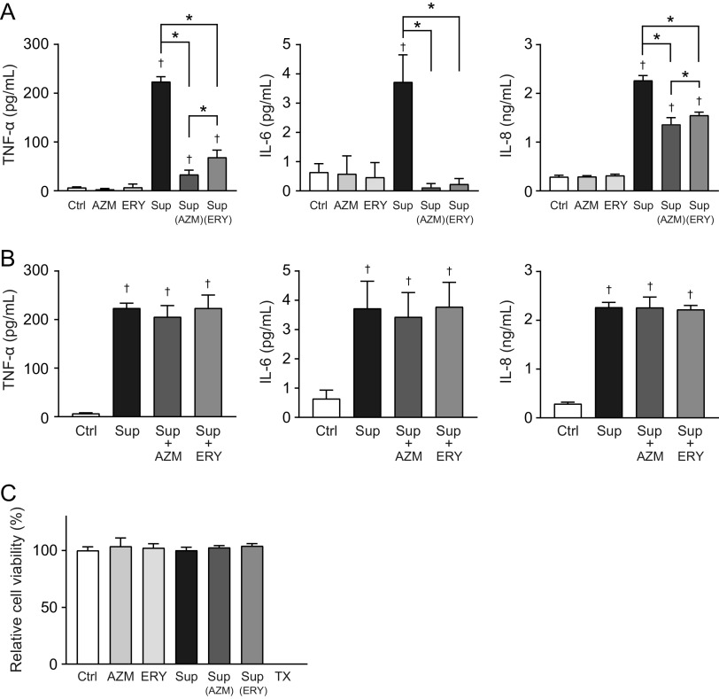
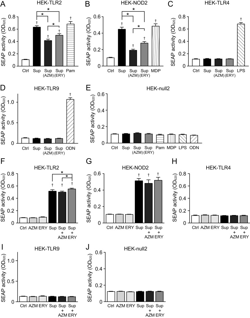
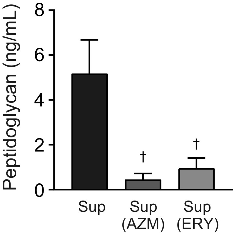
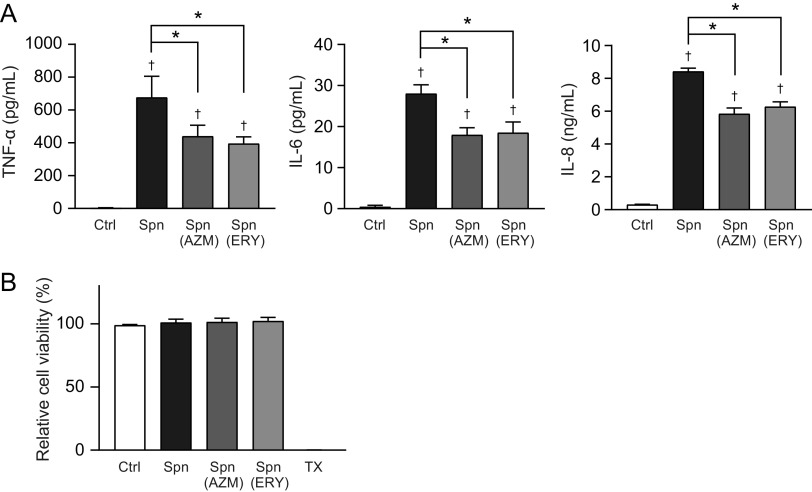
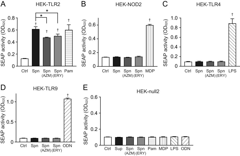
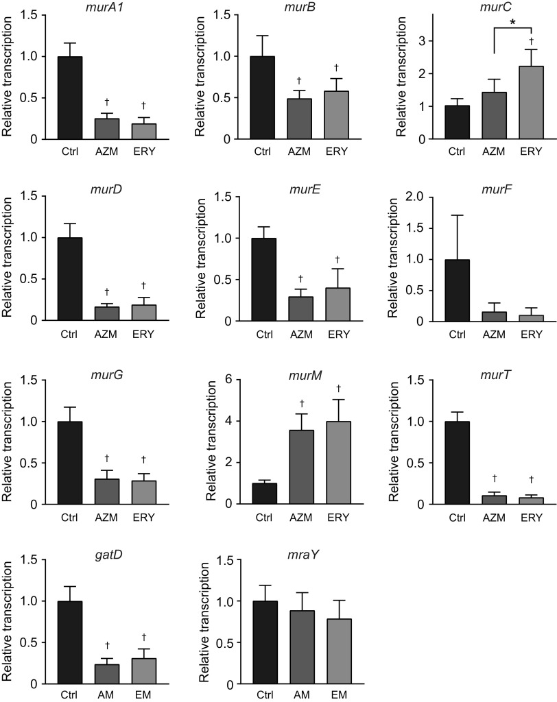
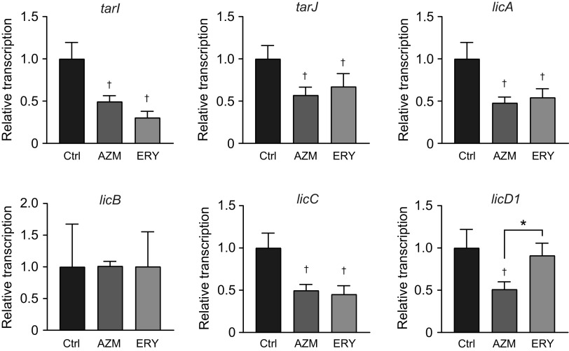
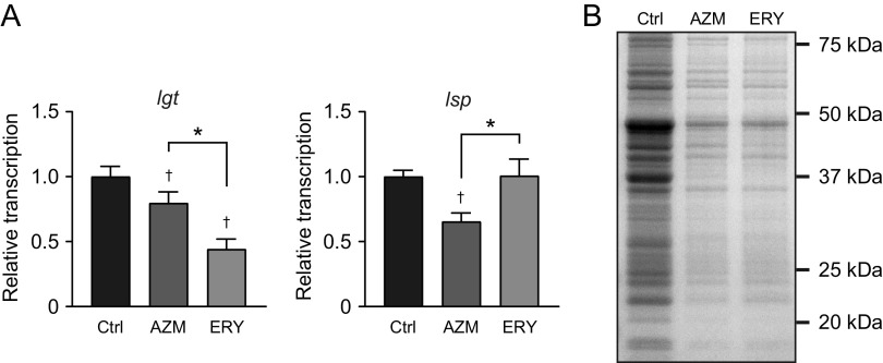
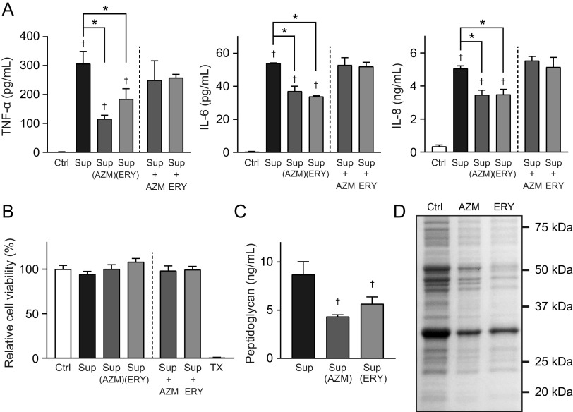
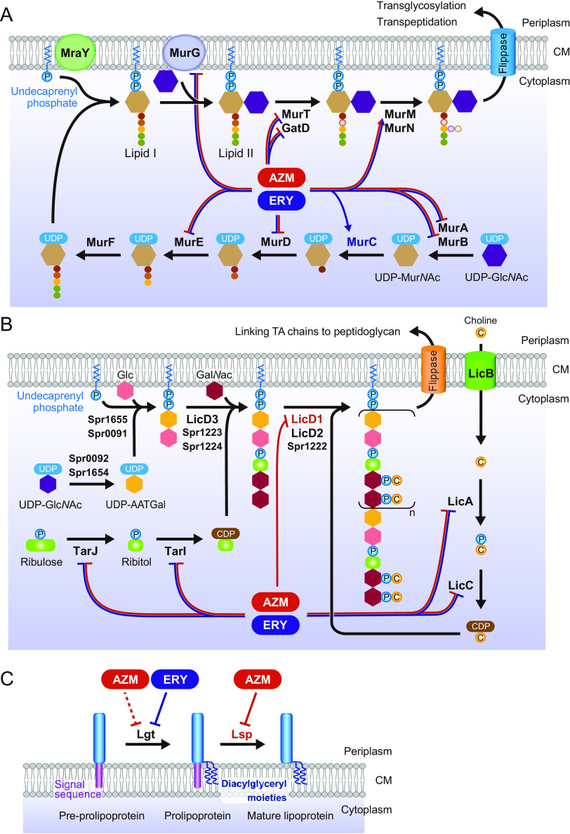
Over the past 2 decades, the prevalence of macrolide-resistant Streptococcus pneumoniae (MRSP) has increased considerably, due to widespread macrolide use. Although macrolide usage has been proposed to be associated with treatment failure in patients with pneumococcal diseases, macrolides may be clinically effective for treating these diseases, regardless of the susceptibility of the causative pneumococci to macrolides. As we previously demonstrated that macrolides downregulate the transcription of various genes in MRSP, including the gene encoding the pore-forming toxin pneumolysin, we hypothesized that macrolides affect the proinflammatory activity of MRSP. Using HEK-Blue cell lines, we found that the supernatants from macrolide-treated MRSP cultures induced decreased NF-κB activation in cells expressing Toll-like receptor 2 and nucleotide-binding oligomerization domain 2 compared to the supernatants from untreated MRSP cells, suggesting that macrolides inhibit the release of these ligands from MRSP. Real-time PCR analysis revealed that macrolides significantly downregulated the transcription of various genes encoding peptidoglycan synthesis-, lipoteichoic acid synthesis-, and lipoprotein synthesis-related molecules in MRSP cells. The silkworm larva plasma assay demonstrated that the peptidoglycan concentrations in the supernatants from macrolide-treated MRSP cultures were significantly lower than those from untreated MRSP cultures. Triton X-114 phase separation revealed that lipoprotein expression decreased in macrolide-treated MRSP cells compared to the lipoprotein expression in untreated MRSP cells. Consequently, macrolides may decrease the expression of bacterial ligands of innate immune receptors, resulting in the decreased proinflammatory activity of MRSP. IMPORTANCE To date, the clinical efficacy of macrolides in pneumococcal disease is assumed to be linked to their ability to inhibit the release of pneumolysin. However, our previous study demonstrated that oral administration of macrolides to mice intratracheally infected with macrolide-resistant Streptococcus pneumoniae resulted in decreased levels of pneumolysin and proinflammatory cytokines in bronchoalveolar lavage fluid samples compared to the levels in samples from untreated infected control mice, without affecting the bacterial load in the fluid. This finding suggests that additional mechanisms by which macrolides negatively regulate proinflammatory cytokine production may be involved in their efficacy in vivo. Furthermore, in this study, we demonstrated that macrolides downregulated the transcription of various proinflammatory-component-related genes in S. pneumoniae, which provides an additional explanation for the clinical benefits of macrolides.

Keywords: inflammation; lipoproteins; lipoteichoic acid; macrolide-resistant Streptococcus pneumoniae; macrolides; peptidoglycan.


Conflict of interest statement

The authors declare no conflict of interest.

Figures

FIG 1
FIG 1
Culture supernatants from macrolide-treated MRSP cells induce lesser inflammatory responses in THP-1-derived macrophages. MRSP strain NU4471 was inoculated into RPMI 1640 medium supplemented with 10% fetal bovine serum and cultured in the presence or absence of 5 μg/mL azithromycin (AZM) or erythromycin (ERY) until the cells reached the stationary phase. (A) THP-1-derived macrophages were exposed to 2-fold-diluted culture supernatant (Sup) from untreated or macrolide-treated MRSP cells or RPMI 1640 medium containing 2.5 μg/mL macrolides (AZM or ERY) for 9 h. TNF-α, IL-6, and IL-8 levels in the macrophage culture supernatants were determined using ELISA. (B) THP-1-derived macrophages were exposed to supernatant from untreated MRSP cells in the presence or absence of AZM or ERY for 9 h. TNF-α, IL-6, and IL-8 levels in the macrophage culture supernatants were determined using ELISA. (C) The MTT assay was performed to quantify the viability of THP-1-derived macrophages. Data represent the mean values ± standard deviations (SD) from quadruplicate experiments and were evaluated using one-way analysis of variance with Tukey’s multiple-comparison test. †, significant difference compared with the control at P < 0.05; *, significant difference between the indicated groups at P < 0.05. Ctrl, control; MRSP, macrolide-resistant S. pneumoniae; MTT, thiazolyl blue tetrazolium bromide; TX, Triton X-100.
FIG 2
FIG 2
Culture supernatants from macrolide-treated MRSP cells induce less TLR2 and NOD2 activation. MRSP strain NU4471 was grown in the presence or absence of 5 μg/mL azithromycin (AZM) or erythromycin (ERY) until it reached the stationary phase. (A to E) HEK-Blue hTLR-2 cells (A), HEK-Blue hNOD2 cells (B), HEK-Blue hTLR4 cells (C), HEK-Blue hTLR9 cells (D), and HEK-Blue null2 negative-control cells (E) were incubated with the culture supernatant (Sup) from untreated or macrolide-treated MRSP cells for 12 to 20 h (final concentrations of macrolides in macrolide-treated groups, 0.5 μg/mL). (F to J) HEK-Blue hTLR-2 cells (F), HEK-Blue hNOD2 cells (G), HEK-Blue hTLR4 cells (H), HEK-Blue hTLR9 cells (I), and HEK-Blue null2 negative-control cells (J) were exposed to supernatant from untreated MRSP cells in the presence or absence of 0.5 μg/mL AZM or ERY for 12 h. As controls, cells were also exposed to RPMI 1640 medium containing 0.5 μg/mL AZM or ERY. (A to J) Secreted alkaline phosphatase (SEAP) levels were quantified using spectrophotometry at 620 nm. Data represent the mean values ± SD from quintuplicate experiments and were evaluated using one-way analysis of variance with Tukey’s multiple-comparison test. †, significant difference compared with the control at P < 0.05; *, significant difference between the indicated groups at P < 0.05. Ctrl, control; LPS, lipopolysaccharide; MDP, muramyl dipeptide; MRSP, macrolide-resistant S. pneumoniae; ODN, CpG ODN; Pam, Pam3CSK4.
FIG 3
FIG 3
Macrolide treatment significantly decreases the peptidoglycan concentration in MRSP culture supernatant. MRSP strain NU4471 was grown in the presence or absence of 5 μg/mL azithromycin (AZM) or erythromycin (ERY) until it reached the stationary phase. The peptidoglycan concentrations in the pneumococcal culture supernatants (Sup) were determined. Data represent the mean values ± SD from quadruplicate experiments and were evaluated using one-way analysis of variance with Tukey’s multiple-comparison test. †, significant difference compared with the control at P < 0.05. MRSP, macrolide-resistant S. pneumoniae.
FIG 4
FIG 4
Treatment of MRSP cells with macrolides induces lesser inflammatory responses in THP-derived macrophages. MRSP strain NU4471 was grown in the presence or absence of 5 μg/mL azithromycin (AZM) or erythromycin (ERY) until it reached the stationary phase. (A) THP-1-derived macrophages were stimulated with ethanol-killed S. pneumoniae cells (Spn) for 9 h. TNF-α, IL-6, and IL-8 levels in macrophage culture supernatants were determined using ELISA. (B) The MTT assay was performed to quantify the viability of THP-1-derived macrophages. Data represent the mean values ± SD from quintuplicate experiments and were evaluated using one-way analysis of variance with Tukey’s multiple-comparison test. †, significant difference compared with the control at P < 0.05; *, significant difference between the indicated groups at P < 0.05. Ctrl, control; MRSP, macrolide-resistant S. pneumoniae; MTT, thiazolyl blue tetrazolium bromide; TX, Triton X-100.
FIG 5
FIG 5
Treatment of bacterial cells with macrolides induces less TLR2 activation. MRSP strain NU4471 was grown in the presence or absence of 5 μg/mL azithromycin (AZM) or erythromycin (ERY) until it reached the stationary phase. Pneumococcal cells were killed by treatment with 70% ethanol. (A) HEK-Blue hTLR-2 cells were stimulated with ethanol-killed S. pneumoniae (Spn) for 12 h. (B) HEK-Blue hNOD2 cells were stimulated with Spn for 18 h. (C) HEK-Blue hTLR4 cells were stimulated with Spn for 20 h. (D) HEK-Blue hTLR9 cells were stimulated with Spn for 20 h. (E) HEK-Blue null2 negative-control cells were stimulated with Spn for 17 h. (A to E) Secreted alkaline phosphatase (SEAP) levels were quantified using spectrophotometry at 620 nm. Data represent the mean values ± SD from quintuplicate experiments and were evaluated using one-way analysis of variance with Tukey’s multiple-comparison test. †, significant difference compared with the control at P < 0.05; *, significant difference between the indicated groups at P < 0.05. Ctrl, control; LPS, lipopolysaccharide; MRSP, macrolide-resistant S. pneumoniae; MDP, muramyl dipeptide; ODN, CpG ODN; Pam, Pam3CSK4.
FIG 6
FIG 6
Treatment of MRSP cells with macrolides affects the balance of peptidoglycan synthesis-related gene transcription. MRSP strain NU4471 was cultured in the presence or absence of 5 μg/mL azithromycin (AZM) or erythromycin (ERY) for 2 h. Real-time PCR was performed to quantify the transcription of genes encoding peptidoglycan synthesis-related molecules in MRSP cells. The relative quantities of target mRNAs were normalized to the relative quantity of 16S rRNA mRNA. Data represent the mean values ± SD from quadruplicate experiments and were evaluated using one-way analysis of variance with Tukey’s multiple-comparison test. †, significant difference compared with the control at P < 0.05; *, significant difference between the indicated groups at P < 0.05. Ctrl, control; MRSP, macrolide-resistant S. pneumoniae.
FIG 7
FIG 7
Treatment of MRSP cells with macrolides decreased the transcription of the genes encoding lipoteichoic acid synthesis-related molecules. MRSP strain NU4471 was cultured in the presence or absence of 5 μg/mL azithromycin (AZM) or erythromycin (ERY) for 2 h. Real-time PCR was performed to quantify the transcription of the genes encoding lipoteichoic acid synthesis-related molecules in MRSP cells. The relative quantities of target mRNAs were normalized to the relative quantity of 16S rRNA mRNA. Data represent the mean values ± SD from quadruplicate experiments and were evaluated using one-way analysis of variance with Tukey’s multiple-comparison test. †, significant difference compared with the control at P < 0.05; *, significant difference between the indicated groups at P < 0.05. Ctrl, control; MRSP, macrolide-resistant S. pneumoniae.
FIG 8
FIG 8
Treatment of MRSP cells with macrolides downregulated the transcription of the genes encoding lipoprotein synthesis-related enzymes and decreased lipoprotein expressions in MRSP cells. (A) MRSP strain NU4471 was cultured in the presence or absence of 5 μg/mL azithromycin (AZM) or erythromycin (ERY) for 2 h. Real-time PCR was performed to quantify the transcription of the genes encoding lipoprotein synthesis-related enzymes in MRSP cells. The relative quantities of target mRNAs were normalized to the relative quantity of 16S rRNA mRNA. Data represent the mean values ± SD from quadruplicate experiments and were evaluated using one-way analysis of variance with Tukey’s multiple-comparison test. †, significant difference compared with the control at P < 0.05; *, significant difference between the indicated groups at P < 0.05. (B) MRSP strain NU4471 was grown in the presence or absence of 5 μg/mL AZM or ERY until it reached the stationary phase. Coomassie blue-stained SDS-PAGE of Triton X-114 extracts prepared from these MRSP cells is shown. Ctrl, control; MRSP, macrolide-resistant S. pneumoniae.
FIG 9
FIG 9
Transition electron microscopy (TEM) analysis of macrolide-treated S. pneumoniae cells. MRSP strain NU4471 was grown in the presence or absence of 5 μg/mL azithromycin (AZM) or erythromycin (ERY) until it reached the stationary phase. Representative TEM images of untreated and macrolide-treated MRSP cells are shown. Scale bars = 500 nm.
FIG 10
FIG 10
Exposure of other clinical MRSP isolates to macrolides results in decreased proinflammatory activity of the culture supernatants. MRSP strain KM2412 was inoculated into RPMI 1640 medium supplemented with 10% fetal bovine serum and cultured in the presence or absence of 4 μg/mL azithromycin (AZM) or erythromycin (ERY) until it reached the stationary phase. (A) THP-1-derived macrophages were exposed to the culture supernatant (Sup) from untreated or macrolide-treated MRSP cells for 9 h. TNF-α, IL-6, and IL-8 levels in the macrophage culture supernatants were determined using ELISA. (B) The MTT assay was performed to quantify the viability of THP-1-derived macrophages. (C) Peptidoglycan concentrations in pneumococcal supernatants were determined. (A to C) Data represent the mean values ± SD from quadruplicate experiments and were evaluated using one-way analysis of variance with Tukey’s multiple-comparison test. †, significant difference compared with the control at P < 0.05; *, significant difference between the indicated groups at P < 0.05. (D) Coomassie blue-stained SDS-PAGE gel of the Triton X-114 extract prepared from MRSP cells. Ctrl, control; MRSP, macrolide-resistant S. pneumoniae; MTT, thiazolyl blue tetrazolium bromide; TX, Triton X-100.
FIG 11
FIG 11
Scheme of macrolide-induced alterations in the transcription of pneumococcal genes encoding cell wall synthesis-related molecules. (A) Transcriptional alterations in peptidoglycan synthesis-related gene transcription in macrolide-treated MRSP cells. (B) Transcriptional alterations in the proposed lipoteichoic acid synthesis-related gene transcription in macrolide-treated MRSP cells. Although the pathway applies to S. pneumoniae generally, the gene/protein names are from S. pneumoniae strain R6. (C) Transcriptional alterations in lipoprotein synthesis-related gene transcription in MRSP cells treated with macrolides. AATGal, 2-acetamido-4-amino-2,4,6-trideoxygalactose; AZM, azithromycin; CM, cytoplasmic membrane; ERY, erythromycin; GalNAc, N-acetylgalactosamine; Glc, glucose; GlcNAc, N-acetylglucosamine; MRSP, macrolide-resistant S. pneumoniae; MurNAc, N-acetylmuramic acid; P, phosphate residue; TA, teichoic acid.

Similar articles

References

    1. Garau J. 2002. Treatment of drug-resistant pneumococcal pneumonia. Lancet Infect Dis 2:404–415. doi:10.1016/s1473-3099(02)00316-x. - DOI - PubMed
    1. Asbell PA, Sanfilippo CM, Sahm DF, DeCory HH. 2020. Trends in antibiotic resistance among ocular microorganisms in the United States from 2009 to 2018. JAMA Ophthalmol 138:439–450. doi:10.1001/jamaophthalmol.2020.0155. - DOI - PMC - PubMed
    1. Nagai K, Kimura O, Domon H, Maekawa T, Yonezawa D, Terao Y. 2019. Antimicrobial susceptibility of Streptococcus pneumoniae, Haemophilus influenzae, and Moraxella catarrhalis clinical isolates from children with acute otitis media in Japan from 2014 to 2017. J Infect Chemother 25:229–232. doi:10.1016/j.jiac.2018.08.018. - DOI - PubMed
    1. Schroeder MR, Stephens DS. 2016. Macrolide resistance in Streptococcus pneumoniae. Front Cell Infect Microbiol 6:98. doi:10.3389/fcimb.2016.00098. - DOI - PMC - PubMed
    1. Rzeszutek M, Wierzbowski A, Hoban DJ, Conly J, Bishai W, Zhanel GG. 2004. A review of clinical failures associated with macrolide-resistant Streptococcus pneumoniae. Int J Antimicrob Agents 24:95–104. doi:10.1016/j.ijantimicag.2004.03.008. - DOI - PubMed

Publication types