

Identification and Analysis of Monoclonal Antibodies with Neutralizing Activity against Diverse SARS-CoV-2 Variants
- PMID: 37191569
- PMCID: PMC10308935
- DOI: 10.1128/jvi.00286-23
Identification and Analysis of Monoclonal Antibodies with Neutralizing Activity against Diverse SARS-CoV-2 Variants
Abstract
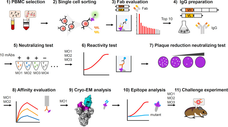
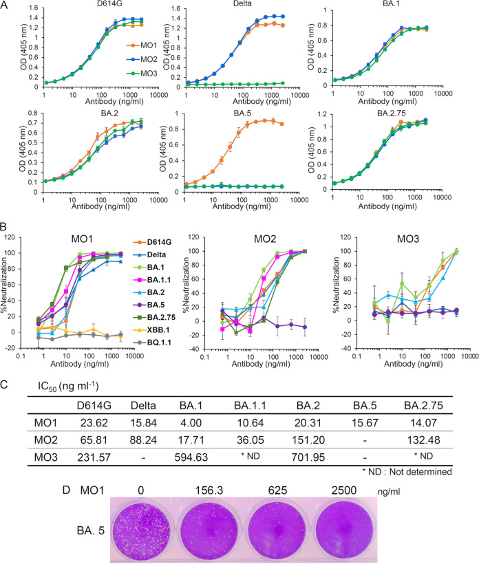
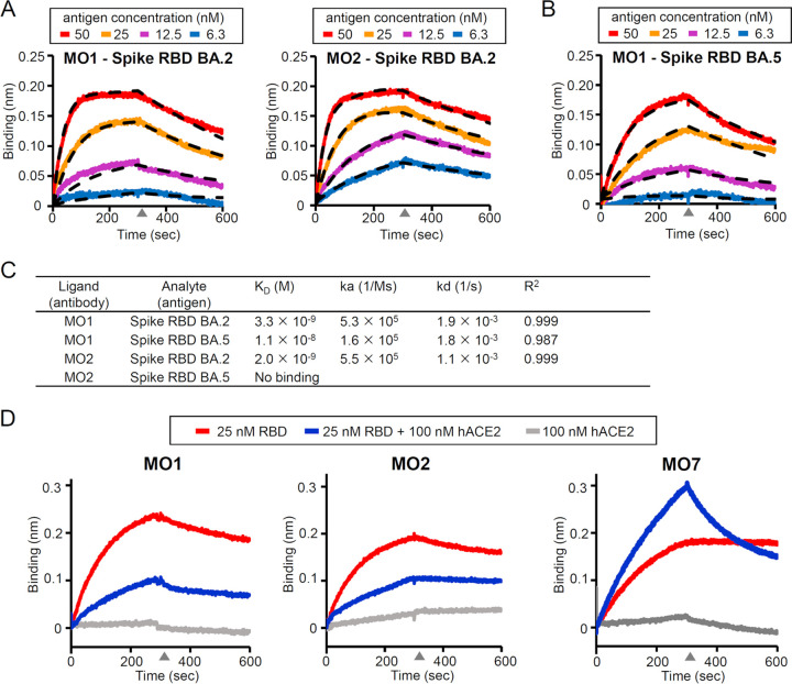
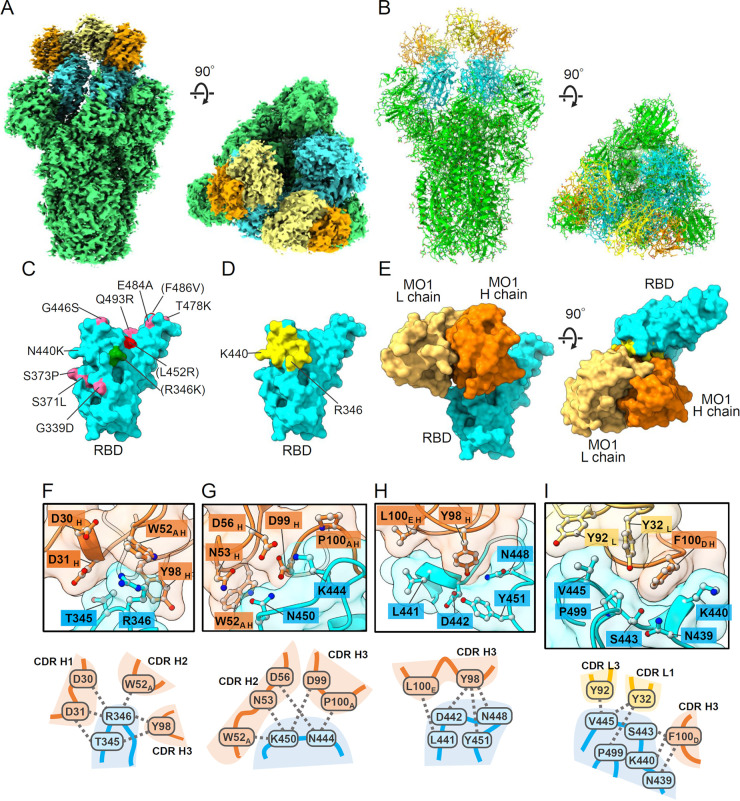
We identified neutralizing monoclonal antibodies against severe acute respiratory syndrome-coronavirus 2 (SARS-CoV-2) variants (including Omicron variants BA.5 and BA.2.75) from individuals who received two doses of mRNA vaccination after they had been infected with the D614G virus. We named them MO1, MO2, and MO3. Among them, MO1 showed particularly high neutralizing activity against authentic variants: D614G, Delta, BA.1, BA.1.1, BA.2, BA.2.75, and BA.5. Furthermore, MO1 suppressed BA.5 infection in hamsters. A structural analysis revealed that MO1 binds to the conserved epitope of seven variants, including Omicron variants BA.5 and BA.2.75, in the receptor-binding domain of the spike protein. MO1 targets an epitope conserved among Omicron variants BA.1, BA.2, and BA.5 in a unique binding mode. Our findings confirm that D614G-derived vaccination can induce neutralizing antibodies that recognize the epitopes conserved among the SARS-CoV-2 variants. IMPORTANCE Omicron variants of SARS-CoV-2 acquired escape ability from host immunity and authorized antibody therapeutics and thereby have been spreading worldwide. We reported that patients infected with an early SARS-CoV-2 variant, D614G, and who received subsequent two-dose mRNA vaccination have high neutralizing antibody titer against Omicron lineages. It was speculated that the patients have neutralizing antibodies broadly effective against SARS-CoV-2 variants by targeting common epitopes. Here, we explored human monoclonal antibodies from B cells of the patients. One of the monoclonal antibodies, named MO1, showed high potency against broad SARS-CoV-2 variants including BA.2.75 and BA.5 variants. The results prove that monoclonal antibodies that have common neutralizing epitopes among several Omicrons were produced in patients infected with D614G and who received mRNA vaccination.
Keywords: Omicron variants; broad neutralizing activity; common epitope; cryoelectron microscopy; human monoclonal antibody; receptor-binding domain; severe acute respiratory syndrome-coronavirus 2 (SARS-CoV-2); spike; vaccine.
Conflict of interest statement
The authors declare a conflict of interest. S.O. is employed by BIKEN foundation. The other authors declare no conflicts of interest related to this research.
Figures

Similar articles
-
Epitopes of an antibody that neutralizes a wide range of SARS-CoV-2 variants in a conserved subdomain 1 of the spike protein.J Virol. 2024 May 14;98(5):e0041624. doi: 10.1128/jvi.00416-24. Epub 2024 Apr 16. J Virol. 2024. PMID: 38624232 Free PMC article.
-
A human monoclonal antibody neutralizing SARS-CoV-2 Omicron variants containing the L452R mutation.J Virol. 2024 Dec 17;98(12):e0122324. doi: 10.1128/jvi.01223-24. Epub 2024 Nov 4. J Virol. 2024. PMID: 39494911 Free PMC article.
-
BA.2.12.1, BA.4 and BA.5 escape antibodies elicited by Omicron infection.Nature. 2022 Aug;608(7923):593-602. doi: 10.1038/s41586-022-04980-y. Epub 2022 Jun 17. Nature. 2022. PMID: 35714668 Free PMC article.
-
Susceptibility of SARS-CoV-2 Omicron Variants to Therapeutic Monoclonal Antibodies: Systematic Review and Meta-analysis.Microbiol Spectr. 2022 Aug 31;10(4):e0092622. doi: 10.1128/spectrum.00926-22. Epub 2022 Jun 14. Microbiol Spectr. 2022. PMID: 35700134 Free PMC article.
-
Comprehensive Overview of Broadly Neutralizing Antibodies against SARS-CoV-2 Variants.Viruses. 2024 Jun 1;16(6):900. doi: 10.3390/v16060900. Viruses. 2024. PMID: 38932192 Free PMC article. Review.
Cited by
-
SARS-CoV-2 BA.2.86 is susceptible to the neutralizing antibody MO11 targeting subdomain 1 despite the E554K mutation near the epitope.J Virol. 2025 Feb 25;99(2):e0138924. doi: 10.1128/jvi.01389-24. Epub 2025 Jan 22. J Virol. 2025. PMID: 39840983 Free PMC article. No abstract available.
-
Epitopes of an antibody that neutralizes a wide range of SARS-CoV-2 variants in a conserved subdomain 1 of the spike protein.J Virol. 2024 May 14;98(5):e0041624. doi: 10.1128/jvi.00416-24. Epub 2024 Apr 16. J Virol. 2024. PMID: 38624232 Free PMC article.
-
Rapid isolation of pan-neutralizing antibodies against Omicron variants from convalescent individuals infected with SARS-CoV-2.Front Immunol. 2024 Mar 6;15:1374913. doi: 10.3389/fimmu.2024.1374913. eCollection 2024. Front Immunol. 2024. PMID: 38510237 Free PMC article.
-
Overcoming antibody-resistant SARS-CoV-2 variants with bispecific antibodies constructed using non-neutralizing antibodies.iScience. 2024 Feb 29;27(4):109363. doi: 10.1016/j.isci.2024.109363. eCollection 2024 Apr 19. iScience. 2024. PMID: 38500835 Free PMC article.
References
-
- Dejnirattisai W, Huo J, Zhou D, Zahradník J, Supasa P, Liu C, Duyvesteyn HME, Ginn HM, Mentzer AJ, Tuekprakhon A, Nutalai R, Wang B, Dijokaite A, Khan S, Avinoam O, Bahar M, Skelly D, Adele S, Johnson SA, Amini A, Ritter TG, Mason C, Dold C, Pan D, Assadi S, Bellass A, Omo-Dare N, Koeckerling D, Flaxman A, Jenkin D, Aley PK, Voysey M, Costa Clemens SA, Naveca FG, Nascimento V, Nascimento F, Fernandes da Costa C, Resende PC, Pauvolid-Correa A, Siqueira MM, Baillie V, Serafin N, Kwatra G, Da Silva K, Madhi SA, Nunes MC, Malik T, Openshaw PJM, Baillie JK, Semple MG, ISARIC4C Consortium ., et al. 2022. SARS-CoV-2 Omicron-B.1.1.529 leads to widespread escape from neutralizing antibody responses. Cell 185:467–484.e15. doi: 10.1016/j.cell.2021.12.046. - DOI - PMC - PubMed
-
- Tegally H, Moir M, Everatt J, Giovanetti M, Scheepers C, Wilkinson E, Subramoney K, Moyo S, Amoako DG, Baxter C, Althaus CL, Anyaneji UJ, Kekana D, Viana R, Giandhari J, Lessells RJ, Maponga T, Maruapula D, Choga W, Matshaba M, Mayaphi S, Mbhele N, Mbulawa MB, Msomi N, Naidoo Y, Pillay S, Sanko TJ, San JE, Scott L, Singh L, Magini NA, Smith-Lawrence P, Stevens W, Dor G, Tshiabuila D, Wolter N, Preiser W, Treurnicht FK, Venter M, Davids M, Chiloane G, Mendes A, McIntyre C, O’Toole A, Ruis C, Peacock TP, Roemer C, Williamson C, Pybus OG, Bhiman J, et al. 2022. Continued emergence and evolution of Omicron in South Africa: new BA.4 and BA.5 lineages. medRxiv. doi: 10.1101/2022.05.01.22274406. - DOI
-
- Muik A, Lui BG, Wallisch A-K, Bacher M, Mühl J, Reinholz J, Ozhelvaci O, Beckmann N, Güimil Garcia RdlC, Poran A, Shpyro S, Finlayson A, Cai H, Yang Q, Swanson KA, Türeci Ö, Şahin U. 2022. Neutralization of SARS-CoV-2 Omicron by BNT162b2 mRNA vaccine-elicited human sera. Science 375:678–680. doi: 10.1126/science.abn7591. - DOI - PMC - PubMed
Publication types
MeSH terms
Substances
Supplementary concepts
Grants and funding
LinkOut - more resources
Full Text Sources
Medical
Molecular Biology Databases
Miscellaneous
