

Radiologic-pathologic correlation of prostatic cancer extracapsular extension (ECE)
- PMID: 37191739
- PMCID: PMC10188796
- DOI: 10.1186/s13244-023-01428-3
Radiologic-pathologic correlation of prostatic cancer extracapsular extension (ECE)
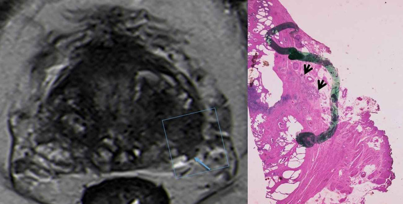
Abstract
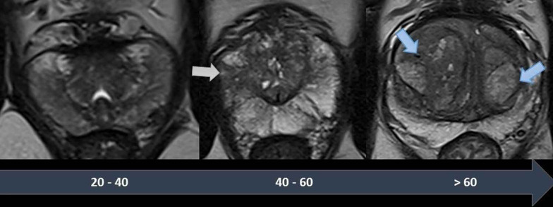
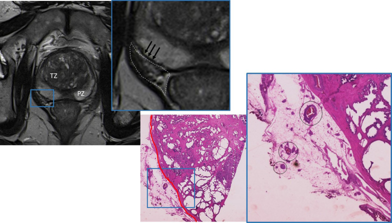
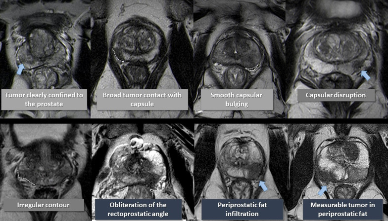
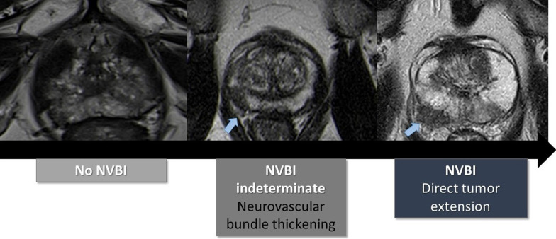
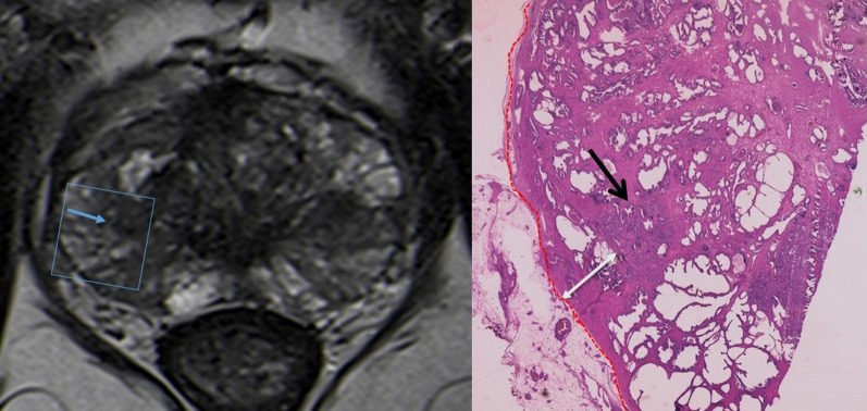
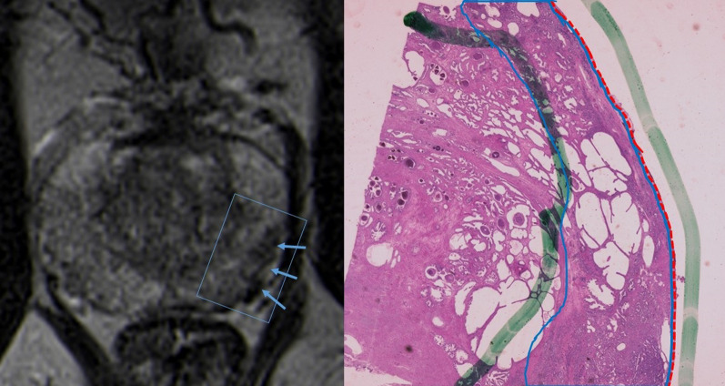
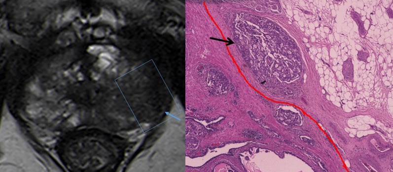
Recent advancements on nerve-sparing robotic prostatectomy allow fewer side effects such as urinary incontinence and sexual dysfunction. To perform such techniques, it is essential for the surgeon to know if the neurovascular bundle is involved. Despite being the gold-standard imaging method for Prostate Cancer (PCa) staging, Magnetic Resonance Imaging (MRI) lacks high specificity for detecting extracapsular extension (ECE). Therefore, it is essential to understand the pathologic aspects of ECE to better evaluate the MRI findings of PCa. We reviewed the normal MRI appearance of the prostate gland and the periprostatic space and correlated them to prostatectomy specimens. The different findings of ECE and neurovascular bundle invasion are exemplified with images of both MRI and histologic specimens.
Keywords: Extracapsular extension; Magnetic resonance imaging; Prostate; Prostatic cancer; Radiologic-pathologic correlation.
© 2023. The Author(s).
Conflict of interest statement
The authors declare that they have no competing interests.
Figures


References
Publication types
LinkOut - more resources
Full Text Sources
