Review
. 2023 Jun:246:154497.
doi: 10.1016/j.prp.2023.154497. Epub 2023 May 3.

COVID-19, post-acute COVID-19 syndrome (PACS, "long COVID") and post-COVID-19 vaccination syndrome (PCVS, "post-COVIDvac-syndrome"): Similarities and differences

Affiliations
Review

COVID-19, post-acute COVID-19 syndrome (PACS, "long COVID") and post-COVID-19 vaccination syndrome (PCVS, "post-COVIDvac-syndrome"): Similarities and differences

Felix Scholkmann et al. Pathol Res Pract. 2023 Jun.

Abstract

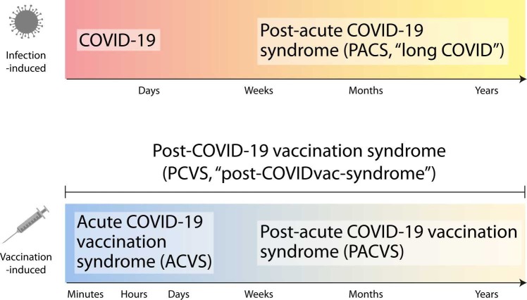
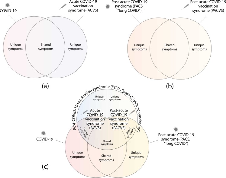
Worldwide there have been over 760 million confirmed coronavirus disease 2019 (COVID-19) cases, and over 13 billion COVID-19 vaccine doses have been administered as of April 2023, according to the World Health Organization. An infection with severe acute respiratory syndrome coronavirus 2 (SARS-CoV-2) can lead to an acute disease, i.e. COVID-19, but also to a post-acute COVID-19 syndrome (PACS, "long COVID"). Currently, the side effects of COVID-19 vaccines are increasingly being noted and studied. Here, we summarise the currently available indications and discuss our conclusions that (i) these side effects have specific similarities and differences to acute COVID-19 and PACS, that (ii) a new term should be used to refer to these side effects (post-COVID-19 vaccination syndrome, PCVS, colloquially "post-COVIDvac-syndrome"), and that (iii) there is a need to distinguish between acute COVID-19 vaccination syndrome (ACVS) and post-acute COVID-19 vaccination syndrome (PACVS) - in analogy to acute COVID-19 and PACS ("long COVID"). Moreover, we address mixed forms of disease caused by natural SARS-CoV-2 infection and COVID-19 vaccination. We explain why it is important for medical diagnosis, care and research to use the new terms (PCVS, ACVS and PACVS) in order to avoid confusion and misinterpretation of the underlying causes of disease and to enable optimal medical therapy. We do not recommend to use the term "Post-Vac-Syndrome" as it is imprecise. The article also serves to address the current problem of "medical gaslighting" in relation to PACS and PCVS by raising awareness among the medical professionals and supplying appropriate terminology for disease.

Keywords: ACVS; Acute COVID-19 vaccination syndrome; COVID-19; Long COVID; PACS; PACVS; PCVS; Post-COVID-19 vaccination syndrome; Post-acute COVID-19 syndrome; Post-acute COVID-19 vaccination syndrome; SARS-CoV-2.


Conflict of interest statement

Declaration of Competing Interest The authors declare that they have no known competing financial interests or personal relationships that could have appeared to influence the work reported in this paper.

Figures

Fig. 1
Fig. 1
Definition of the terminology of syndromes with respect to the causative factor (infection/vaccination) und their general temporal manifestation. The colour gradient shows that it is a spectrum where the initial syndrome can change to the following syndrome.
Fig. 2
Fig. 2
Visualisation of the terminology in the form of Venn diagrams based on overlapping symptoms, in terms of (a) COVID-19 and ACVS, (b) PACS and PACVS, and (c) COVID-19, PCVS and PACS.
Fig. 3
Fig. 3
Latency between COVID-19 disease onset or COVID-19 vaccination and associated death. (a) Distribution of time intervals of COVID-19 symptom onset to death (n = 3478, range: 1–97 days) based on data from South Korea (19 January 2020–10 January 2022, covering the phase of the pandemic where the wild-type (Wuhan-Hu-1), alpha, delta and omicron (BA.1) variants were present) . A double-exponential function is fitted to the data (r2 = 0.9597). (b) Distribution of time intervals between SARS-CoV-2 infection to death (n = 63,855) as a function of sex, age and four time periods during the pandemic based on data from the United Kingdom (1 January 2020–20 January 2021, covering the phase of the pandemic where the wild-type (Wuhan-Hu-1) and alpha variants were present) . (c) Distribution of time intervals between COVID-19 vaccination and associated deaths (n = 33,904) according to data from the US Vaccine Adverse Event Reporting System (VAERS) (based on 1509,410 reports through January 20, 2023). The distribution follows a double-exponential decay function (red) (r2 = 0.9819). However, it should be noted that there is very likely a reporting bias, i.e. the probability of reporting deaths after vaccination is higher the closer the death occurred to the time of vaccination. Therefore, it must be assumed that the exponential decline in reality is slower than the data shows.
Fig. 4
Fig. 4
(a) Amyloid fibrin microclots and (b) hyperactivated platelets in in the blood plasma (platelet poor plasma) in COVID-19 and PACS, compared to healthy controls. Aggregated platelets are indicated by white arrows. Amyloid fibrin microclots are visualized with Thioflavin T (green fluorescence) and platelets with PAC-1 (green fluorescence) and CD62P-PE (purple fluorescence).
Fig. 5
Fig. 5
Circulating SARS-CoV-2 proteins and mRNA in COVID-19 and PACS. (a) Spike protein concentration in blood plasma in individuals with and without PACS. (b) Time-dependent spike and nucleocapsid protein concentration in blood plasma in individuals with PACS and COVID-19. (c) Spike protein concentration in non-classical monocytes in healthy controls and individuals with severe COVID-19 and PACS. (d) Presence of SARS-CoV-2 mRNA in individuals with COVID-19 as a function of the maximum clinical COVID-19 severity. (e) Presence of SARS-CoV-2 mRNA in individuals with PACS as a function of the PACS severity. (f) Presence of SARS-CoV-2 mRNA (obtained with droplet digital-PCR (ddPRC), spike protein and extracellular vesicles (EV) with (with spike protein) in the blood plasma of individuals with PACS. A representative transmission electron microscopy (TEM) micrograph shows EVs (50.000 × magnification). The bar plot depicting the differences in EVs in controls and PACS refers to small EVs.
Fig. 6
Fig. 6
Circulating SARS-CoV-2 proteins and mRNA after COVID-19 vaccination and in PCVS. (a) SARS-CoV-2 spike mRNA in blood plasma as a function of type of vaccine and time after vaccination. Shown is the mapping of trimmed and filtered reads to the coding regions of the specific SARS-CoV-2 spike protein from the two COVID-19 vaccines. (b) Western blot showing the detection of SARS-CoV-2 spike protein S2 subunit in exosomes from blood plasma at 14 days after the first dose, 14 days after the second dose and 4 months after the second dose of the COVID-19 vaccine. (c) Circulating mRNA in blood (plasma and white blood cells) at different time-points after BNT162b2 COVID-19 vaccination. Left shows the group average, right an example from a single individual. (d) SARS-CoV-2 spike and nucleocapsid protein concentration after COVID-19 vaccination. (e) Spike protein concentration in non-classical monocytes in the blood of COVID-19 vaccinated individuals with and without experiencing PCVS symptoms. (f) Free and total full-length SARS-CoV-2 spike protein concentrations in COVID-19 vaccinated individuals who developed myocarditis compared to healthy ones. Shown is also the concentration of free full-length and S1 subunit spike protein as a function of time after vaccination and for the two cohorts (myocarditis and healthy controls).
Fig. 7
Fig. 7
Definition of the terminology of second-order syndromes as a combination of infection- and vaccination-induced syndromes. PACS: post-acute COVID-19 syndrome. ACVS: acute COVID-19 vaccination syndrome, PACVS: post-acute COVID-19 vaccination syndrome.

Similar articles

Cited by

References

    1. WHO Pneumonia of unknown cause – China. 2020.
    1. Wu F., et al. A new coronavirus associated with human respiratory disease in China. Nature. 2020;579(7798):265–269. - PMC - PubMed
    1. Zsichla L., Müller V. Risk factors of severe COVID-19: a review of host, viral and environmental factors. Viruses. 2023;15(1):175. - PMC - PubMed
    1. Gao Y.D., et al. Risk factors for severe and critically ill COVID-19 patients: a review. Allergy. 2021;76(2):428–455. - PubMed
    1. Mercola J., Grant W.B., Wagner C.L. Evidence regarding vitamin D and risk of COVID-19 and its severity. Nutrients. 2020;12(11) - PMC - PubMed

Substances