Case Reports
. 2023 Apr;13(4):30-36.
doi: 10.13107/jocr.2023.v13.i04.3604.

Secondary Chondrosarcoma Arising in Synovial Chondromatosis of Wrist Joint

Affiliations
Case Reports

Secondary Chondrosarcoma Arising in Synovial Chondromatosis of Wrist Joint

Cleofina Furtado et al. J Orthop Case Rep. 2023 Apr.

Abstract

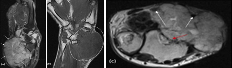
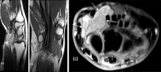
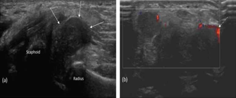
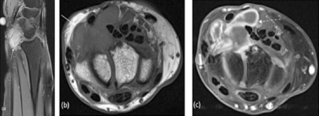
Introduction: Chondrosarcoma of the synovium is a rare and malignant form of cartilaginous tumor that originates in synovial tissue. There have only been a limited number of reported cases of malignant transformation of synovial chondromatosis (SC) into secondary chondrosarcoma (SCH), primarily in the hip and knee, in patients with resistant illness. The occurrence of chondrosarcoma in SC of the wrist is highly uncommon, as evidenced by only a single previous case study that has been documented in the literature.

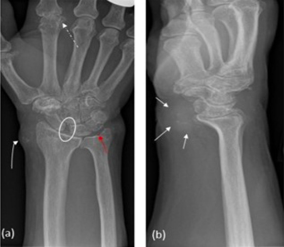
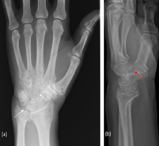
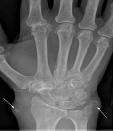
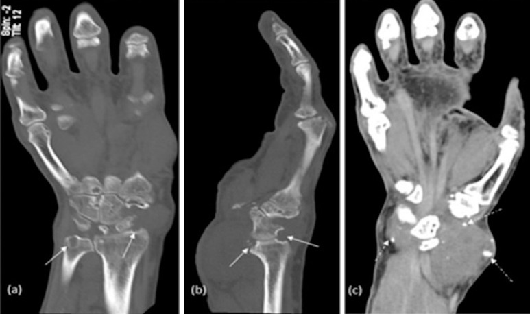
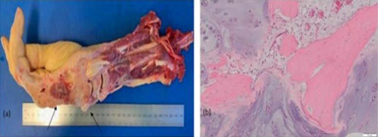
Case report: This study presents a case series of two patients with primary SC who developed SCH at the wrist joint.

Conclusion: Clinicians treating localized swellings of the hand and wrist should be alert to the likelihood of a sarcoma diagnosis to minimize delays to definitive therapy.

Keywords: Secondary chondrosarcoma; malignant transformation; primary synovial chrondromatosis; secondary synovial chondromatosis; synovitis.


Figures

Figure 1
Figure 1
Right hand frontal (a) and lateral (b) X-ray at initial presentation. Periartricular soft-tissue swelling with foci of internal mineralization (between white solid arrows and white curved arrow). Chondrocalcinosis in the region of the triangular fibrocartilage complex (Red arrow) and in the second metacarpophalangeal joint which shows severe arthropathy with erosions, subluxation and secondary osteoarthritis (OA) (white dashed arrow). Severe OA at the trapezioscaphoid joint and scapholunate diastasis (white circle).
Figure 2
Figure 2
LEFT hand frontal (a) and lateral (b) X-ray at initial presentation. Chondrocalcinosis is the triangular fibrocartilage complex (white solid arrow). Multiple carpal cysts are seen (White dashed arrow). There is severe osteoarthritis in the first CMC (red arrow) and Trapezioscaphoid joints and moderate degeneration in the distal radioulnar joint. No erosions at the metacarpophalangeal joint.
Figure 3
Figure 3
RIGHT hand frontal X-ray. Progressive soft-tissue swelling adjacent to the radial and ulnar styloid processes with some mineralization here and in then region of the triangular fibrocartilage (white arrows).
Figure 4
Figure 4
RIGHT wrist plain CT: Bone window Coronal (a), sagittal (b); soft-tissue window coronal (c). Periarticular soft-tissue swelling and periarticular calcification better visualized on soft tissue {white dashed arrow in (c)]. Large erosions with bone destruction at radiocarpal and ulnocarpal joint (solid white arrows).
Figure 5
Figure 5
Right wrist MRI: Sagittal T2 fat sat (a), Coronal T1 W1 (b), (c) Axial T2 W1. Large peri articular 5.6 * 4.5 *3.3 cm soft-tissue signal intensity mass-like lesion seen centred along the volar and eradial aspect of the wrist extednding into the distal forearm. This is hypointense on T1-weightened images and shows heterogeneous T2 hyperintensity (between white solid arrows). The lesion is seen to cause well-defined scalloping and bony erosions of the adjacent ventral aspect of the distal radius {red arrow in (c)}. Mass-effect and splaying of the adjacent flexor tendons (white dashed arrow) with displacement at the myotendinous junction.
Figure 6
Figure 6
Histopathological images: Macroscopic (a), microscopic (b). Right forearm amputation showing a tumour (5×5×6 cm) which is 16 cm from the proximal bone resection margin and 11 cm from the proximal soft tissue margin {between black arrows on (a)}. Tumor composed of nodules of hyaline cartilage with increased cellularity invading through trabecular bone with no evidence of dedifferentiation (b) consistent with Grade 2 secondary chondrosarcoma, on the background of primary synovial chondromatosis.
Figure 7
Figure 7
Right wrist at initial presentation: Sagittal T2 (a), Coronal T1 (b) Axial PD fat sat (c) Prominent soft-tissue mass on the volar surface of the wrist at the radio scaphoid articulation, which is T1 low, T2 isointense and bright on PD fat sat (white arrows).
Figure 8
Figure 8
Right wrist ultrasound: Longitudinal axis (a) transverse axis with power colour doppler (b) 2.8×1.2×1.8 cm (between white solid arrows) solid lobular lesion in the volar-radical aspect of the wrist, displacing flexor tendons. Minimal internal vascularity is evident {white dashed arrow in (b)}.
Figure 9
Figure 9
Right wrist MRI: Sagittal PD fat sat (a), Axial T1 (b) Axial T1 fat sat with contrast (c). A soft tissue deep-seated lesion with lobulated margins is seen within the wrist (white solid arrows), measuring about 1.6×2.6×2.8 cm in AP, transverse, and CC diameters. It is iso-intense to the muscles at T1, intermediate to low signal at T2 Wl and STIR WI and marginal enhancement on the post-contrast images {White dashed arrow in ©}.

References

    1. Leung L. Painful swollen wrist:A case study. Aust Fam Physician. 2013;42:301–3. - PubMed
    1. Tomasevich KM, Moradi S, Lindsay AD. Chondrosarcoma arising in synovial chondromatosis of the hip:A case report. JBJS Case Connect. 2021;11 - PubMed
    1. Wittkop B, Davies AM, Mangham DC. Primary synovial chondromatosis and synovial chondrosarcoma:A pictorial review. Eur Radiol. 2002;12:2112–9. - PubMed
    1. McKenzie G, Raby N, Ritchie D. A pictorial review of primary synovial osteochondromatosis. Eur Radiol. 2008;18:2662–9. - PubMed
    1. Boninsegna E, Fassio A, Testoni M, Gatti D, Viapiana O, Mansueto G, et al. Radiological features of knee joint synovial chondromatosis. Reumatismo. 2019;71:81–4. - PubMed

Publication types

LinkOut - more resources