. 2022 Sep 29;1(5):100057.
doi: 10.1016/j.cellin.2022.100057. eCollection 2022 Oct.

Trim25 restricts rabies virus replication by destabilizing phosphoprotein

Affiliations

Trim25 restricts rabies virus replication by destabilizing phosphoprotein

Yueming Yuan et al. Cell Insight. .

Erratum in

Abstract

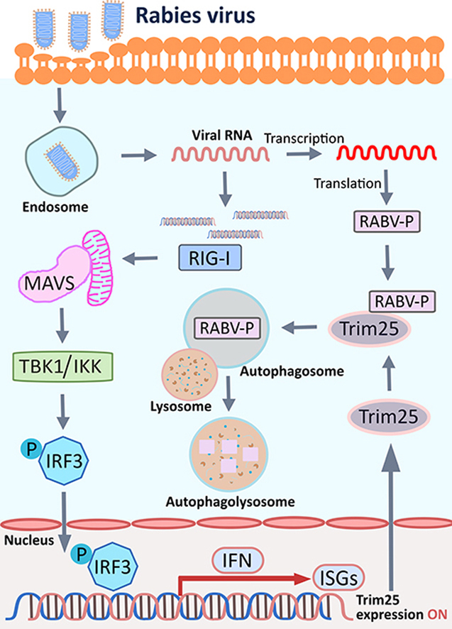
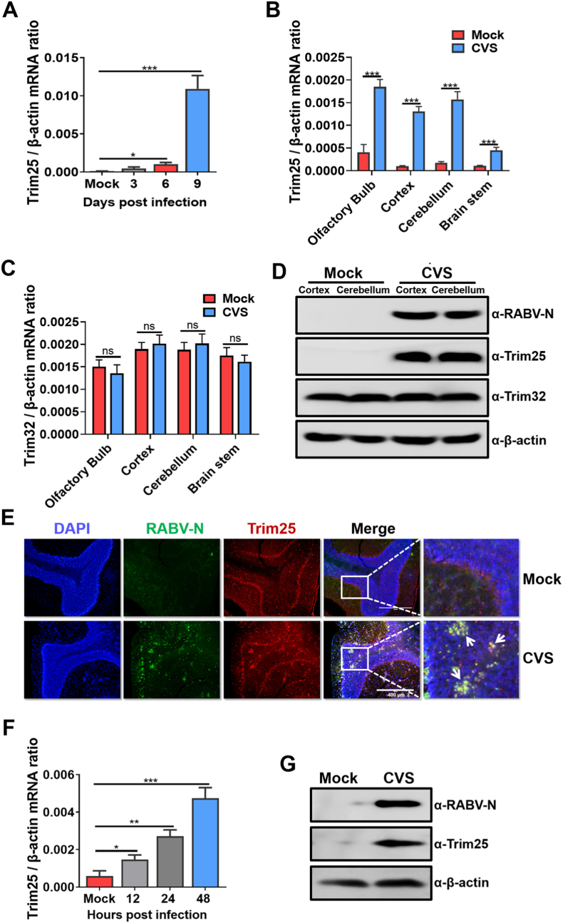
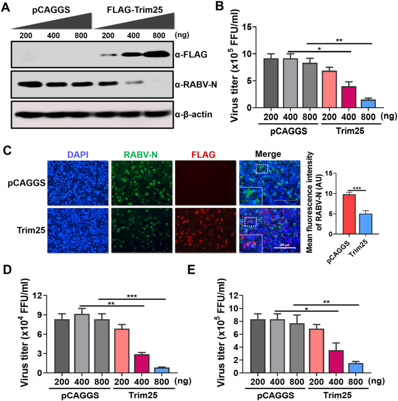
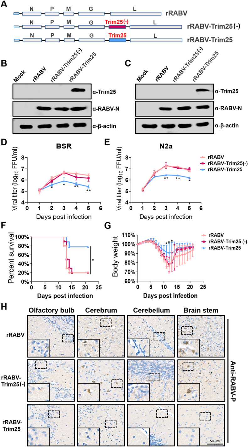
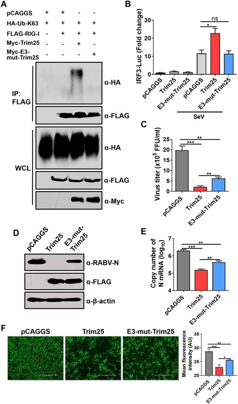
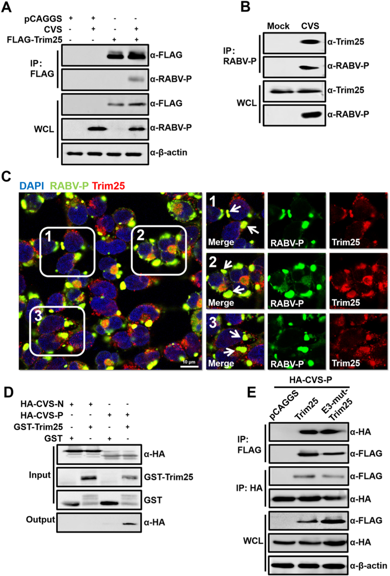
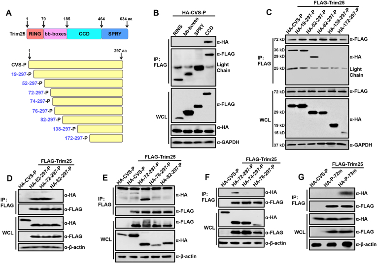
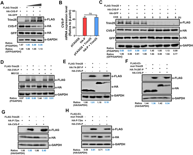
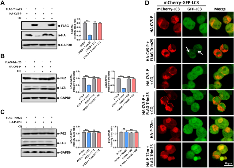
Tripartite motif-containing protein 25 (Trim25) is an E3 ubiquitin ligase that activates retinoid acid-inducible gene I (RIG-I) and promotes the antiviral interferon response. Recent studies have shown that Trim25 can bind and degrade viral proteins, suggesting a different mechanism of Trim25 on its antiviral effects. In this study, Trim25 expression was upregulated in cells and mouse brains after rabies virus (RABV) infection. Moreover, expression of Trim25 limited RABV replication in cultured cells. Overexpression of Trim25 caused attenuated viral pathogenicity in a mouse model that was intramuscularly injected with RABV. Further experiments confirmed that Trim25 inhibited RABV replication via two different mechanisms: an E3 ubiquitin ligase-dependent mechanism and an E3 ubiquitin ligase-independent mechanism. Specifically, the CCD domain of Trim25 interacted with RABV phosphoprotein (RABV-P) at amino acid (AA) position at 72 and impaired the stability of RABV-P via complete autophagy. This study reveals a novel mechanism by which Trim25 restricts RABV replication by destabilizing RABV-P, which is independent of its E3 ubiquitin ligase activity.

Keywords: Autophagy; Phosphoprotein; Protein stability; Rabies virus; Trim25.


Figures

Image 1
Graphical abstract
Fig. 1
Fig. 1
Trim25 expression is upregulated after RABV infection. C57BL/6 mice (n = 3) were intracerebrally (i.c.) inoculated with a lab-attenuated RABV strain CVS-B2c (CVS) at 100 FFU or the same volume of DMEM (mock). At 3-, 6-, and 9-days post-infection (d.p.i.), mouse brains were harvested and used for further analysis. (A) Total RNA was isolated from brain tissues, and the mRNA level of Trim25 was analyzed by qPCR. (B) Total RNA from different brain sections (harvested at 9 d.p.i.) was isolated, and the mRNA level of Trim25 was analyzed by qPCR. (C) Total RNA from different brain sections (harvested at 9 d.p.i.) was isolated, and the mRNA level of Trim32 was analyzed by qPCR. (D) The protein levels of Trim25, Trim32, and RABV-N in the cortex and cerebellum of mouse brains harvested at 9 d.p.i. were assessed by Western blotting, and β-actin was used as the control. (E) Mouse cerebellums harvested at 9 d.p.i. were fixed and sectioned. In situ Trim25 expression was analyzed by immunofluorescence assay (IFA). Scale bar, 400 μm. (F) BV2 cells were infected with CVS at an MOI of 1 for the indicated time points, and the Trim25 mRNA levels were measured using qPCR. (G) At 48 h post-RABV infection, BV2 cells were harvested, and the Trim25 protein levels were measured by Western blotting. Error bars represent standard deviation (SD), n = 3. Statistical differences between virus-infected cells and mock infected cells were determined by using Student’s t-test and are denoted as follows: ∗, P ​< ​0.05; ∗∗, P ​< ​0.01; ∗∗∗, P ​< ​0.001. Western blotting data are representative of at least two independent experiments.
Fig. 2
Fig. 2
Trim25 restrains RABV infection in 293T cells. (A–C) 293T cells were transfected with the expression vector pCAGGS or pCAGGS to express the FLAG-Trim25 (FLAG-Trim25) at the indicated volumes. The cells were infected with CVS at an MOI of 0.01 at 12 h post-transfection. After 36 h incubation, the cells were harvested to measure the protein levels of Trim25, RABV-N, and β-actin by Western blotting (A). The supernatants were harvested for virus titration (B). The same treated 293T cells were fixed and stained to measure RABV-N and Trim25 expression using IFA. The merged pictures are shown in the right panels, and the enlarged pictures are placed inside the white dotted box (C). Scale bar, 200 μm. (D, E) 293T cells were transfected with the expression vector pCAGGS or pCAGGS to express FLAG-Trim25 (FLAG-Trim25) at the indicated volumes. At 12 h post-transfection, the cells were infected with a wildtype RABV strain DRV (D) or SHBRV (E) at an MOI of 0.01. The supernatants were harvested for virus titration. Statistical analysis of comparisons between groups was carried out by Student’s t-test (∗, P ​< ​0.05; ∗∗, P ​< ​0.01; ∗∗∗, P ​< ​0.001). The bar graph shows the mean ​± ​SD, n ​= ​3. Western blotting data are representative of at least two independent experiments. See also Fig. S1.
Fig. 3
Fig. 3
Trim25 attenuates RABV pathogenicity in mice. (A) Schematic diagram of the construction of rRABV, rRABV-Trim25(−), and rRABV-Trim25. (B, C) BSR (B) and N2a (C) cells were infected with rRABV, rRABV-Trim25(−), or rRABV-Trim25 at an MOI of 0.01 for 48 h, and cells were collected for measurement of the protein levels of Trim25, RABV-P, and β-actin by Western blotting. (D, E) BSR (D) and N2a (E) cells were infected with rRABV, rRABV-Trim25(−), or rRABV-Trim25 at an MOI of 0.01 for the indicated time points, and the cell culture supernatants were harvested for virus titration. (F, G) Female C57BL/6 mice (n = 10) were intramuscularly (i.m.) infected with 8 × 105 FFU rRABV, rRABV-Trim25(−), or rRABV-Trim25. The survival ratio (F) and body weight change (G) was monitored daily for 21 continuous days (mean ​± ​SD; ∗, P ​< ​0.05; survival ratio was analyzed by Log-rank test). (H) Female C57BL/6 mice (n = 3) were inoculated as in (F). At 12 d.p.i., the mouse brains were harvested, prepared as paraffin sections, and analyzed by immunohistochemical staining with antibodies against RABV-P. Scale bar, 50 μm. Statistical analysis of comparisons between groups was carried out by Student’s t-test (∗, P ​< ​0.05; ∗∗, P ​< ​0.01; ∗∗∗, P ​< ​0.001). The bar graph shows the mean ​± ​SD, n ​= ​3.
Fig. 4
Fig. 4
Trim25 lacking its RIG-I activation ability, still inhibits RABV replication. (A) 293T cells were co-transfected with the expression vector pCAGGS or pCAGGS to express the Myc-Trim25 (Myc-Trim25) or with the expression, vector pCAGGS to express the Myc-E3-mut-Trim25 (Myc-E3-mut-Trim25). With HA-ubiquitin-K63 (HA-Ub-K63), FLAG-RIG-I for 48 h, Co-immunoprecipitation (Co-IP) was then performed using magnetic beads conjugated with antibodies against the FLAG-tag. The expression of the indicated proteins was measured by Western blotting (B) 293T cells were co-transfected with the vector plasmid or plasmids encoding Trim25 or its mutant, along with the IRF3 reporter plasmid and pRL-TK. The cells were mock infected or infected with Sendai virus (SeV) at 24 h post-transfection (h.p.t.); after infection for 16 h, the luciferase activities were measured. (C–F) 293T cells were transfected with Trim25 or their mutant and then infected with CVS at an MOI of 0.01 at 12 h.p.t. After 36 h of incubation, the cell culture supernatants were harvested for virus titration (C). The cells were either harvested for Western blotting (D) and qPCR (E) or fixed for measurement of RABV-P expression by using IFA. Scale bar, 200 μm. (F). The mean fluorescence intensity of RABV-P in the respective groups was calculated using ImageJ software (F). Data are presented as mean ± SD, n = 3. Western blotting data are representative of at least two independent experiments. See also Fig. S2.
Fig. 5
Fig. 5
Trim25 directly interacts with RABV-P. (A) 293T cells were transfected with the plasmid vector pCAGGS or FLAG-Trim25. The cells were infected with CVS at an MOI of 1 at 12 h.p.t. and incubated for an additional 36 h. Then, the cells were lysed with NP-40 lysis buffer. Co-immunoprecipitation (Co-IP) was then performed using magnetic beads conjugated with antibodies against the FLAG-tag. Western blotting was applied to measure the expression of the indicated proteins. (B) 293T cells were infected with CVS at an MOI of 1 for 36 h, the cells were lysed, and IP was performed using magnetic beads conjugated with antibodies against the RABV-P protein. The expression of the indicated proteins was measured by Western blotting. (C) 293T cells were infected with CVS at an MOI of 1 for 36 h, fixed with 4% paraformaldehyde, stained with antibodies against Trim25, RABV-P, or DAPI, and observed under confocal fluorescence microscopy. The white arrow indicates the co-localization of Trim25 and RABV-P. Scale bar, 10 μm. (D) GST or GST-Trim25 was expressed in E. coli, and purified lysates containing GST or GST-Trim25 were incubated with HA-CVS-P or HA-CVS-N (expressed in 293T cells) to assess binding. (E) HA-CVS-P was expressed alone or co-expressed with FLAG-Trim25 or FLAG-E3-mut-Trim25 in 293T cells. Cell lysates were subjected to Co-IP and analyzed by Western blotting. Western blotting data are representative of at least two independent experiments. See also Fig. S3.
Fig. 6
Fig. 6
Amino acid 72 of RABV-P is critical for its interaction with Trim25. (A) Schematic diagram of different domains of Trim25 and different HA-CVS-P truncations. (B) 293T cells were co-transfected with HA-CVS-P, and different truncations of Trim25 (RING, bb-boxes, CCD, SPRY domains) fused with a FLAG-tag. At 48 h.p.t., cell lysates were subjected to Co-IP and analyzed by Western blotting. (C–F) 293T cells were co-transfected with FLAG-Trim25, HA-CVS-P (HA-P), or pCAGGS expressing different HA-CVS-P truncations. At 48 h.p.t., Co-IP and Western blotting were performed with the indicated antibodies. (G) 293T cells were co-transfected with FLAG-Trim25 or pCAGGS and HA-CVS-P, HA-P-72m, or HA-P-73m. At 48 h.p.t., Co-IP and Western blotting were performed with the indicated antibodies. Western blotting data are representative of at least two independent experiments. See also Fig. S4.
Fig. 7
Fig. 7
Trim25 disrupts the stability of RABV-P. (A) 293T cells were co-transfected with FLAG-Trim25 (at the indicated volumes), HA-CVS-P, and HA-GFP for 48 h, and cell lysates were analyzed by Western blotting. (B) 293T cells were transfected with pCAGGS, HA-CVS-P, FLAG-Trim25 with pCAGGS, or FLAG-Trim25 with HA-CVS-P for 36 h. Then, cellular RNA was extracted, and CVS-P mRNA levels were measured. (C) 293T cells were co-transfected with FLAG-Trim25, HA-CVS-P, and HA-GFP for 48 h, and 10 μM CHX was added to the culture medium at 0, 3, 6, and 9 h before cell lysis. The cell lysates were analyzed by Western blotting. (D) 293T cells were co-transfected with FLAG-Trim25 and HA-CVS-P for 48 h and treated with 10 μM MG132 10 h before cell lysis. The cell lysates were analyzed by Western blotting. (E, F) 293T cells were co-transfected with HA-74-297-P, HA-74-297-P with FLAG-Trim25 (E) or FLAG-E3-mut-Trim25 (F), HA-CVS-P, and HA-CVS-P with FLAG-Trim25 (E) or FLAG-E3-mut-Trim25 (G) for 48 h. Then, cell lysates were analyzed by Western blotting. (G, H) 293T cells were co-transfected with HA-72m-P, HA-72m-P with FLAG-Trim25 (G) or FLAG-E3-mut-Trim25 (H), HA-CVS-P, and HA- CVS-P with FLAG-Trim25 (G) or FLAG-E3-mut-Trim25 (H) for 48 h. Then, cell lysates were analyzed by Western blotting. Western blotting data are representative of at least two independent experiments. See also Fig. S5.
Fig. 8
Fig. 8
Trim25 mediated P degradation through autophagy. (A) 293T cells were co-transfected with FLAG-Trim25 and HA-CVS-P for 48 h and treated with 1 μM CQ 24 h before cell lysis. The cell lysates were analyzed by Western blotting. (B) 293T cells were co-transfected with FLAG-Trim25 and HA-CVS-P for 48 h and treated with 1 μM CQ 24 h before cell lysis; cell lysates were analyzed by Western blotting. (C) 293T cells were co-transfected with FLAG-Trim25 and HA-P-72m for 48 h and treated with 1 μM CQ 24 h before cell lysis; cell lysates were analyzed by Western blotting. (D) 293T cells were co-transfected with mCherry-GFP-LC3 and HA-CVS-P, or FLAG-Trim25, or HA-P-72m for 36 h and then analyzed for the co-localization of mCherry with GFP. The white arrow indicates the fluorescence reduction of GFP-LC3. Scale bar, 10 μm. Data are presented as mean ± SD, n = 3. Western blotting data are representative of at least three independent experiments.

References

    1. Atkinson S.C., Heaton S.M., Audsley M.D., Kleifeld O., Borg N.A. TRIM25 and DEAD-box RNA helicase DDX3X cooperate to regulate RIG-I-mediated antiviral immunity. Int. J. Mol. Sci. 2021;22 - PMC - PubMed
    1. Brzózka K., Finke S., Conzelmann K.K. Identification of the rabies virus alpha/beta interferon antagonist: phosphoprotein P interferes with phosphorylation of interferon regulatory factor 3. J. Virol. 2005;79:7673–7681. - PMC - PubMed
    1. Brzózka K., Finke S., Conzelmann K.K. Inhibition of interferon signaling by rabies virus phosphoprotein P: activation-dependent binding of STAT1 and STAT2. J. Virol. 2006;80:2675–2683. - PMC - PubMed
    1. Carthagena L., Bergamaschi A., Luna J.M., David A., Uchil P.D., Margottin-Goguet F., Mothes W., Hazan U., Transy C., Pancino G., et al. Human TRIM gene expression in response to interferons. PLoS One. 2009;4 - PMC - PubMed
    1. Castello A., Fischer B., Eichelbaum K., Horos R., Beckmann B.M., Strein C., Davey N.E., Humphreys D.T., Preiss T., Steinmetz L.M., et al. Insights into RNA biology from an atlas of mammalian mRNA-binding proteins. Cell. 2012;149:1393–1406. - PubMed

LinkOut - more resources