

Addressing uncertainties in correlative imaging of exogenous particles with the tissue microanatomy with synchronous imaging strategies
- PMID: 37193667
- PMCID: PMC10243979
- DOI: 10.1093/mtomcs/mfad030
Addressing uncertainties in correlative imaging of exogenous particles with the tissue microanatomy with synchronous imaging strategies
Abstract
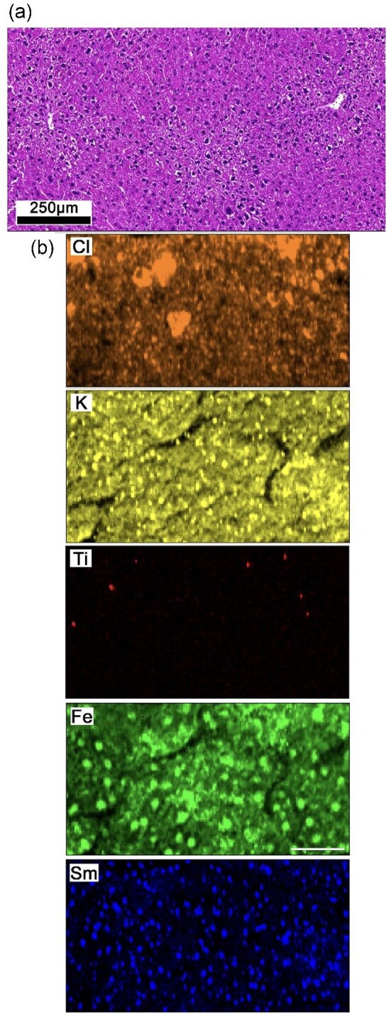
Exposure to exogenous particles is of increasing concern to human health. Characterizing the concentrations, chemical species, distribution, and involvement of the stimulus with the tissue microanatomy is essential in understanding the associated biological response. However, no single imaging technique can interrogate all these features at once, which confounds and limits correlative analyses. Developments of synchronous imaging strategies, allowing multiple features to be identified simultaneously, are essential to assess spatial relationships between these key features with greater confidence. Here, we present data to first highlight complications of correlative analysis between the tissue microanatomy and elemental composition associated with imaging serial tissue sections. This is achieved by assessing both the cellular and elemental distributions in three-dimensional space using optical microscopy on serial sections and confocal X-ray fluorescence spectroscopy on bulk samples, respectively. We propose a new imaging strategy using lanthanide-tagged antibodies with X-ray fluorescence spectroscopy. Using simulations, a series of lanthanide tags were identified as candidate labels for scenarios where tissue sections are imaged. The feasibility and value of the proposed approach are shown where an exposure of Ti was identified concurrently with CD45 positive cells at sub-cellular resolutions. Significant heterogeneity in the distribution of exogenous particles and cells can be present between immediately adjacent serial sections showing a clear need of synchronous imaging methods. The proposed approach enables elemental compositions to be correlated with the tissue microanatomy in a highly multiplexed and non-destructive manner at high spatial resolutions with the opportunity for subsequent guided analysis.
Keywords: correlative imaging; endogenous elemental imaging; exogenous metal imaging; lanthanide X-ray fluorescence; metal-labelled antibodies; synchrotron X-ray fluorescence spectroscopy; synchrotron confocal X-ray fluorescence spectroscopy.
© The Author(s) 2023. Published by Oxford University Press.
Conflict of interest statement
The authors declare that the research was conducted in the absence of any commercial or financial relationships that could be construed as a potential conflict of interest.
Figures


References
-
- Jalava P. I., Aakko-Saksa P., Murtonen T., Happo M. S., Markkanen A., Yli-Pirilä P., Hakulinen P., Hillamo P., Maki-Paakkanen J., Salonen R. O., Jokiniemi J., Toxicological properties of emission particles from heavy duty engines powered by conventional and bio-based diesel fuels and compressed natural gas, Part. Fibre Toxicol., 2012, 9 (1), 1–14. - PMC - PubMed
-
- Navel A., Uzu G., Spadini L., Sobanska S., Martins J. M., Combining microscopy with spectroscopic and chemical methods for tracing the origin of atmospheric fallouts from mining sites, J. Hazard. Mater., 2015, 30, 538–545. - PubMed
Publication types
MeSH terms
Substances
LinkOut - more resources
Full Text Sources
Research Materials
Miscellaneous
