. 2023 Oct;94(4):1355-1364.
doi: 10.1038/s41390-023-02652-9. Epub 2023 May 16.

FGF21 modulates hippocampal cold-shock proteins and CA2-subregion proteins in neonatal mice with hypoxia-ischemia

Affiliations

FGF21 modulates hippocampal cold-shock proteins and CA2-subregion proteins in neonatal mice with hypoxia-ischemia

Jeremy R Herrmann et al. Pediatr Res. 2023 Oct.

Abstract

Background: Fibroblast growth factor 21 (FGF21) is a neuroprotectant with cognitive enhancing effects but with poorly characterized mechanism(s) of action, particularly in females. Prior studies suggest that FGF21 may regulate cold-shock proteins (CSPs) and CA2-marker proteins in the hippocampus but empirical evidence is lacking.

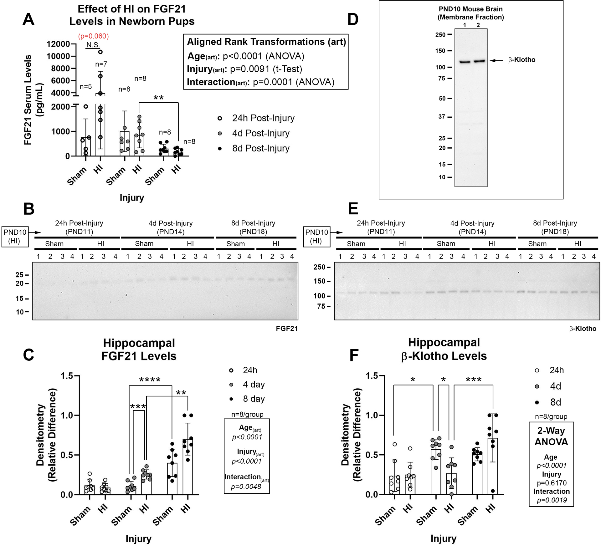
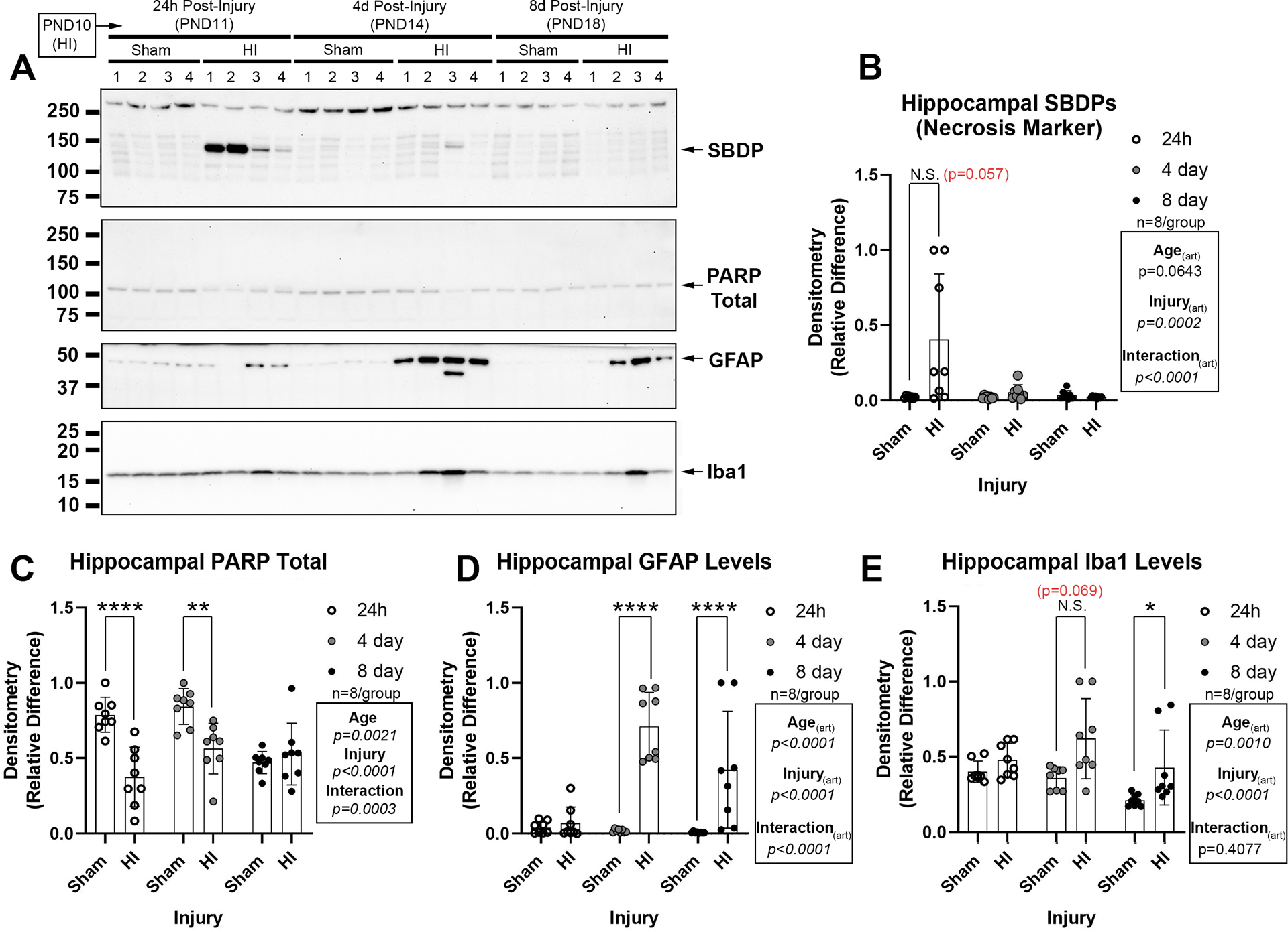
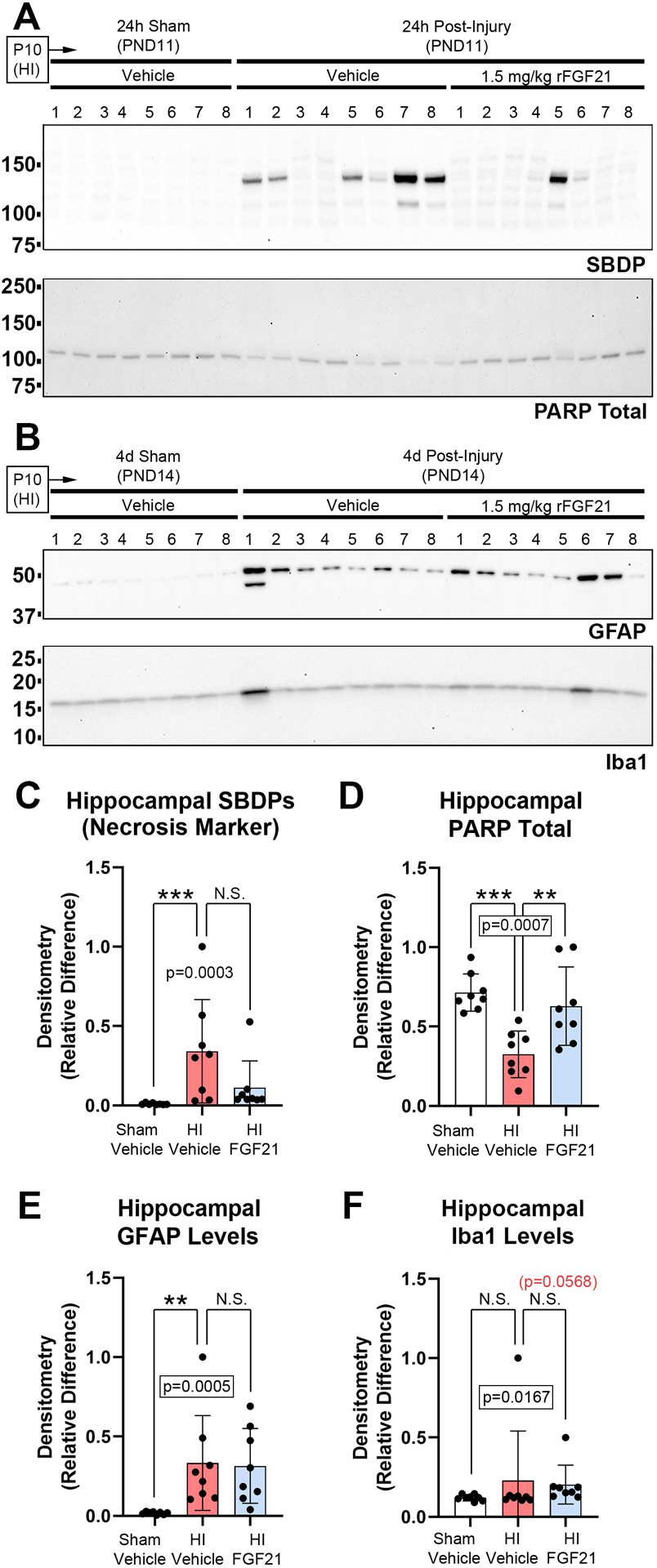
Methods: We assessed in normothermic postnatal day (PND) 10 female mice, if hypoxic-ischemic (HI) brain injury (25 min 8% O2/92% N2) altered endogenous levels of FGF21 in serum or in the hippocampus, or its receptor β-klotho. We also tested if systemic administration of FGF21 (1.5 mg/kg) modulated hippocampal CSPs or CA2 proteins. Finally, we measured if FGF21 therapy altered markers of acute hippocampal injury.

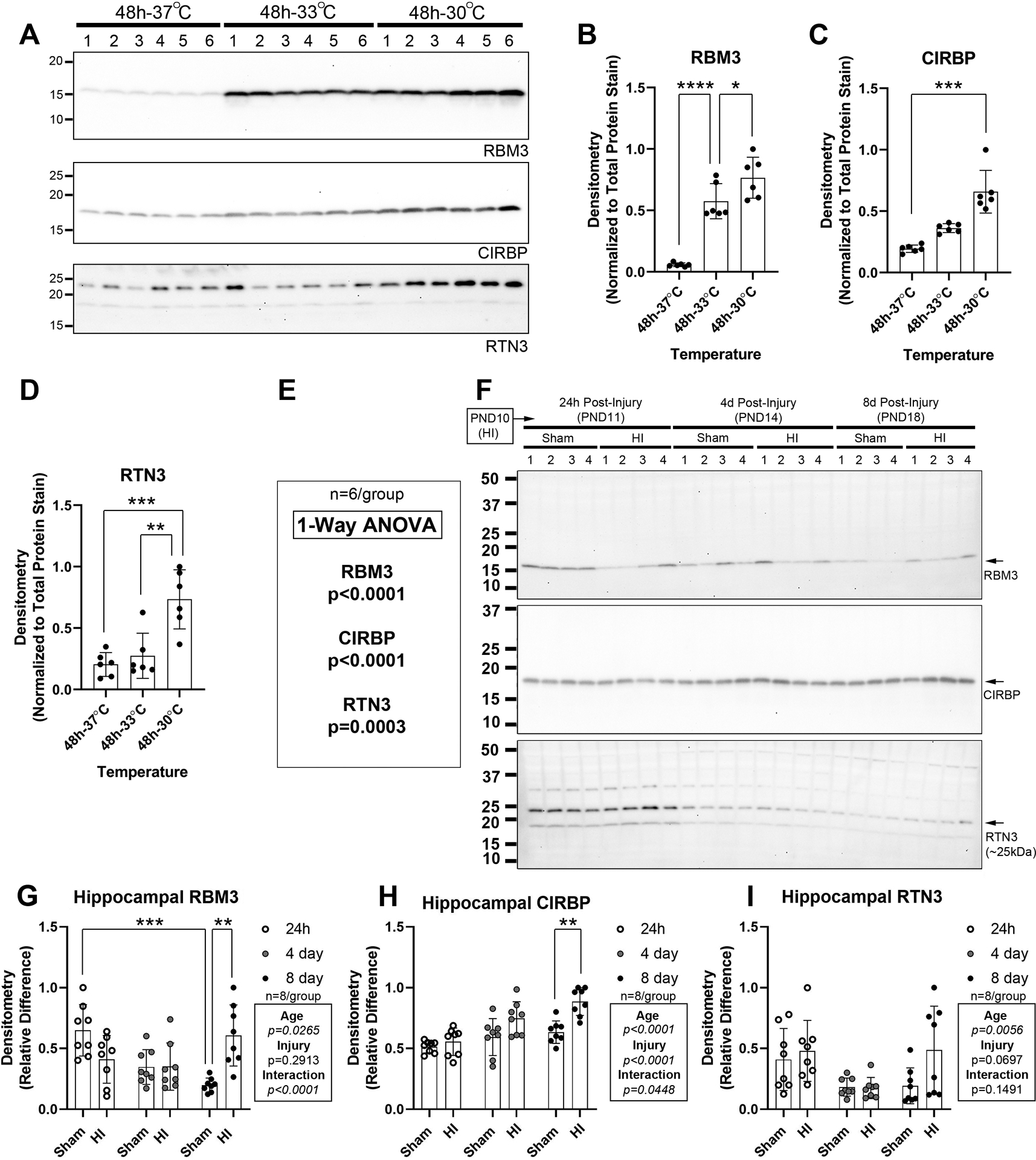
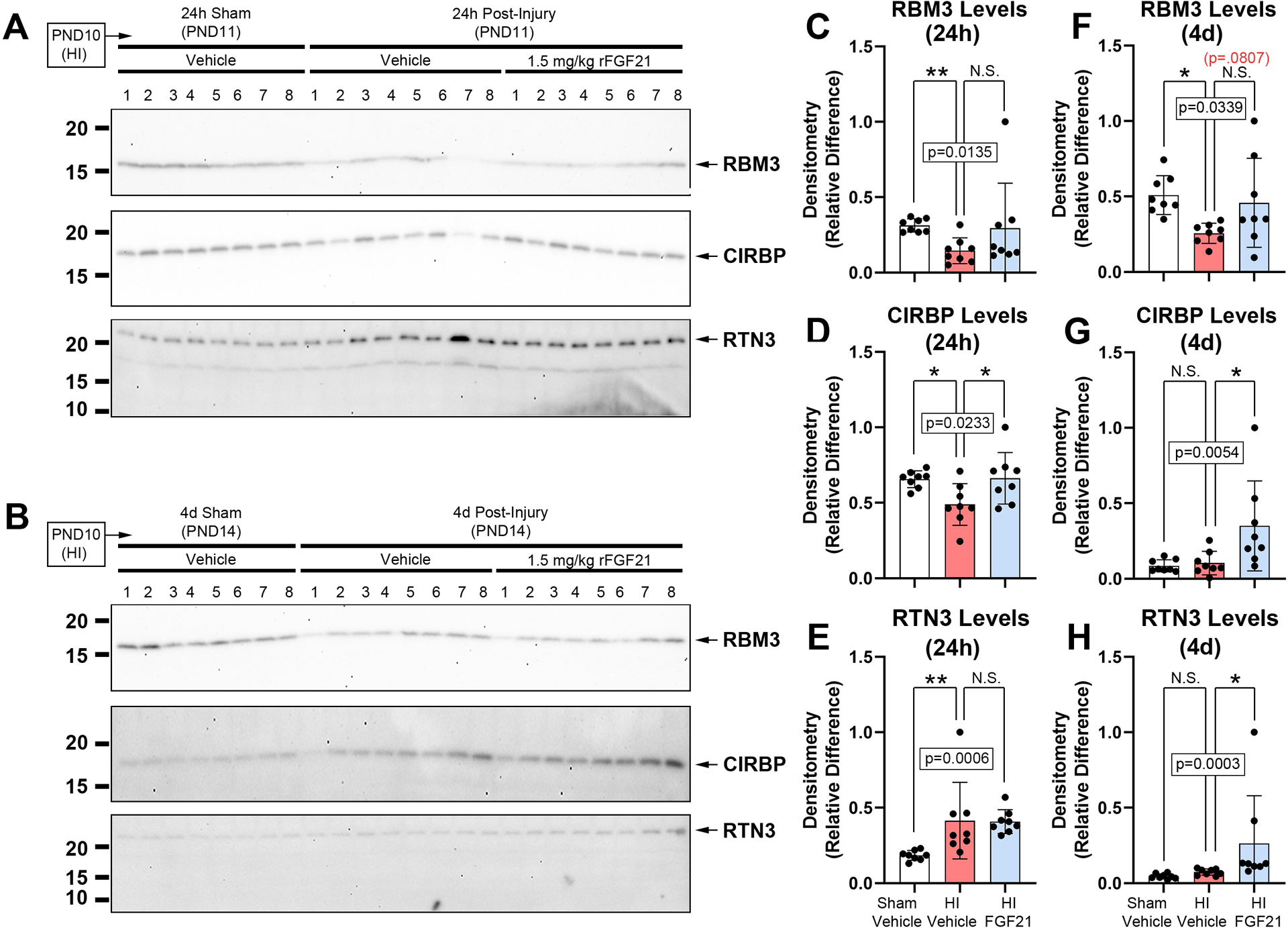
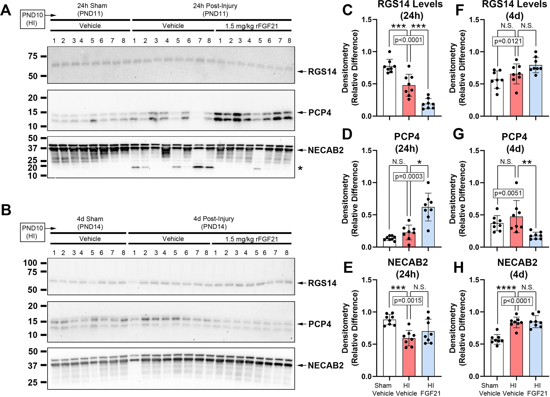
Results: HI increased endogenous serum FGF21 (24 h), hippocampal tissue FGF21 (4d), and decreased hippocampal β-klotho levels (4d). Exogenous FGF21 therapy modulated hippocampal CSP levels, and dynamically altered hippocampal CA2 marker expression (24 h and 4d). Finally, FGF21 ameliorated neuronal damage markers at 24 h but did not affect GFAP (astrogliosis) or Iba1 (microgliosis) levels at 4d.

Conclusions: FGF21 therapy modulates CSP and CA2 protein levels in the injured hippocampus. These proteins serve different biological functions, but our findings suggest that FGF21 administration modulates them in a homeostatic manner after HI.

Impact: Hypoxic-ischemic (HI) injury in female post-natal day (PND) 10 mice decreases hippocampal RNA binding motif 3 (RBM3) levels in the normothermic newborn brain. HI injury in normothermic newborn female mice alters serum and hippocampal fibroblast growth factor 21 (FGF21) levels 24 h post-injury. HI injury in normothermic newborn female mice alters hippocampal levels of N-terminal EF-hand calcium binding protein 2 (NECAB2) in a time-dependent manner. Exogenous FGF21 therapy ameliorates the HI-mediated loss of hippocampal cold-induced RNA-binding protein (CIRBP). Exogenous FGF21 therapy modulates hippocampal levels of CA2-marker proteins after HI.


Figures

Fig. 1:
Fig. 1:. The Effect of HI on Endogenous FGF21 and Hippocampal β-Klotho Levels.
(A) Serum FGF21 levels in pups at 24h- (n=5/group sham and n=7/group HI), 4d- (n=8/group sham and n=8/group HI), and 8d-post-injury (n=8/group sham and n=8/group HI). (B) Representative Western blot (n=4/group) of hippocampal FGF21 in shams vs. HI-injured pups 24h-, 4d-, and 8d-post-injury. (C) Densitometry of FGF21 (n=8/group). (D) Specificity of β-klotho antibody to detect a ~120 kDa band in membrane-enriched protein fractions from neonatal brain. (E) Representative Western blot (n=4/group) of hippocampal β-klotho in shams vs. HI-injured pups 24h-, 4d-, and 8d-post-injury. (F) Densitometry of β-klotho (n=8/group). Images of total protein stains used to normalize protein loading are available in the supplementary. Data were significant at p <0.05. Asterisks in graphs indicate post hoc significance. (*) p<0.05, (**) p<0.01, (***) p<0.001, (****) p<0.0001, (N.S.) = non-significant. Red highlighted p-values correspond to a N.S. trend on post-hoc.
Fig. 2:
Fig. 2:. Validation of CSP detecting Antibodies and the Effect of HI on Neurodevelopmental Expression of Hippocampal CSP Proteins.
Hypothermia experiments in human neuronal SHSY5Y cells were done to validate antibody reagent fidelity to detect CSPs. (A) Western blots (n=6/group) show RNA-binding motif 3 (RBM3), cold-inducible RNA-binding protein (CIRBP), and reticulon 3 (RTN3) levels after 48h at 37°C, 33°C, or 30°C. (B-E) Densitometry of CSPs (n=6/group). (F) Representative Western blot (n=4/group) of hippocampal RBM3, CIRBP, and RTN3 levels in shams vs. HI-injured pups 24h-, 4d-, and 8d-post-injury. (G, H, & I) Densitometry of RBM3, CIRBP, and RTN3 levels (n=8/group). Data were significant at p <0.05. Asterisks in graphs indicate post hoc significance. (*) p<0.05, (**) p<0.01, (***) p<0.001, (****) p<0.0001.
Fig 3.
Fig 3.. The Effect of FGF21 Administration in HI-Injured Pups on Hippocampal CSPs.
Pups were administered vehicle or 1.5mg/kg FGF21 subcutaneously every 24h (maximum 4 injections) starting 5min-post-injury. (A and B) Western blot (n=8/group) of hippocampal RBM3, CIRBP, and RTN3 levels in sham-vehicle vs. HI-vehicle vs. FGF21-treated pups at 24h- and 4d post-injury, respectively. (C, D, E and F, G, H) Densitometry of RBM3, CIRBP, and RTN3 levels (n=8/group) at 24h- and 4d post-injury, respectively. Data were significant at p <0.05. Asterisks in graphs indicate post hoc significance. RNA-binding motif 3 (RBM3), cold inducible RNA-binding protein (CIRBP), reticulon 3 (RTN3). (*) p<0.05, (**) p<0.01, (N.S.) = non-significant. Red highlighted p-value corresponds to a N.S. trend on post-hoc.
Fig 4.
Fig 4.. The Effect of FGF21 Administration in HI-Injured Pups on Hippocampal CA2 Marker Proteins.
Pups were administered vehicle or 1.5mg/kg FGF21 subcutaneously every 24h (maximum 4 injections) starting 5-min post-injury. (A and B) Western blot (n=8/group) of hippocampal RGS14, PCP4, and NECAB2 levels in sham-vehicle vs. HI-vehicle vs. FGF21-treated pups at 24h- and 4d post-injury, respectively. The asterisk on the NECAB2 blot denotes a potential uncharacterized cleavage product. (C, D, E and F, G, H) Densitometry of RGS14, PCP4, and NECAB2 levels (n=8/group) at 24h- and 4d post-injury, respectively. Data were significant at p <0.05. Asterisks in graphs indicate post hoc significance. (*) p<0.05, (**) p<0.01, (***) p<0.001, (****) p<0.0001, (N.S.) = non-significant. Regulator of G-protein signaling 14 (RGS14), purkinje cell protein 4 (PCP4), N-terminal EF-hand calcium binding protein 2 (NECAB2).
Fig. 5:
Fig. 5:. Acute Brain Injury Markers in Normothermic PND10 Female Mice Subjected to HI.
(A) Representative western blot of brain injury biomarkers. (B) The necrosis marker α-II-spectrin breakdown product (SBDP) is increased in the ipsilateral hippocampus at 24h-post-injury. (C) Total levels of poly(ADP-ribose)polymerase (PARP) are decreased at 24h- and 4d-post-injury (decreased levels suggest increased cell death). (D) The astrogliosis marker glial fibrillary acidic protein (GFAP) is increased at 4d- and 8d-post-injury. (E) The microgliosis marker ionized Ca+2-binding adapter protein 1 (Iba1) is increased at 4d post-injury. Western blots are representative images (n=4/group). Graphs of densitometry comprise n=8/group. Data were significant at p <0.05. Asterisks in graphs indicate post hoc significance. (*) p<0.05, (**) p<0.01, (****) p<0.0001, (N.S.) = non-significant. Red highlighted p-value correspond to a N.S. trend on post-hoc.
Fig 6.
Fig 6.. The Effect of FGF21 Administration on Acute Brain Injury Markers.
Pups were administered vehicle or 1.5mg/kg FGF21 subcutaneously every 24h (maximum 4 injections) starting 5min-post-injury. (A) Western blot (n=8/group) of hippocampal SBDP and total PARP at 24h-post-injury. (B) Western blot (n=8/group) of GFAP and IbA1 at 4d-post injury. (C & D) Densitometry of SBDPs and total PARP at 24h-post-injury (n=8/group). (E & F) Densitometry of GFAP and IbA1 at 4d-post-injury (n=8/group). Data were significant at p <0.05. Asterisks in graphs indicate post hoc significance. (**) p<0.01, (***) p<0.001, (N.S.) = non-significant. Red highlighted p-value correspond to a N.S. trend on post-hoc.

Similar articles

Cited by

References

    1. Lawn JE et al. Every Newborn: Progress, Priorities, and Potential Beyond Survival. Lancet 384, 189–205 (2014). - PubMed
    1. Lee AC et al. Intrapartum-Related Neonatal Encephalopathy Incidence and Impairment at Regional and Global Levels for 2010 with Trends from 1990. Pediatr Res 74 Suppl 1, 50–72 (2013). - PMC - PubMed
    1. Acun C et al. Trends of Neonatal Hypoxic-Ischemic Encephalopathy Prevalence and Associated Risk Factors in the United States, 2010 to 2018. Am J Obstet Gynecol (2022). - PubMed
    1. Sandoval Karamian AG et al. Neonatal Encephalopathy: Etiologies Other Than Hypoxic-Ischemic Encephalopathy. Semin Fetal Neonatal Med 26, 101272 (2021). - PubMed
    1. Schiering IA et al. Correlation between Clinical and Histologic Findings in the Human Neonatal Hippocampus after Perinatal Asphyxia. J Neuropathol Exp Neurol 73, 324–334 (2014). - PubMed