. 2023 May 16;6(1):524.
doi: 10.1038/s42003-023-04900-4.

GDE7 produces cyclic phosphatidic acid in the ER lumen functioning as a lysophospholipid mediator

Affiliations

GDE7 produces cyclic phosphatidic acid in the ER lumen functioning as a lysophospholipid mediator

Keisuke Kitakaze et al. Commun Biol. .

Abstract

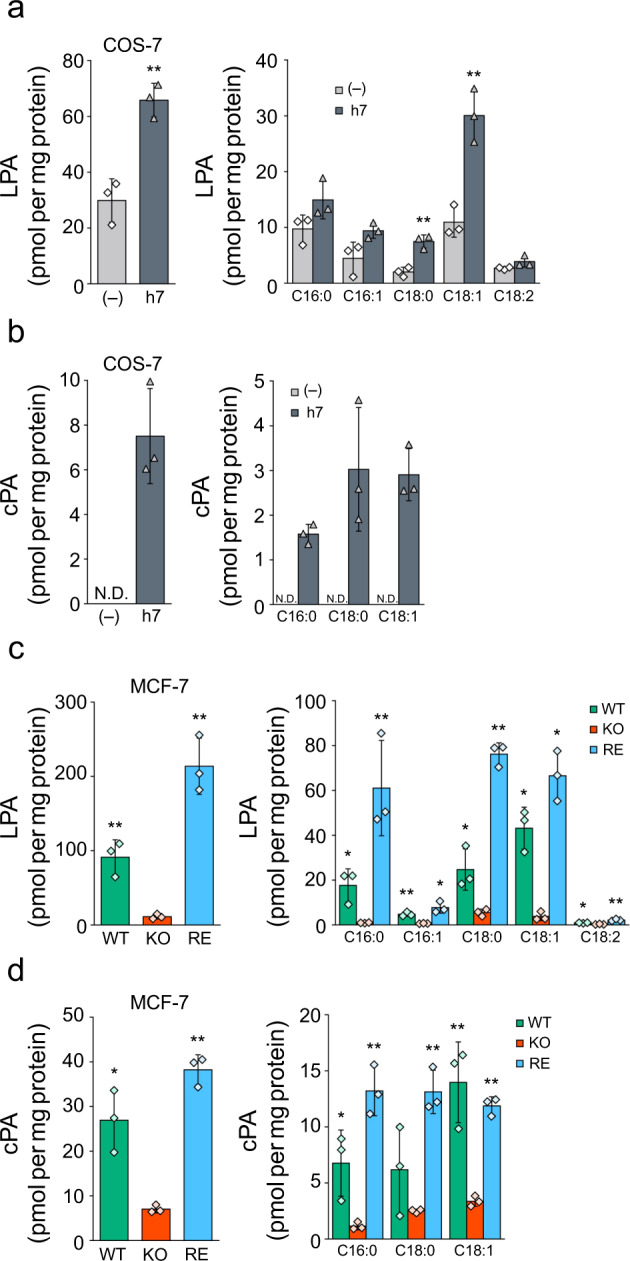
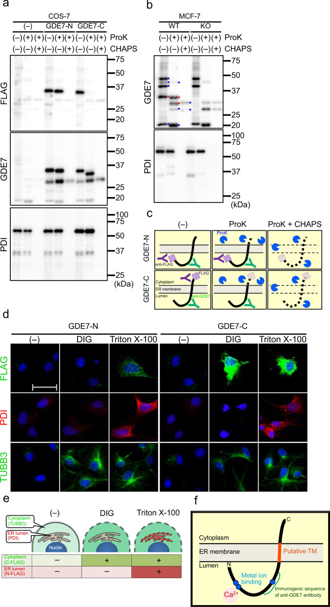
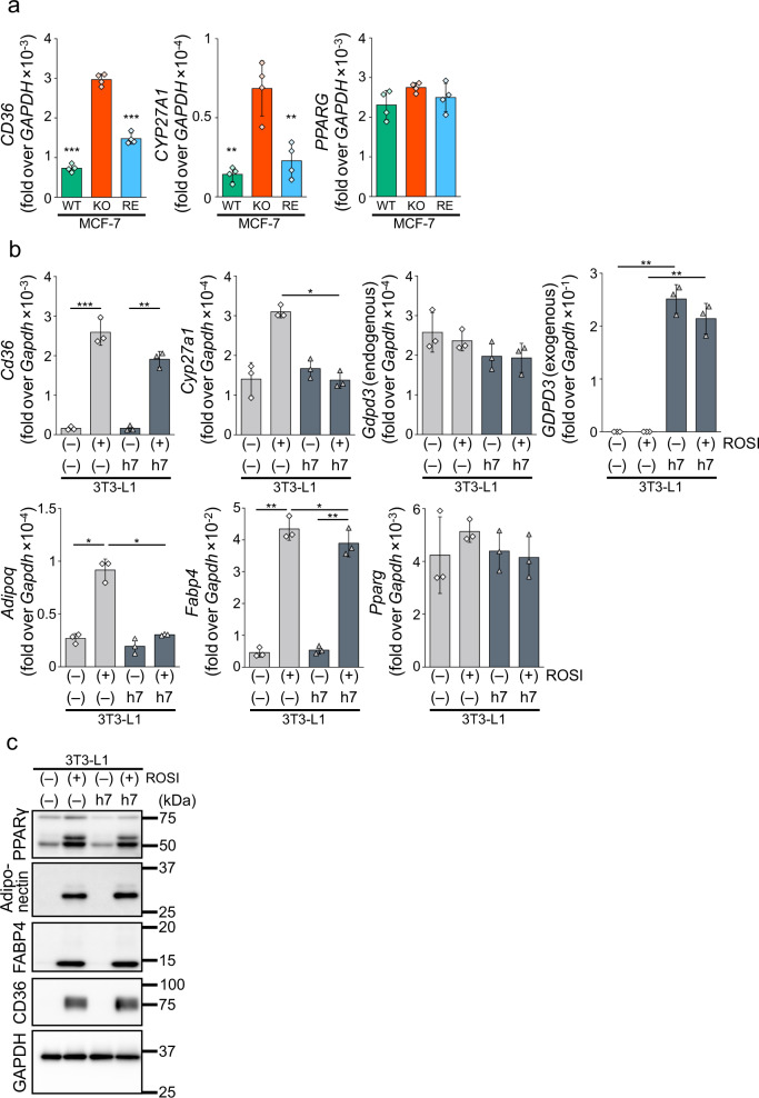
Cyclic phosphatidic acid (cPA) is a lipid mediator, which regulates adipogenic differentiation and glucose homeostasis by suppressing nuclear peroxisome proliferator-activated receptor γ (PPARγ). Glycerophosphodiesterase 7 (GDE7) is a Ca2+-dependent lysophospholipase D that localizes in the endoplasmic reticulum. Although mouse GDE7 catalyzes cPA production in a cell-free system, it is unknown whether GDE7 generates cPA in living cells. Here, we demonstrate that human GDE7 possesses cPA-producing activity in living cells as well as in a cell-free system. Furthermore, the active site of human GDE7 is directed towards the luminal side of the endoplasmic reticulum. Mutagenesis revealed that amino acid residues F227 and Y238 are important for catalytic activity. GDE7 suppresses the PPARγ pathway in human mammary MCF-7 and mouse preadipocyte 3T3-L1 cells, suggesting that cPA functions as an intracellular lipid mediator. These findings lead to a better understanding of the biological role of GDE7 and its product, cPA.


Conflict of interest statement

The authors declare the following competing interests: K.K. and Y.O. received grants from Teijin Pharma Limited, Bayer Yakuhin, Ltd., Kyowa Kirin Co., Ltd., Teijin Nakashima Medical Co., Ltd., Otsuka Pharmaceutical Co., Ltd., Asahi Kasei Pharma Corporation, EA Pharma Co., Ltd., Senko Medical Instrument Mfg. Co., Ltd., and CSL Behring K.K. H.I. is an employee of Maruho Co., Ltd. All other authors declare no conflicts of interest.

Figures

Fig. 1
Fig. 1. cPA-producing activity of human GDE7 in cell-free systems.
The membrane fractions of COS-7 cells (left graphs and panels) overexpressing mGDE7 (m7) and hGDE7 (h7) and control cells (–) were prepared. The membrane fractions were also prepared from wild-type (WT) cells and GDE7-deficient (KO) MCF-7 cells (right graphs and panels) and KO cells overexpressing mGDE7 (RE). a, e The membrane fractions were incubated with 5 µM FS-3 for 3 h at 37 °C, and FS-3-degrading activities were presented. Bars represent mean values ± S.D. (n = 3). b, f Equal amounts of membrane fraction proteins were analyzed by immunoblotting with anti-FLAG- and anti-GDE7 antibodies. Anti-GAPDH antibody was used as a loading control. Different blots were used for each antibody. c, d, g, h The membrane fractions were incubated with 25 μM 1-[14C]oleoyl LPC for 30 min at 37 °C. Radiolabeled lipids were then extracted and separated by TLC (c). The positions of the origin, LPA, LPC, cPA, and the solvent front on the TLC plates are indicated. LPA- and cPA-producing activities are shown in d and h (mean values ± S.D., n = 3 or 4). Dunnett’s test was used for analysis. *P < 0.05, **P < 0.01 (COS-7, vs. (–); MCF-7, vs. KO). All the experiments were repeated at least twice.
Fig. 2
Fig. 2. cPA-producing activity of human GDE7 in cells.
The levels of LPA (a) and cPA (b) in control (–) and hGDE7-overexpressing (h7) COS-7 cells analyzed by LC-MS/MS. The levels of LPA (c) and cPA (d) analyzed with wild-type (WT) and GDE7-deficient (KO) MCF-7 cells and KO cells overexpressing mGDE7 (RE). The graphs present the total levels (left) and the levels of each molecular species (right). Bars represent mean values ± S.D (n = 3). Log transformed data were used for Dunnett’s test analysis. *P < 0.05, **P < 0.01 (COS-7, vs. (–); MCF-7, vs. KO). N.D., not detected.
Fig. 3
Fig. 3. Prediction of TM domains and active site of hGDE7.
a The estimated TM domains, Ca2+ binding site, and immunogenic sequence of anti-GDE7 antibody were determined with the indicated TM domain prediction software. Numbers indicate the positions of amino acid residues. b, c The predicted conformation of hGDE7 is shown as a cartoon (b) or surface (c) representation. The estimated TM domains, immunogenic sequence of the anti-GDE7 antibody, Ca2+ binding site, and glycerol 3-phosphate (G3P) binding site are shown.
Fig. 4
Fig. 4. hGDE7 Topology.
a Equal amounts of particulate fraction proteins from COS-7 cells overexpressing N- or C-terminally FLAG-tagged hGDE7 (GDE7-N and GDE7-C, respectively) or the control cells (–) were treated with proK and CHAPS as indicated, and analyzed by immunoblotting using anti-FLAG-, anti-GDE7-, and anti-PDI antibodies. Different blots were used for each antibody. b Equal amounts of particulate fraction proteins from wild-type (WT) and GDE7-deficient (KO) MCF-7 cells were treated with proK, and CHAPS as indicated, and analyzed by immunoblotting using anti-GDE7- and anti-PDI antibodies. Red and blue arrowheads show GDE7-specific and nonspecific bands, respectively. Different blots were used for each antibody. c Schematic illustrations of the proK protection assay. d Representative immunofluorescence images of COS-7 cells overexpressing GDE7-N or GDE7-C using anti-FLAG-, anti-PDI-, and anti-TUBB3 antibodies. The cells were permeabilized with digitonin (DIG), Triton X-100, or without them (–). The scale bar represents 50 μm. e Schematic illustration of the permeabilization with digitonin and Triton X-100. f Schematic illustration of hGDE7 topology. All the experiments were repeated at least twice.
Fig. 5
Fig. 5. hGDE7 mutations.
a Amino acid sequences of hGDE4 (h4), hGDE7 (h7), mGDE4 (m4), and mGDE7 (m7). Sequences were aligned using the Smith-Waterman algorithm. Blue and red letters indicate hydrophilic and hydrophobic amino acids, respectively. Green asterisks indicate amino acids on the surface of the predicted GDE7 active pocket. Open boxes indicate amino acids used for mutations (F227 and Y238). be Analysis of membrane fractions from COS-7 cells overexpressing GDE7 wild-type (WT), each GDE7 mutant, and control COS-7 cells (–). b The membrane fractions were subjected to immunoblotting with anti-FLAG- and anti-GDE7 antibodies. Anti-GAPDH antibody was used as a loading control. Different blots were used for each antibody. c The membrane fractions of COS-7 cells overexpressing each mutant GDE7 were incubated with FS-3, and FS-3-degrading activities were shown. Bars represent mean values ± S.D. (n = 3). d and e The membrane fractions were incubated with 1-[14C]oleoyl LPC. Radiolabeled lipids were then extracted and separated by TLC (d). The positions of the origin, LPA, LPC, cPA, and the solvent front on the TLC plate are indicated. e LPA- and cPA-producing activities (mean values ± S.D., n = 3). The amounts of membrane fractions prepared from GDE7-expressing cells are shown in bd. For each experiment, the total amounts were adjusted to 10 µg (a and c) or 500 ng (d) by adding the membrane fraction from the control cells. Dunnett’s test was conducted for analysis. *P < 0.05, **P < 0.01, ***P < 0.001 (vs. WT). All the experiments were repeated at least twice.
Fig. 6
Fig. 6. Suppression of the PPARγ pathway by intracellular cPA.
a Wild-type (WT), GDE7-deficient (KO) MCF-7 cells, and KO cells overexpressing mGDE7 (RE) were cultured in serum-free medium for 20 h. Total RNA was isolated and analyzed by reverse transcription qPCR for mRNA levels of CD36, CYP27A1, and PPARG. Values are expressed as the ratios to the GAPDH levels (mean values ± S.D., n = 4). Dunnett’s test was conducted for analysis. **P < 0.01, ***P < 0.001 (vs. KO). b 3T3-L1 cells overexpressing hGDE7 (h7) and control 3T3-L1 cells (–) were cultured in serum-free medium with or without 1 μM rosiglitazone (ROSI) for 20 h. Total RNA was isolated and analyzed by reverse transcription qPCR for mRNA levels of Cd36, Cyp27a1, Gdpd3 (endogenous GDE7), GDPD3 (exogenous GDE7), Adipoq, Fabp4, and Pparg. Values are expressed as the ratios to the Gapdh levels (mean values ± S.D., n = 3). Tukey test was conducted for analysis. *P < 0.05, **P < 0.01, ***P < 0.001. c 3T3-L1 cells overexpressing hGDE7 (h7) and control 3T3-L1 cells (–) were treated with or without 1 μM ROSI in the presence of 10% calf serum for 1 week (medium change every other day). The cell lysates containing equal amounts of proteins were subjected to immunoblotting with anti-PPARγ, -adiponectin, -FABP4, and -CD36 antibodies. GAPDH was used as a loading control. Different blots were used for each antibody. All the experiments were repeated at least twice.
Fig. 7
Fig. 7. A proposed model of cPA production by GDE7.
GDE7 is activated by Ca2+ in the ER lumen and produces cPA which inhibits the PPARγ pathway.

Similar articles

Cited by

References

    1. Tokumura A. Metabolic pathways and physiological and pathological significances of lysolipid phosphate mediators. J. Cell. Biochem. 2004;92:869–881. doi: 10.1002/jcb.20147. - DOI - PubMed
    1. Fujiwara Y. Cyclic phosphatidic acid—a unique bioactive phospholipid. Biochim. Biophys. Acta. 2008;1781:519–524. doi: 10.1016/j.bbalip.2008.05.002. - DOI - PMC - PubMed
    1. Geraldo LHM, et al. Role of lysophosphatidic acid and its receptors in health and disease: Novel therapeutic strategies. Signal. Transduct. Target. Ther. 2021;6:45. doi: 10.1038/s41392-020-00367-5. - DOI - PMC - PubMed
    1. Kano K, Aoki J, Hla T. Lysophospholipid mediators in health and disease. Annu. Rev. Pathol. 2022;17:459–483. doi: 10.1146/annurev-pathol-050420-025929. - DOI - PMC - PubMed
    1. Baker DL, et al. Carba analogs of cyclic phosphatidic acid are selective inhibitors of autotaxin and cancer cell invasion and metastasis. J. Biol. Chem. 2006;281:22786–22793. doi: 10.1074/jbc.M512486200. - DOI - PMC - PubMed

Publication types