. 2023 May 16;13(1):7935.
doi: 10.1038/s41598-023-35020-y.

Differences in optic nerve head structure between acute angle-closure glaucoma and open-angle glaucoma

Affiliations

Differences in optic nerve head structure between acute angle-closure glaucoma and open-angle glaucoma

Jeong Han Kong et al. Sci Rep. .

Abstract

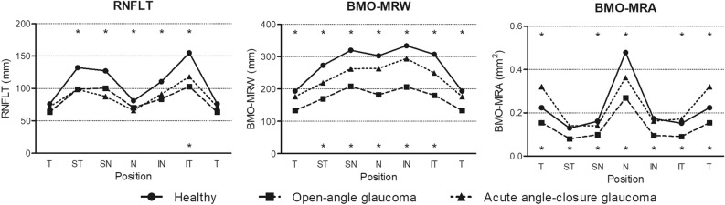
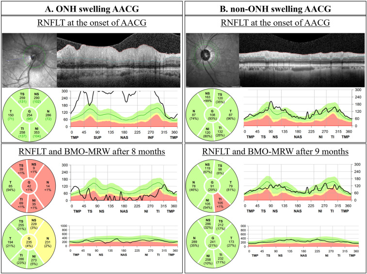
This study aimed to compare the optic nerve head (ONH) structure in acute angle-closure glaucoma (AACG) and open-angle glaucoma (OAG) to investigate the differences in glaucomatous damage. The AACG and OAG eyes were matched with regard to global retinal nerve fiber layer thickness (RNFLT). AACG eyes were divided into two subgroups based on the presence of ONH swelling at the onset of AACG. RNFLT, Bruch's membrane opening-minimum rim width (BMO-MRW), and Bruch's membrane opening-minimum rim area (BMO-MRA) were analyzed. Global RNFLT values were similar in AACG and OAG groups, but lower than in the healthy group (P < 0.001). The global BMO-MRW and total BMO-MRA were significantly higher in AACG than in OAG group (P < 0.001, respectively). AACG showed similar global BMO-MRW and total BMO-MRA, irrespective of the presence or absence of ONH swelling, while AACG with ONH swelling was associated with significantly thinner global RNFLT compared to AACG without ONH swelling (P < 0.006). The result of differences in ONH structure between the OAG and AACG, especially the AACG with ONH swelling at the onset of AACG, suggests that the mechanisms of optic nerve damage in the two diseases are different.


Conflict of interest statement

The authors declare no competing interests.

Figures

Figure 1
Figure 1
Comparison of peripapillary retinal nerve fiber layer thickness (RNFLT), Bruch’s membrane opening-minimum rim width (BMO-MRW), and Bruch’s membrane opening-minimum rim area (BMO-MRA) in each sector among the three subject groups. For the sectors marked with an asterisk above the dots, the one-way analysis of variance with Duncan’s post hoc test revealed a significant difference (P < 0.05) between the two higher values; in contrast, for the sectors marked with an asterisk below the dots, a significant difference (P < 0.05) was found between the two lower.
Figure 2
Figure 2
Comparison of peripapillary retinal nerve fiber layer (RNFLT), Bruch’s membrane opening-minimum rim width (BMO-MRW), and Bruch’s membrane opening-minimum rim area (BMO-MRA) in each sector among the two subgroups in acute angle-closure glaucoma. For the sectors marked with an asterisk above the dots, there is a significant difference (P < 0.05) in the Mann–Whitney test.
Figure 3
Figure 3
Representative cases of optic nerve head (ONH) swelling acute angle-closure glaucoma (AACG), and non-ONH swelling AACG groups. ONH swelling AACG eye, which shows retinal nerve fiber layer thickness (RNFLT) swelling at the onset of AACG, shows no difference in Bruch’s membrane opening-minimum rim width (BMO-MRW) compared to that of the non-ONH swelling AACG eye, but a relatively large decrease in RNFLT.
Figure 4
Figure 4
Optic nerve head analysis using spectral-domain optical coherence tomography. (A) Bruch’s membrane opening-minimum rim width (BMO-MRW) is defined as the shortest distance from the Bruch’s membrane opening (BMO) point to the inner limiting membrane (ILM). (B) Bruch’s membrane opening-minimum rim area is defined as the area of a trapezium at angle θ above the BMO plane.

References

    1. Casson RJ, et al. Prevalence of glaucoma in rural Myanmar: the meiktila eye study. Br. J. Ophthalmol. 2007;91:710–714. doi: 10.1136/bjo.2006.107573. - DOI - PMC - PubMed
    1. Yip LW, Yong VK, Hoh ST, Wong HT. Optical coherence tomography of optic disc swelling in acute primary angle-closure glaucoma. Arch. Ophthalmol. 2005;123:567–569. doi: 10.1001/archopht.123.4.567. - DOI - PubMed
    1. Chai CH, Sng CC, Lin HA, Chew PT. Transient optic nerve swelling after acute primary angle closure. Clin. Exp. Ophthalmol. 2013;41:611–614. doi: 10.1111/ceo.12039. - DOI - PubMed
    1. Choudhari NS, George R, Kankaria V, Sunil GT. Anterior ischemic optic neuropathy precipitated by acute primary angle closure. Indian J. Ophthalmol. 2010;58:437–440. doi: 10.4103/0301-4738.67070. - DOI - PMC - PubMed
    1. Nahum Y, Newman H, Kurtz S, Rachmiel R. Nonarteritic anterior ischemic optic neuropathy in a patient with primary acute angle-closure glaucoma. Can. J. Ophthalmol. 2008;43:723–724. doi: 10.3129/i08-149. - DOI - PubMed

Publication types