. 2023 May 16;13(1):7936.
doi: 10.1038/s41598-023-35139-y.

CTLA-4 suppresses hapten-induced contact hypersensitivity in atopic dermatitis model mice

Affiliations

CTLA-4 suppresses hapten-induced contact hypersensitivity in atopic dermatitis model mice

Hiroe Tetsu et al. Sci Rep. .

Erratum in

Abstract

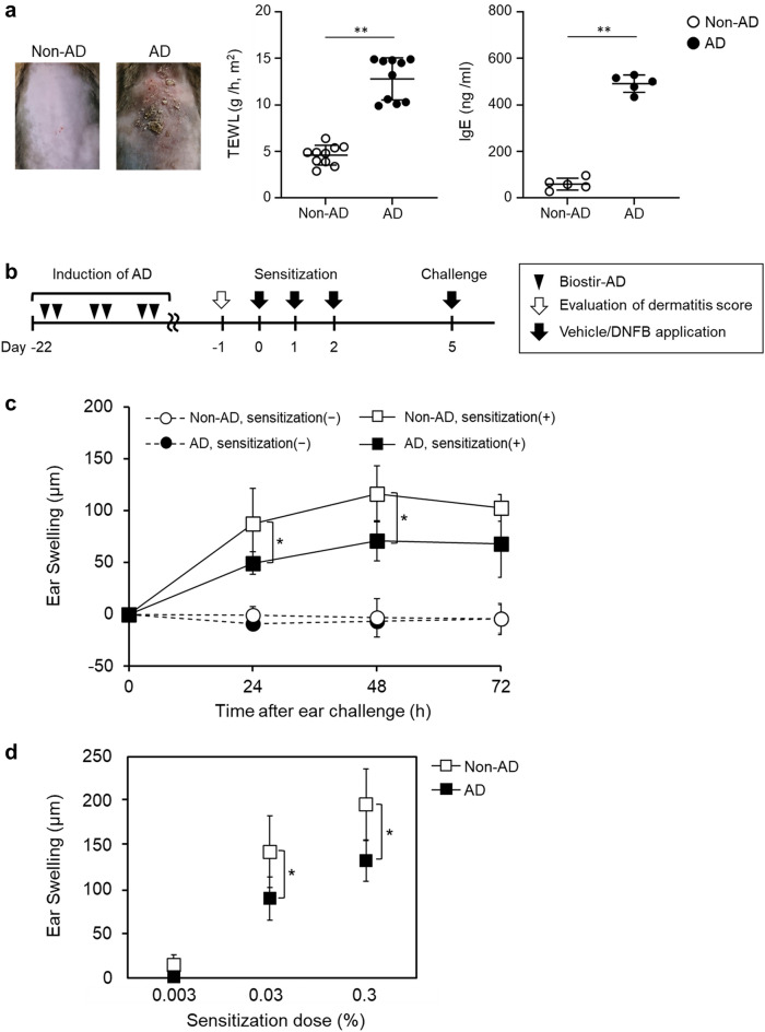
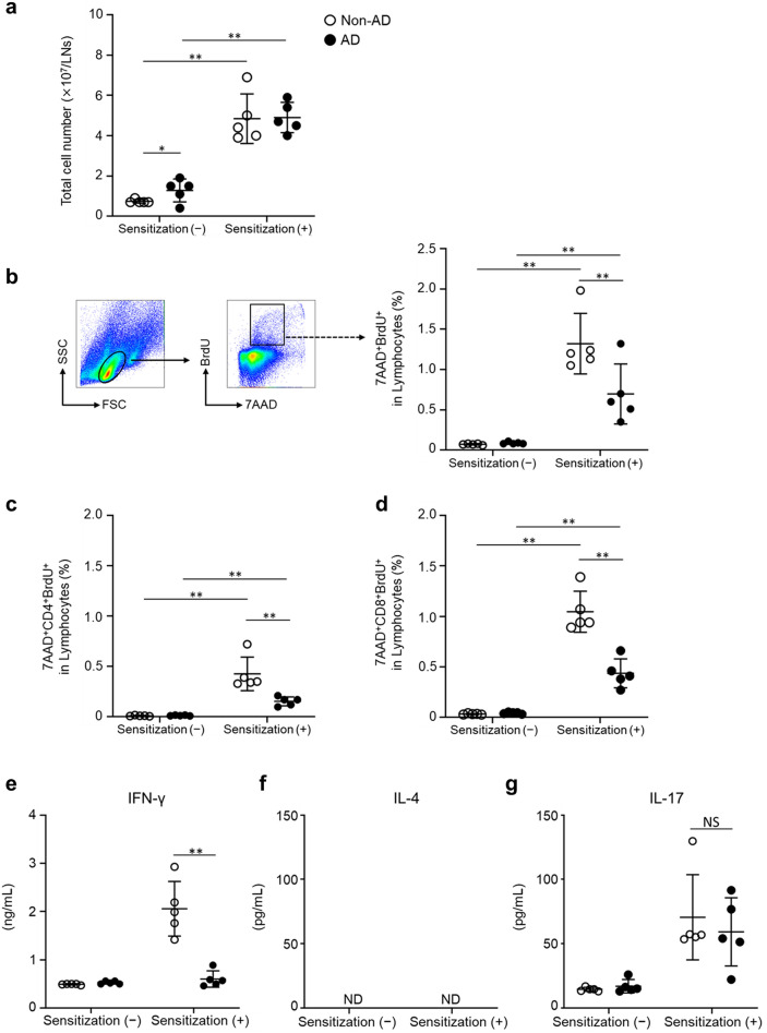
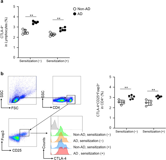
Atopic dermatitis (AD) patients with skin barrier dysfunction are considered to be at a higher risk of allergic contact dermatitis (ACD), although previous studies showed that attenuated ACD responses to strong sensitizers in AD patients compared to healthy controls. However, the mechanisms of ACD response attenuation in AD patients are unclear. Therefore, using the contact hypersensitivity (CHS) mouse model, this study explored the differences in CHS responses to hapten sensitization between NC/Nga mice with or without AD induction (i.e., non-AD and AD mice, respectively). In this study, ear swelling and hapten-specific T cell proliferation were significantly lower in AD than in non-AD mice. Moreover, we examined the T cells expressing cytotoxic T lymphocyte antigen-4 (CTLA-4), which is known to suppress T cell activation, and found a higher frequency of CTLA-4+ regulatory T cells in draining lymph node cells in AD than in non-AD mice. Furthermore, the blockade of CTLA-4 using a monoclonal antibody eliminated the difference in ear swelling between non-AD and AD mice. These findings suggested that CTLA-4+T cells may contribute to suppressing the CHS responses in AD mice.


Conflict of interest statement

The authors declare no competing interests.

Figures

Figure 1
Figure 1
Decreased CHS response induced by DNFB in AD model mice. AD-like skin lesions in NC/Nga mice were induced by Biostir-AD. (a) Features of the back skin after AD induction. Transepidermal water loss (TEWL) through dorsal skin. Total serum IgE concentrations of non-AD and AD mice were measured by ELISA. (b) CHS responses were induced, as shown in the scheme. (c) Mice were sensitized with or without 0.3% DNFB on the dorsal skin, and ear swelling was measured after the challenge. Data represents the change in ear thickness at 24, 48 and 72 h. (d) Mice were sensitized with DNFB at the following concentrations: 0.003%, 0.03%, and 0.3%. Data represents the change in ear thickness at 48 h. Data were expressed as the mean ± SD (n = 5, TEWL data; n = 10) and represented three independent experiments with similar findings. *p < 0.05, **p < 0.01 between indicated groups.
Figure 2
Figure 2
Impaired DNFB-specific T cell proliferation and cytokine production after DNFB sensitization in AD mice. Draining LNs were collected five days after sensitization of non-AD and AD mice with or without 0.3% DNFB. (a) The total number of draining LN cells. (b–g) DNBS-induced lymphocyte proliferation and cytokine production. Draining LN cells were collected from non-AD and AD mice five days after 0.3% DNFB application and cultured for two days with or without 50 µg/mL DNBS. Cell proliferation was evaluated using the BrdU assay. (b) Representative flow cytometry dot plots (left) and the frequency of 7AAD+BrdU+ cells per lymphocyte with DNBS. (c) 7AAD+CD4+BrdU+ cells, (d) 7AAD+CD8+BrdU+ cells per lymphocyte with DNBS. (e–g) The amount of IFN-γ, IL-4, and IL-17 in the culture medium was measured by ELISA. Data were expressed the mean values ± SD (n = 5) and represented two independent experiments with similar results. *p < 0.05, **p < 0.01 between indicated groups, ND; not detected, NS; not significant.
Figure 3
Figure 3
CTLA-4+Treg cells increased in AD mice. Draining LNs were collected from non-AD and AD mice one day after 0.3% DNFB sensitization. (a) CTLA-4+cells per lymphocyte (b) Representative flow cytometry histogram results (left) and the frequency of CTLA-4+CD25+FoxP3+Treg cells per CD4+. Data were expressed as the mean values ± SD (n = 5) and represented two independent experiments with similar results. *p < 0.05, **p < 0.01 between indicated groups.
Figure 4
Figure 4
Ear swelling and expression of CD86 on migratory DCs between non-AD and AD mice are comparable after treatment with anti-CTLA-4 mAb. Mice were treated with anti-CTLA-4 mAb or control anti-IgG mAb 1 day before 0.3% DNFB or 1.0% FITC sensitization. (a) Treatment of mice was performed as per the schematic. (b) Ear swelling was measured after the challenge with 0.3% DNFB. Data represents the change in ear thickness at 24 h. (c) Mice were sensitized to FITC and the expression level of CD86 on MHC class IIhighFITC+migratory DCs. Data were expressed as mean ± SD (n = 5) and represented two independent experiments with similar results. *p < 0.05, **p < 0.01 between the indicated groups.

References

    1. Beck LA, et al. Type 2 inflammation contributes to skin barrier dysfunction in atopic dermatitis. JID Innov. 2022;2:100131. doi: 10.1016/j.xjidi.2022.100131. - DOI - PMC - PubMed
    1. Vocanson M, Hennino A, Rozieres A, Poyet G, Nicolas JF. Effector and regulatory mechanisms in allergic contact dermatitis. Allergy. 2009;64:1699–1714. doi: 10.1111/j.1398-9995.2009.02082.x. - DOI - PubMed
    1. Yang G, et al. Skin barrier abnormalities and immune dysfunction in atopic dermatitis. Int. J. Mol. Sci. 2020 doi: 10.3390/ijms21082867. - DOI - PMC - PubMed
    1. Kohli N, Nedorost S. Inflamed skin predisposes to sensitization to less potent allergens. J. Am. Acad. Dermatol. 2016;75:312–317. doi: 10.1016/j.jaad.2016.03.010. - DOI - PubMed
    1. Newell L, et al. Sensitization via healthy skin programs Th2 responses in individuals with atopic dermatitis. J. Investig. Dermatol. 2013;133:2372–2380. doi: 10.1038/jid.2013.148. - DOI - PubMed